


Actinomycetes of Marine Mammals
Nocardiosis is commonly reported in debilitated marine mammals. Several species of Nocardia have been described from both captive and free-ranging marine mammals of many species.
Disease can manifest in numerous ways, including cutaneous and subcutaneous abscess formation or pyogranulomatous disease in various organs or systemically.
Diagnosis may be difficult and is often made after death due to systemic disease, although cytologic or histologic evaluation may show acid-fast, branching filamentous organisms.
Treatment is also challenging, and successful outcome likely depends on characteristics of the infection and early diagnosis. Infections due to Actinomyces or Arcanobacterium spp have also have been diagnosed in many marine mammal species. Arcanobacterium phocae has been implicated in disease in stranded California sea lions, common dolphins, gray seals, harbor seals, northern elephant seals, and sea otters. Arcanobacterium animalium has been isolated from harbour porpoise. Most cases are diagnosed after death, but infection may be underreported.
Brucellosis of Marine Mammals
Since the 1990s, previously unknown strains of Brucella have been found in both captive and free-ranging pinnipeds and cetaceans from many countries. Two species have been classified: Brucella ceti (cetaceans) and Brucella pinnipedialis (seals). There appears to be host preferences. Lesion distribution and clinical signs also appear to differ between hosts.
In cetaceans, B ceti–associated pathological findings include placentitis, orchitis, abortion, mastitis, pneumonia, subcutaneous lesions, osteomyelitis, arthritis, meningoencephalitis, meningitis, and hepatic and splenic necrosis. Pinnipeds have less Brucella-associated pathological changes by comparison, although B pinnipedialis has been associated with various organs and disease processes. Transmission may be horizontal and vertical, and as Brucella species have been isolated from lungworm and can survive in fish, ingestion may also be an important mode of transmission. Brucella ceti has also been documented in a traumatic nonhealing wound in a sea otter.
The bacteria possess the same surface antigens commonly used for diagnosis in livestock. Diagnosis is generally by culture, PCR assay, or immunohistochemical analysis.
The prevalence of marine mammal brucellosis is not known, but cases appear to be widespread given that serologic surveys have documented nearly worldwide exposure. The role of environmental factors in the emergence of marine mammal disease is unknown.
Treatment depends on the location and extent of the infection. Resolution may be very difficult and generally requires long-term antimicrobial therapy targeting this intracellular pathogen; antimicrobial resistance may develop.
There is some evidence of potential for zoonotic infections, and there have been a few cases of human infections in those with contact with tissues from marine mammals. In one case, the organism cultured from the blood of a laboratory worker matched a marine mammal strain she was working with. Three other human cases in which marine mammal–associated Brucella strains were isolated had no documented contact with marine mammals but histories of eating raw shellfish or fish and contact with raw fish bait.
Clostridial Myositis of Marine Mammals
Severe myositis due to infections with Clostridium spp has been diagnosed in captive killer whales, pilot whales, bottlenose dolphins, California sea lions, and manatees. All marine mammals are probably susceptible.
Disease is characterized by acute swelling, muscle necrosis, and accumulations of gas in affected tissues, accompanied by severe leukocytosis. It can also be found in the gastrointestinal tract and cause enterotoxemia. Untreated, it can be fatal.
Diagnosis is based on detection of gram-positive bacilli in aspirates of the lesions and is confirmed by anaerobic culture and identification of the organism. Some enterotoxins can also be detected in feces.
Treatment includes systemic and local antimicrobial therapy and surgical drainage of abscessed areas. Commercially available inactivated clostridial bacterins are used routinely in some facilities, although efficacy in marine mammals has not been studied.
Botulism has been reported in captive California sea lions during an endemic outbreak of the disease in waterfowl. Affected animals stopped eating and appeared unable to swallow several days before dying.
Erysipelas (Diamond Skin Disease) of Marine Mammals

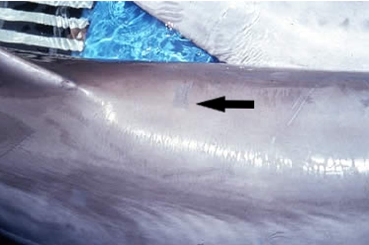
Courtesy of Dr. James McBain.
Erysipelas can be a serious infectious disease of captive cetaceans and pinnipeds and has been recognized in wild harbour porpoises, bottlenose dolphins, and harbor seals. Erysipelothrix rhusiopathiae, which causes erysipelas in pigs and other domestic species, is a common contaminant that persists in the slime layer of fish. A septicemic form of the disease in marine mammals can be peracute or acute; affected animals die suddenly either with no prodromal signs or with sudden onset of lethargy, inappetence, or fever. A cutaneous form that causes typical rhomboidal skin lesions is a more chronic form of the disease. Animals with this form usually recover with timely antimicrobial therapy.
In peracute cases, necropsy generally fails to reveal grossly discernible lesions other than widespread petechiation. Diagnosis is based on culture of the organism from the blood, spleen, or body cavities. Arthritis has been found in animals that have died with the chronic form.
Treatment of the peracute and acute forms has rarely been attempted because the absence of prodromal signs obscures the diagnosis. Animals with the dermatologic form usually recover with administration of penicillins, tetracyclines, or chloramphenicol and supportive care.
Control seems primarily related to provision of high-quality fish that is properly stored and handled. Vaccination is controversial, and vaccine breaks can occur. No marine mammal–specific vaccine exists, but vaccination with commercial swine vaccines has been performed for many years, particularly in cetaceans. Vials of killed erysipelas bacterin should be cultured for surviving organisms before use in marine mammals. Modified-live bacterins should be avoided for the initial vaccination. Fatal anaphylaxis can occur on revaccination, although this is less common with more modern swine vaccines. For this reason, some vaccination programs have been reduced to one-time administration, even though antibody titers fall below the presumed effective level.
If cetaceans are to be revaccinated, sensitivity tests should be performed by injecting a small amount of bacterin submucosally on the lower surface of the tongue. Hypersensitive animals develop swelling and redness at the injection site within 30 minutes. Because the vaccine is extremely irritating, no more than 3–5 mL should be used at any one site, even in nonsensitive mammals. A sufficiently long needle (≥5 cm [2 in.] and longer in larger cetaceans) should be used to assure that the vaccine is deposited in the muscle and not between muscle and blubber, or a sterile abscess can result. Bacterin should be administered in the dorsal musculature anterior and lateral to the dorsal fin. Administration posterior to the dorsal fin can result in a severe tissue reaction, immobilizing the animal for several days due to involvement of muscles critical for locomotion. The optimal interval between vaccines required to maintain high antibody titers is not known; however, a booster after 6 months and annual revaccination are frequently administered.
Leptospirosis of Marine Mammals
Leptospirosis is widespread and has been diagnosed in pinnipeds and polar bears.
In pinnipeds, disease is characterized by lethargy, dehydration, anorexia, signs of abdominal pain, reluctance to move, polydipsia, pyrexia, vomiting, and icterus. Ulceration of mucosal tissues and flipper edges, gastroenteritis, and pulmonary disease can also occur. It may also cause abortions and neonatal deaths in California sea lions and northern fur seals. Lesions include severe, diffuse, interstitial nephritis, with dense accumulation of spirochetes in the renal tubules. The gallbladder may contain inspissated black bile, but hepatitis may not be apparent grossly. Hyperplasia of Kupffer cells, erythrophagocytosis, and hemosiderosis are seen histologically.
Bloodwork changes indicative of renal disease are noted, including elevated concentrations of serum creatinine, BUN, phosphorus, and sodium, as well as marked leukocytosis. Dark-field microscopy and bacteriologic culture often do not yield positive diagnostic results. While PCR assay is increasingly useful, serologic testing remains the most rapid and widely used diagnostic technique. Antibodies to various Leptospira serovars (including L Canicola, L Icterohaemorrhagiae, L Autumnalis, and L Pomona) have been identified in many species, including sea otters and manatee. Leptospira interrogans serovar Pomona is associated with cyclical epizootics in free-ranging California sea lions, resulting in high morbidity and mortality. Cases can occur year-round but peak in the late summer and fall, with most cases in juvenile and subadult males.
Treatment in pinnipeds is similar to that in dogs and primarily involves supportive care, particularly parenteral fluid therapy, which can usually be administered subcutaneously. Control in captive animals requires serologic evaluation of new animals during quarantine. Captive animals can be vaccinated in endemic areas, and measures to control rodent populations should be instituted.
Leptospirosis is zoonotic, and appropriate precautions should be taken.
Streptothricosis (Dolphin Pseudopox, Cutaneous Dermatophilosis) of Marine Mammals
Streptothricosis, a subcutaneous bacterial infection with Dermatophilus congolensis, has been reported in pinnipeds and polar bears. It must be distinguished from sealpox. Simultaneous infections of streptothricosis and pox have been recorded in sea lions. Cutaneous streptothricosis can cause severe pruritic dermatitis in polar bears, which usually manifests as sharply delineated nodules distributed over the entire body and usually progresses to death.
Diagnosis is based on demonstration of the organism in biopsies or culture.
Treatment with prolonged high dosages of systemic antimicrobials can be successful.
Sporothrix schenckii, the cause of a subcutaneous mycosis, has been reported in Pacific white-sided dolphins (Laegenorhynchus obliquidens).
Mycobacteriosis of Marine Mammals
Marine mammals are susceptible to various mycobacteria. Evidence points to mycobacterial disease being possibly endemic in free-ranging otariids off the coast of Australia. Originally thought to be caused by Mycobacterium bovis, subsequent molecular assessment places the isolates from free-ranging southern hemisphere pinnipeds in a unique cluster assigned its own species in the M tuberculosis complex. Subantarctic fur seals (Arctocephalus tropicalis) are thought to be the common link in the spread of M pinnipedii to other pinniped species because they cohabit with the other known affected species: Australian sea lions (Neophoca cinerea) and New Zealand fur seals (Arctocephalus forsteri). Otherwise, mycobacteriosis has been a disease of captivity. Pinnipeds, cetaceans, and sirenians have developed disease due to M bovis, M smegmatis, M chitae, M fortuitum, M chelonae, and M marinum. Cutaneous and systemic forms are seen. There are strong indications that immunosuppression may be involved in development of infections by the atypical mycobacteria.
Intradermal testing with high concentrations of bovine or avian purified protein derivative tuberculin can be used to screen exposed animals; however, anergy occurs and the usefulness is controversial. In pinnipeds, injections in the webbing of the rear flippers should be read at 48 and 72 hours. Antibodies have been identified in seals by means of ELISA, but this requires further evaluation before it can be considered a screening test. Diagnosis is made by culture and identification of the organism from lesion biopsies, tracheal washes, or feces.
Treatment is controversial given the nature of the disease and zoonotic and cross-species infection potential, but most isolates of M pinnipedii are susceptible to isoniacide, rifampicin, streptomycin, ethambutol, and pyrazinamide.
Mycobacteriosis in marine mammals is an emerging disease and is possibly of public health significance. (Also see Tuberculosis in Various Animals.)
Mycoplasmosis of Marine Mammals
Mycoplasma spp have been isolated from the teeth, wounds, and respiratory tracts of pinnipeds. They have been associated with respiratory disease historically but can be found in healthy animals. They are frequently cultured from animals concurrently infected with respiratory viruses and are the etiologic agent of seal finger, a common zoonosis transmitted from pinnipeds to humans through bite wounds.
Miscellaneous Bacterial Diseases of Marine Mammals
Marine mammals are probably susceptible to the entire range of pathogenic bacteria. Pasteurella multocida has caused several outbreaks of hemorrhagic enteritis associated with lethargy and abdominal distress leading to acute death in dolphins and pinnipeds. It has also been reported to cause pneumonia in pinnipeds, as have Klebsiella spp. In dolphins, Mannheimia haemolytica has been incriminated in hemorrhagic tracheitis that responded to chloramphenicol therapy.
Plesiomonas shigelloides has been responsible for gastroenteritis in harbor seals. Burkholderia pseudomallei has caused serious fatal outbreaks of disease (melioidosis) in various marine mammals in captivity throughout Asia and in the South Pacific and manifests clinically in a manner similar to that of terrestrial animals with high morbidity and mortality. Salmonella spp have caused fatal gastroenteritis in manatees, beluga whales, and pinnipeds and are often isolated from the GI tract of marine mammals. Staphylococcal septicemia has caused the death of a dolphin with osteomyelitis of the spine (pyogenic spondylitis). Another case of intradiscal osteomyelitis, due to Staphylococcus aureus, was treated successfully with a prolonged course of cefazolin sodium and cephalexin; S aureus also has been incriminated in a fatal case of pneumonia in a killer whale and is commonly isolated from cetaceans and pinnipeds with bacterial pneumonia. Vibrio spp infect slow-healing wounds of cetaceans managed in open sea pens, and increased anthropogenic activity along with chronic stress may be causing more frequent infection in near-shore species such as sea otters.


