


Courtesy of Dr. Ronald Green.
Degenerative joint disease or osteoarthritis of the tarsometatarsal, distal intertarsal (and less commonly the proximal intertarsal joint), colloquially known as “bone spavin,” is a common cause of lameness or poor performance in horses from all disciplines. Lameness may be unilateral or bilateral, and pathology may develop in one joint only, or two or even three concurrently. Distal hock joint pain may be a sequela of incomplete ossification of the central and third tarsal bones; certain conformational abnormalities (sickle hock, cow hocked, or excessively straight hock conformation) are also believed to be predisposing. It has been proposed that degenerative joint disease of the distal tarsal joints may be caused by excessive compression and rotation of these joints as the horse jumps or stops. In Icelandic horses, bone spavin is thought to be heritable.
In most horses, few clinical signs are evident on physical examination, although in horses with more chronic distal hock pathology, the soft tissues over the medial aspect of the distal hock joints may be appreciably thickened. Lameness varies from subtle loss of performance without overt lameness to moderate or severe lameness. A characteristic gait related to lameness from the distal hock joints has been described as adduction of the hindlimb with an abrupt abduction occurring just before the limb contacting the ground; this has been referred to as a "stabbing" gait, which, although frequently evident, is not pathognomonic. Horses can exhibit this gait with lameness originating from other causes, and distal hock pain may also present with a different gait. Lameness may be exacerbated when the horse is on a circle, with some horses showing more lameness with the affected limb on the inside and some with the limb on the outside. A proximal limb flexion test will exacerbate lameness in some, but not all, horses with distal hock joint pain.
Diagnostic analgesia should be used to localize the source of pain. Most, but not all, horses with degenerative joint disease of the distal tarsal joints will improve after intra-articular analgesia of the distal tarsal joints. A negative response to intra-articular analgesia of the distal tarsal joints does not preclude distal hock joint pain. Perineural analgesia of the superficial and deep fibular and tibial nerves can be useful to confirm hock pain.
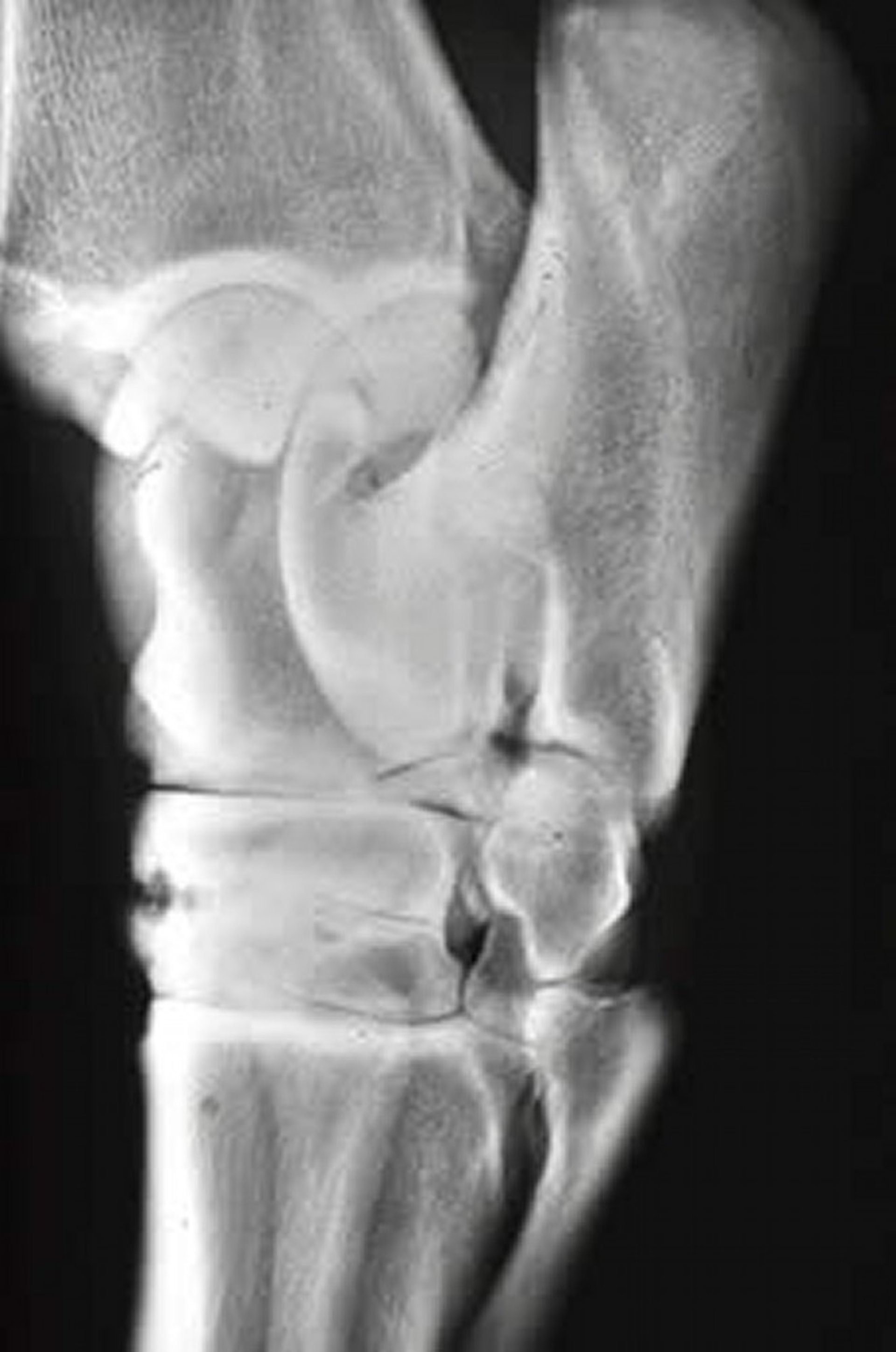
Radiography is useful to confirm the diagnosis. Radiologic changes may be evident in only one or two projections; therefore, a minimum of four standard orthogonal radiographic projections of the hock should be taken. Radiographic changes include narrowing or loss of joint space, sclerosis of the subchondral bone, lysis of the subchondral bone, periarticular osteophyte formation, and periosteal new bone formation. The severity of lameness and degree of radiologic change are poorly correlated. In those horses in which distal hock joint pain is suspected but there is little radiologic change, scintigraphy of the tarsus may reveal an increased focal uptake of radionuclide in the distal tarsal bones.
The aim of treatment is to provide pain relief so that the horse may remain in work. It has been suggested that by maintaining the horse in work, the distal hock joints will eventually ankylose and the horse will become pain free. However, progressive radiologic ankylosis is rarely observed and has not occurred in lame horses without intervention.
Conservative treatment involves systemic NSAIDs, intra-articular medication with corticosteroids, with or without hyaluronan, remedial trimming and farriery, and adaptation of the work program. Systemic treatment with polysulfated glycosaminoglycan (PSGAG), hyaluronan, or oral nutraceuticals may be used as adjunctive therapy.
Tiludronate, a bisphosphonate compound that inhibits osteoclastic activity, has been advocated for use in horses with distal hock pain. There are few controlled studies with this medication and, in a small double-blind clinical study, only one of eight horses improved with administration of tiludronate for distal tarsal inflammation or arthritis.
Extracorporeal shockwave treatment (ESWT) has also been used in treatment of distal tarsitis. Both focused and radial portable units are available. In one retrospective study of 74 horses with osteoarthritis of the distal tarsal joints treated with ESWT, 80% of the horses showed improvement in lameness but only 18% of horses became sound.
Intra-articular injection of sodium monoiodoacetate (MIA) has been used for chemical arthrodesis of the distal intertarsal and tarsometatarsal joints. MIA inhibits chondrocyte glycolysis and causes chondrocyte death. Contrast arthrography is necessary to ensure accurate needle placement and that no communication with proximal joints is present before injection of MIA. After injection, pain control is important, because severe pain is often present for 4–18 hr. Several studies have shown that 75%–85% of horses have been free of lameness with radiographic evidence of joint fusion at ~6 mo after treatment. However, when horses were followed for several years after treatment, the success rate decreased and a significant number of horses developed arthritis of the proximal intertarsal and tarsocrural joints. Other complications, including persistent swelling, septic arthritis, skin sloughing, and increased lameness, have been reported. Therefore, this technique is difficult to recommend because of the initial severe pain and significant possible complications. Intra-articular injection of ethyl alcohol to achieve arthrodesis has also been reported, although more longterm follow-up studies are required to evaluate its usefulness and safety.
In horses that do not respond to conservative treatment, a number of surgical techniques have been reported. These include cunean tenectomy, subchondral forage, neurectomy, and several different techniques of surgical arthrodesis.
Cunean tenectomy may result in a temporary improvement in lameness but is unlikely to restore soundness. It is believed to reduce the pressure over the medial aspect of the distal tarsus and cunean bursa and to reduce the rotational and shear stress over these joints during contraction of the tibialis cranialis muscle.
The most common surgical technique used in treatment of distal tarsal osteoarthritis is facilitation of fusion of the affected joints. Drilling across the distal intertarsal and tarsometatarsal joints is the most frequently used procedure to promote distal tarsal arthrodesis. Initial techniques described a more aggressive procedure, with 60% of the articular cartilage removed. Significant postoperative pain was associated with this procedure, and a more conservative approach using three drill tracts is currently recommended. Return to full athletic performance usually takes 10–12 mo. Retrospective studies evaluating this technique report ~60% of horses successfully return to their previous level of performance.
Both neodymium:yttrium aluminium garnet (Nd:YAG) and 980-nm diode laser have also been used to facilitate arthrodesis of the distal tarsal joints. Several studies have shown that horses treated with laser-facilitated arthrodesis are more comfortable after the procedure than when surgical drilling or MIA is used. It has been theorized that the superheating that occurs with the laser may cause thermal damage to nerve endings and diminish postoperative pain. Intra-articular drilling with diode-laser treatment has also been recommended, because the drilling should stimulate more bone production and fusion, whereas the laser reduces the postoperative pain.
Surgical stabilization in addition to intra-articular drilling of the distal tarsal joints has been proposed to have a superior clinical result versus intra-articular drilling alone. Lag screws or a screw and plate combination have been used. One study reported a success rate of 89% of horses returning to soundness versus 60% with drilling alone; however, only a small number of cases were reported.
Neurectomy of the deep fibular nerve and a partial neurectomy of the tibial nerves may be performed to relieve pain associated with the distal hock joints, with ~60% of horses undergoing surgery returning to full athletic function.


