


Some joint diseases, such as arthritis, affect the joint membranes themselves. Other types of joint conditions affect the tendons, cartilage, bursae, and fluid within the joint. Joint disorders may be congenital (present at birth) or may be the result of injury to the joint, degeneration (deterioration with loss of function), abnormal development, immune-related conditions, cancers, or infections.
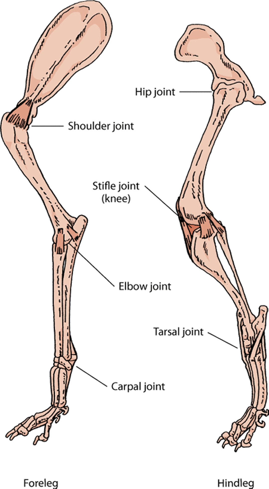
Leg joints, dog

Aseptic Necrosis of the Femoral Head (Legg-Calvé-Perthes Disease)
This deterioration of the top of the femur (femoral head) seen in young miniature and small breeds of dogs is characterized by a lack of blood supply and destruction of blood vessels of the bone. It is also known by the technical term, aseptic necrosis of the femoral head. The cause is unknown, although the condition may be hereditary in Manchester Terriers. The sudden loss of blood supply to the femur leads to collapse of the top of the bone. The condition often affects both hip joints.
Signs include hindlimb lameness, wasting away of the thigh muscles, and pain during movement of the hip joint. Longterm cases have evidence of degenerative joint disease. X-rays can help identify characteristic changes of this condition and may be used to confirm the diagnosis. Treatment involves surgical removal of the affected femoral head and neck and physical therapy to stimulate limb usage. If these procedures are followed, most animals with this condition recover.
Displacement of the Kneecap
This hereditary disorder is caused by abnormal development of the kneecap (patella). Displacement of the kneecap is often associated with multiple deformities of the hindlimb, involving the hip joint, femur, and tibia. The condition can also lead to cartilage and ligament injuries in the knee. Affected animals are lame or walk with a skipping gait. Dogs of any age may be affected.
Signs vary widely based on the severity of the displacement. In mild cases, the kneecap can be manually displaced but easily returned to normal position. These cases show infrequent and mild signs. As displacement becomes more severe, the dislocated kneecap is more often out of place, the limb is consistently lame, and bone deformities may be seen. X-rays can help your veterinarian see how severely the kneecap is displaced and what effects this has had on the limb.
There are several surgical options for treatment. The type of surgery performed is based on the severity of the displacement and can include minor procedures and bone surgeries. Mild or moderately affected dogs generally recover fully.
Osteochondrosis
Osteochondrosis is a disturbance in cartilage and bone formation of medium and large dogs that grow quickly. In this condition, the immature joint cartilage cracks and separates from the underlying bone. Fluid enters the space, and cysts may form under the cartilage. Fragments of cartilage may separate from the end of a bone and float loose in the joint cavity. This results in inflammation of the affected joint, and it can lead to arthritis and continued cartilage breakdown, severely affecting joint motion. The cause is unknown, but possible factors include high-growth diets, as well as rapid growth, trauma, and heredity.
Signs of osteochondrosis include lameness, fluid buildup within the joint, and joint stiffness. Affected areas include the head of the humerus (shoulder joint), the inside of the elbow joint, the stifle (knee) joints, and the ridges of the hock (ankle) joints. Your veterinarian may use x-rays to determine the extent of the damage. Surgery using an endoscope (arthrography) can also be performed to identify cartilage or joint lesions. A computed tomography (CT) scan can also identify bone changes.
Treatment involves surgical removal of cartilage flaps or the free-floating fragments of cartilage ("joint mice"). Dogs with degenerative joint disease may benefit from nonsteroidal anti-inflammatory drugs and joint fluid modifiers. Your veterinarian is best able to advise you on the appropriate use of any relevant medication. The outlook for recovery is excellent for the shoulders, good for the stifle joint, and fair for the elbow and hock (tarsal) joints. If the affected dog also has signs of degenerative joint disease, other joint conditions, or instability of the hock joint, the chances of recovery are reduced.
Elbow Dysplasia
Elbow dysplasia is an abnormal development of the elbow joint in young, large, rapidly growing dogs. It involves abnormal bone growth, cartilage development, or joint stresses. It is considered to be one of the most common causes of osteoarthritis of the canine elbow.
Lameness can develop slowly between 4 and 8 months of age; however, some cases may not be diagnosed until the dogs are more than 1 year old. The joint may appear stiff or unable to move freely. Advanced cases develop osteoarthritis, fluid buildup within the joint, and a grating or crackling sound. Physical examination and the presence of the characteristic signs suggest the diagnosis, and x-rays can confirm it. Both elbows should be examined because the condition can develop in both at the same time.
Surgery should be performed before the degenerative changes of osteoarthritis occur. The outlook for recovery after surgery is good if degenerative joint disease has not developed in the joint. Nonsteroidal anti-inflammatory drugs (as prescribed by your veterinarian) can reduce pain and inflammation. Joint fluid modifiers may also be helpful.
Septic Arthritis
Infectious, or septic, arthritis is usually caused by bacteria that spread through the blood or enter the body as a result of trauma (with penetrating wounds) or surgery. Other causes of septic arthritis include rickettsia (Rocky Mountain spotted fever, ehrlichiosis) and spirochetes (Lyme disease). See also Introduction to Infections
Signs of septic arthritis include lameness, swelling, pain of affected joint(s), fever, listlessness, loss of appetite, and stiffness. Laboratory tests on fluid removed from the joint may be useful in confirming the diagnosis.
Treatment consists of antibiotics administered orally or intravenously, flushing of the joint cavity, and surgical removal of dead, damaged or infected tissue in severe cases.
Immune-mediated Arthritis
Arthritis caused by the body’s own immune system can cause inflammation of joints. It generally affects several joints. Immune-mediated arthritis can destroy the joint cartilage and bone beneath the cartilage or cause inflammation around the joint (periarticular inflammation). Rheumatoid arthritis and Greyhound polyarthritis are examples of arthritis that destroys joint cartilage and bone beneath the cartilage. Systemic lupus erythematosus is the most common form of immune-mediated arthritis.
Signs include lameness, pain, and swelling in multiple joints, fever, a generalized illness, and loss of appetite. These signs commonly come and go. In addition to signs, the diagnosis is aided by x-rays, biopsy of joint tissue, blood tests, and examination of joint fluid (commonly called a joint tap).
Treatment involves anti-inflammatory medications and chemotherapeutic agents. The outlook for recovery is uncertain. Relapses are relatively common and the cause of the reactions is often unknown.
Cancerous Arthritis
This type of arthritis is most commonly caused by a tumor known as a synovial cell sarcoma. Signs include lameness and joint swelling. X-rays show soft-tissue swelling and a reaction around the bone. A biopsy reveals evidence of a soft-tissue tumor. By the time of diagnosis, spread of the cancer to the lungs has already occurred in about 25% of animals. Treatment involves amputation of the limb.
Joint Trauma
There are many types of joint trauma that can contribute to the development of joint disorders. Some of the more common types of trauma that can affect the joints are discussed below.
Cranial Cruciate Ligament Tear
Tearing of the cranial cruciate ligament of the stifle (knee) joint is a very common injury in dogs. However injuries are more likely to occur when the joint structure is already weakened by degeneration, the animal’s own immune system, or defects in conformation (such as those seen in straight-legged dogs). A tear of this type can make the knee joint unstable and can lead to cartilage injury, buildup of joint fluid, bony outgrowths, and hardening and thickening of the joint membrane.
Signs include those generally seen in joint disorders, such as lameness, pain, joint swelling, fluid buildup, and a grating sound when the joint is moved. In addition, the joint may appear to be abnormally loose. Partial cranial crucial ligament tears are characterized by a reduced ability to move the joint, especially bending it. Cartilage injury may be identified by a clicking sound during movement or when the joint is bent and extended. Veterinarians perform special tests during orthopedic exams to identify torn cruciate ligaments. X-rays are taken to evaluate the condition and anatomic angle of the joint, to aid treatment planning.
Both medical and surgical treatment options are available. Physical therapy, weight reduction, and nonsteroidal anti-inflammatory drugs ease discomfort from inflammation and degenerative joint disease. For active dogs, surgery to stabilize the knee joint is recommended. Physical therapy following surgery is critical for recovery. The outlook after surgery is good as long as degenerative joint disease has not progressed too far.
Dislocation and Fracture of the Ankle
Injury to the ankle (tarsus) is often seen in dogs that have been hit by a car. The ankle in dogs includes several bones that connect the lower leg to the foot. Injuries may include fracture or dislocation of these bones or tearing of the ligaments that hold them together. Affected dogs will hold the injured leg up and refuse to put any weight on it. The foot may swing in unusual directions because of its loose attachment following the injury. The extent of the injury is confirmed by physical examination and x-rays. Treatment is surgery to repair the bones and ligaments. The outlook for recovery is good.
Dislocation of the Elbow
Elbow dislocation is usually the result of trauma. Pain is variable, and the dog will usually hold the injured leg up and refuse to put any weight on it. Physical examination and x-rays are used to diagnose the condition. Treatment generally requires surgery, but the outlook for recovery is excellent.
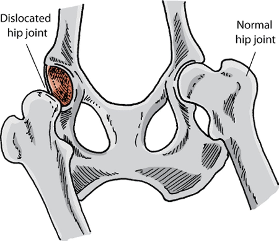
Dislocation of the Hip
Hip dislocation is usually the result of injury or trauma that displaces the head of the femur from the socket of the hip joint. Signs of hip dislocation include lameness, pain during movement of the hip joint, and a shortened limb. X-rays are useful in confirming the dislocation and revealing the presence of fractures. Nonsurgical treatment involves forcefully moving the joint back into place (closed manipulation) and using slings to keep the hip in its normal position. Surgical treatment involves stabilization using sutures or pins. Surgical resection of the bones involved or total hip replacement may be performed if more conservative treatment has not succeeded. The outlook for recovery is usually excellent.
Normal and dislocated hip, dog

Joint Fractures
The shoulder, elbow, carpal (wrist), hip, stifle (knee), and tarsal (ankle) joints are those most commonly involved in fractures due to injury. In young animals, the portion of the bone where growth occurs—called the growth plate and usually located at the ends of the bones—is weak compared with adjacent bones, ligaments, and joint membranes, making this area more prone to injury.
Signs of joint fractures include lameness, pain, and joint swelling. If the injury affects an active growth plate, limb deformities can result. X-rays and computed tomography (CT) scans are used to confirm and locate the fracture.
The goal of treatment is to allow the fracture to heal in proper alignment while maintaining joint and limb functions. This is usually done by holding the fracture in place internally with pins, wires, or screws in order to stabilize it. The outlook for recovery is good as long as damage to the joint is not severe.
Palmar Carpal Ligament Breakdown
Injuries sustained when falling or jumping can cause hyperextension, in which the limb extends beyond its normal range of motion. This produces excessive force on the wrist (carpus), which can cause tearing of the palmar carpal ligaments and fibrocartilage, leading to collapse of the joints. Signs include lameness, swelling of the carpal joint, and a characteristic stance in which the heel is touching the ground. For mild cases a splint or cast may be sufficient, but surgery is usually required. Surgery involves fusing the affected joints using a bone plate and screws, pins and wires, or an external system. The outlook for recovery is good.
For More Information
Also see professional content regarding other joint disorders.


