


Congenital abnormalities are conditions that an animal is born with; they are often called “birth defects.” Some of these conditions are inherited and tend to occur within particular families or breeds, while others are caused by chemicals or injury during pregnancy. For still others, the cause is unknown. Some of the most common congenital abnormalities of the digestive tract in dogs are described below.
Mouth
A cleft palate or cleft lip (harelip) is caused by a defect in the formation of the jaw and face during embryonic development. It leads to a gap or cleft in the center of the lip, the roof of the mouth (hard palate), or both. Often this condition leaves an open space through the roof of the mouth into the breathing passages. These conditions have a wide range in severity. Usually the upper lip and palate are affected; a cleft in the lower lip is rare.
Certain breeds are more prone to cleft palate than others. The defect is more common in Beagles, Cocker Spaniels, Dachshunds, German Shepherds, Labrador Retrievers, Schnauzers, and Shetland Sheepdogs. Dog breeds with short heads (brachycephalic breeds) can have up to a 30% risk of the disorder. Most cases are inherited, although nutritional deficiencies during pregnancy, drug or chemical exposure, injury to the fetus, and some viral infections during pregnancy have also been suggested as causes.
Cleft palate or lip will usually be noticed shortly after birth when the puppy might have problems nursing. For example, milk might be seen dripping from the nostrils or the puppy might have difficulty suckling and swallowing. The veterinarian can readily identify the problem by examining the puppy’s mouth. Affected puppies require intensive nursing care, including hand or tube feeding and possibly antibiotics to treat respiratory infections. Surgical correction is necessary and is most successful when puppies are at least 12 weeks old. A variety of surgical techniques are used, and the success rate in dogs is improving. The decision to perform surgery should be made carefully, and the affected animal should be spayed or neutered to prevent passing the defect on to its offspring.
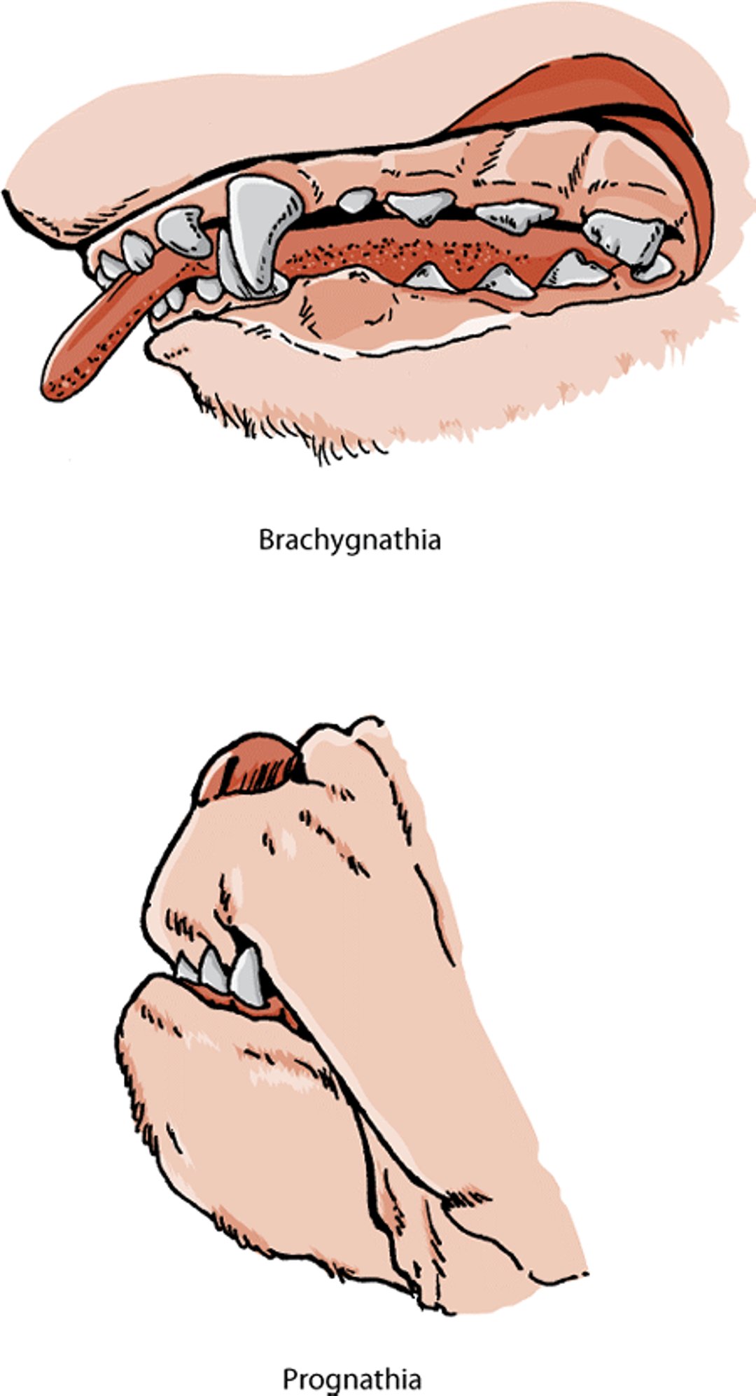
Brachygnathia (also called an overbite) occurs when the lower jaw is shorter than the upper jaw. It can be a minor problem or a serious defect depending on the degree of abnormality. Mild cases may cause no problems. More severe cases can cause damage to the hard palate (roof of the mouth) or restriction of normal jaw growth. The lower canine teeth are often removed or shortened to prevent this damage. For the best chance of success, this treatment should be done early in life.
Prognathia (also called an underbite) occurs when the lower jaw is longer than the upper jaw. This characteristic is normal in some breeds (for example, Boxers, Bulldogs, Pugs, and other breeds with shortened heads) and does not usually require treatment.
Ankyloglossia (also called "tongue-tie") occurs in Anatolian Shepherd dogs. These dogs have a notched or W-shaped tongue that causes difficulty eating, drinking, suckling, or licking. Corrective surgery can be performed. Affected dogs should be spayed or neutered so as not to pass on the condition to offspring.
Microglossia refers to incomplete or abnormal development of the tongue. The condition in dogs is often referred to as “bird tongue.” Affected puppies have difficulty nursing and do not grow properly. Examination of the mouth reveals missing or underdeveloped portions of the tongue. This condition is generally fatal.
Macroglossia, or large tongue, can occur with other birth defects in Dachshunds.
Some Chinese Shar-Peis have a condition called tight-lip syndrome in which the lower lip covers the lower front teeth and folds over the teeth toward the tongue. Contact between the upper front teeth and the lower lip worsens the lip position and may cause the lower front teeth to shift. Repeatedly biting the lower lip is also painful for the dogs. This condition can be corrected by surgery. Affected dogs should be spayed or neuter so that they do not pass on the condition to their puppies.
Brachygnathia and prognathia, dog

Teeth
In most animals, having too few teeth is rare, although in dogs, molars and premolars may fail to develop or erupt. In dogs, extra teeth are seen most often in the upper jaw. Although rare, sometimes a single tooth bud will split to form 2 teeth. The result may be crowding and rotation of the teeth; this condition requires tooth extraction to prevent or correct abnormalities of the bite that can lead to further dental problems.
Delayed loss of deciduous (“baby”) teeth in dogs is common, especially in small-breed dogs. The teeth that do not fall out get in the way of the permanent teeth that are starting to erupt beneath them, altering the position of the permanent teeth within as little as 2 to 3 weeks. This results in bite problems or entrapment of food, leading to tooth and gum disease. For these reasons, retained deciduous teeth should be removed by your veterinarian as soon as possible.
Abnormalities in placement or shape of teeth are reported in various breeds of dogs. The effect on an animal’s health is variable and based on severity. In certain dog breeds with short, flattened heads (brachycephalic breeds), the upper third premolar, lower first molar, and occasionally other teeth may rotate. Usually, this does not cause any problems, but it may require extraction of some teeth if crowding or bite abnormalities occur.
Abnormal development of tooth enamel (the hard outer surface of the tooth) can be caused by fever, trauma, malnutrition, poisoning, birth defects, or infections, such as distemper virus. The damage to the enamel depends on the severity and duration of the cause and can range from pitting to the absence of enamel with incomplete tooth development. Affected teeth are prone to plaque and tartar accumulation, which lead to tooth decay. Resin restoration is sometimes used to cover defects, although careful dental hygiene and home care is critical in reducing the incidence of complications. Discoloration of the enamel may also occur. Giving tetracycline antibiotics to pregnant females or to puppies less than 6 months old may result in permanent brownish-yellow stains on the teeth.
Cysts of the Head and Neck
Cysts (lumps) in the head and neck can be caused by defects during fetal development. These cysts are rare. Your veterinarian needs to distinguish them from abscesses or lumps caused by infection or other disease. These cysts tend to occur in specific locations and may have a characteristic feel to them, which can help the veterinarian to diagnose their cause. Tests, such as x-rays, ultrasound, video endoscopy, and computed tomography (CT), may also be necessary for diagnosis. Treatment involves surgical removal of the cyst(s).
Esophagus
The muscular tube that leads from the back of the mouth to the stomach is known as the esophagus. Some congenital abnormalities of the esophagus seen in dogs include megaesophagus, vascular ring anomalies, and crichopharyngeal achalasia ( see Table: Congenital Esophageal Disorders of Dogs). Signs of defects in the esophagus generally include regurgitation and problems with swallowing. These signs are especially noticeable when your dog starts to eat solid food. Regurgitated food can enter the lungs and cause frequent and severe pneumonia. These conditions are typically diagnosed with x-rays, usually done after the dog swallows a liquid dye that shows up on the x-ray. Other specialized tests may also be necessary. Surgical correction of some esophageal abnormalities (for example, vascular ring anomalies, in which abnormal blood vessels surround and restrict the esophagus) is effective if done early. If not, the esophagus can become permanently damaged by the stretching caused by trapped food. Feeding small amounts of soft foods in an elevated bowl can help affected dogs. Although mildly affected dogs may improve over time, the outlook is usually poor.
Small pouches in the lining of the esophagus, called esophageal diverticula, will sometimes form. Signs depend on severity and are seen in only 10 to 15% of cases. When they do occur, they may cause accumulation of food or become inflamed. In rare cases they rupture. Treatment (if necessary) is by surgical removal of the pouch. This disorder may be more common in English Bulldogs.
Hernias
A hernia is the protrusion of a portion of an organ or tissue through an abnormal opening. One common congenital type involves an abnormal opening in the wall of the diaphragm (the sheet of muscle that separates the chest from the abdomen) or abdomen. The defect may allow abdominal organs to pass into the chest or bulge beneath the skin. Hernias may be congenital (present at birth) or result from injury. Signs of a hernia vary from none to severe and depend on the amount of herniated tissue and its effect on the organ involved. Hiatal hernias involve extension of part of the stomach through the diaphragm. These hernias may be “sliding” and result in signs (such as loss of appetite, drooling, or vomiting) that come and go. Hernias are diagnosed using x-rays; contrast studies (x-rays that include special dyes to outline organs) are often needed. Endoscopy may be used to diagnose sliding hiatal hernias. In many cases, correction of a hernia involving the diaphragm requires surgery. However, the use of antacid preparations and dietary modification may control signs of a hiatal hernia, if they are mild.
Hernias involving the abdominal wall are called umbilical, inguinal, or scrotal, depending on their location ( see Table: Types of Hernias in Horses). Diagnosis of umbilical hernias is usually simple, especially if the veterinarian is able to push the hernia back through the abdominal wall (called “reducing the hernia”). These hernias are corrected by surgery. Small hernias are often corrected at the same time that the dog is spayed or neutered. The tendency to develop hernias may be inherited.
Stomach
Besides hiatal hernia (see above), another common abnormality involving the stomach is pyloric stenosis. It is likely that pyloric stenosis is inherited. This condition results from muscular thickening of the pyloric sphincter (the “exit” of the stomach). The thickening of this opening slows or blocks the flow of digested food from the stomach to the small intestine. Affected breeds include smaller breeds and those with flattened, shortened heads, especially Boxers, Bull Dogs, and Boston Terriers. Because the flow of food out of the stomach is restricted, dogs with this condition will often vomit food for several hours after a meal. Although dietary modification and medication may help, surgery is usually recommended.
Small and Large Intestine
Maldigestion is a condition in which certain foods are not properly digested. Malabsorption occurs when nutrients are not properly absorbed into the bloodstream. These conditions often cause persistent digestive system problems, including vomiting, weight loss, diarrhea, or a combination of these signs. There are many potential causes of maldigestion and malabsorption. Some are in-herited; some are acquired. Most are associated with inflammation of the intestines (called inflammatory bowel disease). Inherited conditions may occur more often in specific breeds. For example, Irish Setters have a family tendency for sensitivity to wheat protein (gluten), with signs beginning as early as 6 months of age. The wheat sensitivity is both confirmed and treated through the use of gluten-free diets. Malabsorption and maldigestion are often treated with a combination of dietary changes and medication; the exact treatment will depend on the condition being treated. In certain conditions in which protein loss is severe (for example, in Soft-coated Wheaten Terriers with protein-losing enteropathy and nephropathy), neither dietary changes nor treatment have been proven effective, and the outlook is poor.
Basenji enteropathy causes severe inflammation of the intestines and sometimes in the stomach in Basenjis. Signs include diarrhea and weight loss. Diagnosis of the disease is based on tissue biopsies. Treatment includes drugs to suppress the immune system and special diets, but they are often ineffective unless started early in the course of the disease.
Lymphangiectasia occurs due to an abnormality in lymphatic vessels of the digestive tract. The disorder causes protein loss from the digestive tract in Norwegian Lundehunds, Yorkshire Terriers, Maltese, Rottweilers, and Chinese Shar-Pei. It can be present at birth or acquired later in life. It is diagnosed with tissue biopsies and by ruling out other intestinal diseases that cause protein loss. Treatment includes medications and the use of specialized diets. Although it is possible to improve the signs of the disease, the longterm outlook is poor.
Exocrine pancreatic insufficiency (EPI) is a syndrome caused by insufficient production and secretion of digestive enzymes from the pancreas. It occurs in many breeds but especially in German Shepherds and Rough Collies. For more information on EPI, see Exocrine Pancreatic Insufficiency.
Granulomatous colitis (previously called histiocytic ulcerative colitis) has been seen in Boxers, French Bulldogs, and a few other breeds. It is suspected that an inherited defect in a dog's immune system leads to inflammation of the large intestine and a bacterial infection. Signs include bloody diarrhea, straining to defecate, and weight loss. These signs typically occur in dogs less than 4 years old. Tissue biopsies are necessary to diagnose the condition. Antibiotics are typically successful in treating the condition.
Various malformations of the intestines can occur as birth defects, including duplication of sections of the intestine or rectum, failure of the rectum to connect with the anus, and openings between the rectum and other structures such as the urethra or vagina. Surgical correction is usually needed. The success rate depends on the extent of the malformation.
Liver
The most common liver defect present at birth is portosystemic shunt. In a healthy animal, blood coming from the intestines is processed by the liver, which removes toxins from the bloodstream before they reach the brain or other organs. In an animal with a portosystemic shunt, however, blood bypasses the liver through one or more “shortcuts” (shunts) and enters directly into the general circulatory system. Breeds with an increased risk of this defect include Yorkshire Terriers, Miniature Schnauzers, Cairn Terriers, Maltese, Havanese, Scottish Terriers, Pugs, Irish Wolfhounds, Golden Retrievers, Labrador Retrievers, German Shepherds, Cattle Dogs, Old English Sheepdogs, and Poodles. Signs of a portosystemic shunt include nervous system disturbances and a failure to grow and thrive. In the late stages, protein-containing fluid may accumulate in the abdomen, a condition called ascites. Your veterinarian may also notice enlargement of the kidneys and kidney stones. A definite diagnosis is made by using an opaque dye to highlight the blood vessels, followed by x-rays. This procedure can identify the location of the shunt and determine whether it is single or multiple. It also allows the veterinarian to assess whether surgical correction is possible. Animals with multiple shunts tend to do poorly.
Hepatoportal microvascular dysplasia is another disorder that results in blood entering the circulatory system without being detoxified by the liver. In this disease, the shunting occurs within the liver itself. The syndrome occurs with some frequency in Cairn and Yorkshire Terriers and has also been reported in Maltese, Dachshunds, Toy and Miniature Poodles, Bichon Frise, Pekingese, Shih Tzus, Norfolk and Norwich Terriers, Tibetan Spaniels, Havanese, and Lhasa Apsos. It generally causes no signs, but when seen, signs are similar to those in dogs with a portosystemic shunt. Dogs that do exhibit signs may be treated with medication. Surgery is not an option because the shunting is caused by many small blood vessels, not a single prominent one that can easily be corrected.
Copper-associated hepatopathy is a defect that causes copper accumulation in the liver. This results in the development of chronic hepatitis and cirrhosis of the liver. The condition is found in Bedlington Terriers and leads to chronic liver failure in older dogs. Carrier dogs, which have no signs of the disease but can pass it on to their puppies, are also seen. Elevated copper levels have also been observed as part of the inherited liver disease of West Highland White Terriers, Skye Terriers, Dalmatians, and Doberman Pinschers. There are apparent variations even within breeds; for example, liver copper levels are worse in Bedlington and West Highland White Terriers of North American descent than in the same breeds from Europe or other regions. Treatment involves the use of drugs that bind copper (chelators) or prevent its absorption, low-copper diets, and other supportive measures directed at helping animals with liver disease. If your dog has copper-associated hepatopathy, follow your veterinarian’s directions for medication, diet, and other treatment carefully and fully.
Other liver developmental anomalies include hepatic (liver) cysts, which generally cause no signs of illness. They are of significance mainly because they must be differentiated from abscesses in the liver. A veterinarian who finds a hepatic cyst will often want to examine the kidneys, because hepatic cysts often occur along with polycystic kidney disease.
Hyperlipidemia is an elevated level of certain types of fats in the blood. These elevations can occur on their own or as a result of another disease, Elevated levels of triglycerides (hypertriglyceridemia) occurs frequently in Miniature Schnauzers, especially as they age. Elevated levels of cholesterol (hypercholesterolemia) has been seen in Briards, Rough Collies, Shetland Sheepdogs, Doberman Pinschers, and Rottweilers. Signs of hyperlipidemia are not always present, but when seen they include vomiting, diarrhea, pancreatitis, seizures, neurologic disturbances, and abdominal pain. Treatment includes diet changes and the management of the underlying condition, if present.
For More Information
Also see professional content regarding congenital and inherited disorders of the digestive system.


