

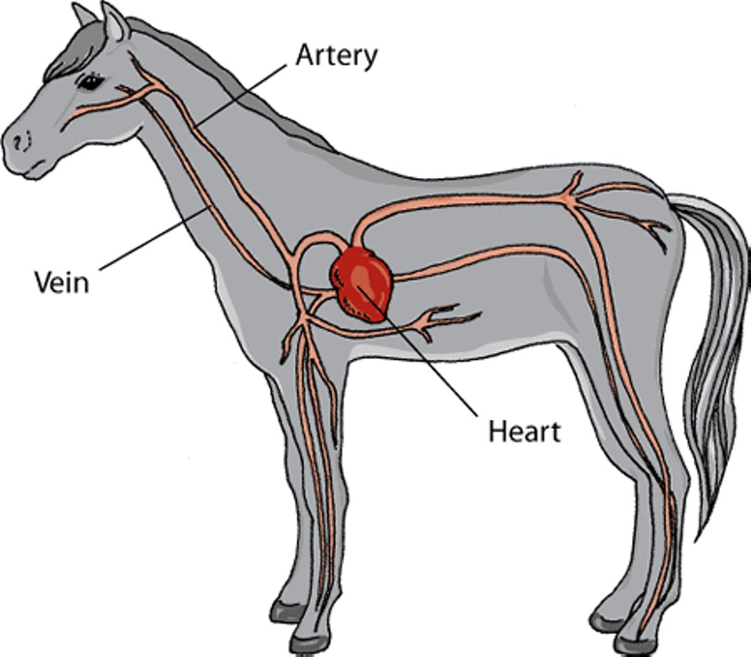
The cardiovascular system includes the heart and the blood vessels—the veins and the arteries. The function of the heart is to pump blood. The right side of the heart pumps blood to the lungs, where oxygen is added to the blood and carbon dioxide is removed from it. The left side pumps blood to the rest of the body, where oxygen and nutrients are delivered to tissues, and waste products (such as carbon dioxide) are removed. In horses, the cardiovascular system must not only efficiently supply blood to all parts of a large animal, but must also function well during strenuous racing or training, in many cases.
Cardiovascular system of a horse

The heart is a hollow, muscular organ which, in mammals, is divided into 4 chambers. The muscular tissue is called the myocardium. There are upper chambers on both the left and right sides of the heart called the left and right atria (the plural form of atrium). There are also 2 lower chambers called the left and right ventricles.
A series of valves keep blood flowing in one direction through the heart. The atrioventricular valves are valves between the atria and the ventricles. The semi-lunar valves are valves between the heart and the aorta and between the heart and the pulmonary artery. Each ventricle has an inlet and an outlet valve. In the left ventricle, the inlet valve is called the mitral valve, and the outlet valve is called the aortic valve. In the right ventricle, the inlet valve is called the tricuspid valve, and the outlet valve is called the pulmonary valve.
Horse heart

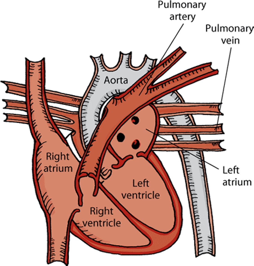
Blood from the body flows through the 2 largest veins, called the venae cavae, into the right atrium. When the right ventricle relaxes, blood in the right atrium pours through the tricuspid valve into the right ventricle. When the right ventricle is nearly full, the right atrium contracts, pushing additional blood into the right ventricle. The right ventricle then contracts, pushing blood through the pulmonary valve into the pulmonary arteries, which lead to the lungs. In the lungs, blood absorbs oxygen and gives up carbon dioxide. The blood then flows through the pulmonary veins into the left atrium. When the left ventricle relaxes, the blood in the left atrium pours through the mitral valve into the left ventricle. When the left ventricle is nearly full, the left atrium contracts, pushing additional blood into the left ventricle. The left ventricle then contracts, pushing blood through the aortic valve into the aorta, the largest artery in the body. This blood carries oxygen to all of the body except to the lungs.
Each heartbeat consists of two parts: diastole and systole. One half of a heartbeat is the sound of the mitral and tricuspid valves closing. The other half is the sound of the aortic and pulmonary valves closing. During diastole, the ventricles relax and fill with blood. During systole they contract and pump blood out to the body.
The rate and force of contraction of the heart and the degree of narrowing or widening of blood vessels are controlled by different hormones and the autonomic nervous system, the part of the nervous system that controls involuntary activity.
Heart Rate in Horses
The heart beats because of a tiny electrical current that originates in the heart’s pacemaker, called the sinoatrial node. Rhythmic electrical impulses or discharges cause the contraction of muscle fibers in the heart. While a horse is at rest, its sinoatrial node discharges about 30 times per minute. Large animals, such as horses, tend to have a slower heart rate than smaller animals (such as cats or birds). With exercise, the heart rate increases. A horse's heart can beat 250 times in a minute during maximal exercise.
Heart Sounds and Murmurs in Horses
Heart sounds are produced by the rapid acceleration and deceleration of blood and the resulting vibrations in the heart due to the circulation of blood. They can be heard using a stethoscope. In healthy horses, 4 heart sounds can possibly be heard. An absence or abnormality of one of these sounds may indicate a heart abnormality.
Heart murmurs are vibrations that can be heard coming from the heart or major blood vessels and generally are the result of turbulent blood flow or vibrations of heart structures such as part of a valve. Murmurs are typically described by their timing (that is, whether they occur during diastole, systole, or continuously), intensity (that is, whether they can be heard easily or with difficulty), and location. Not every murmur indicates a heart disorder. In horses, early systolic and diastolic murmurs can be noted in the absence of heart disease or anemia (having too few red blood cells). A short, high-pitched, squeaking, early diastolic cardiac murmur is sometimes seen in healthy young horses.
Arrhythmias in Horses
Arrhythmias are abnormalities of the rate, regularity, or site of heartbeat formation. An arrhythmia does not necessarily indicate heart disease. Many arrhythmias have no functional significance and require no specific treatment. Some arrhythmias, however, may cause severe signs, such as loss of consciousness due to lack of blood flow to the brain, or lead to sudden death. Many disorders are associated with abnormal heart rhythms. Examples of arrhythmias are a rate that is too slow (bradycardia), a rate that is too fast (tachycardia), premature beats (a beat that is heard too early), an irregular rhythm, and heart that pauses within the rhythm. Whenever an abnormal rhythm is heard, your veterinarian may recommend an electrocardiogram to help discover its cause.
Atrial fibrillation is a type of arrhythmia that commonly occurs in horses. In atrial fibrillation, the electric current running through the atria is not coordinated, stimulation of the atrioventricular node is frequent but random, and the heart rate is rapid and irregular. The horse may or may not have a normal heart rate when resting. Atrial fibrillation can occur due to heart disease, but most of the time it occurs in horses without an underlying heart disorder. Most horses that do not have underlying heart disease will show no signs during rest or moderate exercise; the arrhythmia may only become apparent during strenuous exercise. Your veterinarian can help determine whether the arrhythmia is due to heart disease. If appropriate, your veterinarian may use medication or electricity to convert the arrhythmia to a normal rhythm.
Pulse in Horses
A pulse is the rhythmic expansion of an artery that can be felt with the fingertips during physical examination. A horse's pulse is easiest to feel on the facial artery, which is located under the lower jawbone. A jugular pulse in the lower neck can be noted in healthy animals, but excessive pulsing or distension of the jugular vein can be seen in horses with heart failure. A pulse may be absent, increased (strong), or decreased (weak)—each of which may indicate a specific type of heart disease or defect.
For More Information
Also see professional content regarding the cardiovascular system.


