


Illustration by Dr. Gheorghe Constantinescu.
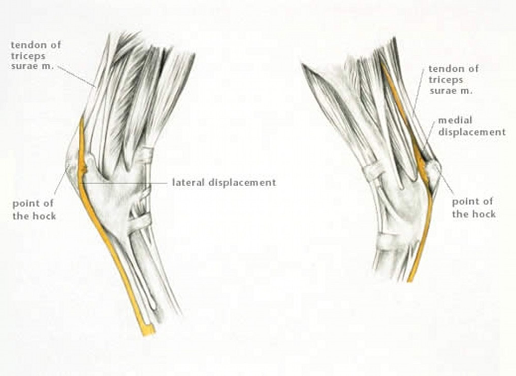
Luxation or subluxation of the superficial digital flexor tendon from the point of the hock may occur after disruption to the retinaculum that attaches the tendon to the calcaneus. Luxation or subluxation more commonly occurs laterally but may occur medially. Rarely, the superficial digital flexor tendon may split sagitally, and part of the tendon luxates medially and part laterally.
Lameness is usually acute and severe in onset and associated with marked swelling over the point of the hock. Horses may become panic-stricken in the acute stages if the tendon is moving on and off the os calcis, and they may kick out frequently with the affected limb. In some horses, the tendon will resume its normal position when the horse is standing but luxate or subluxate as the horse moves; in other horses, the tendon remains permanently displaced. Diagnosis is based on careful observation and palpation and can be confirmed by ultrasonography.
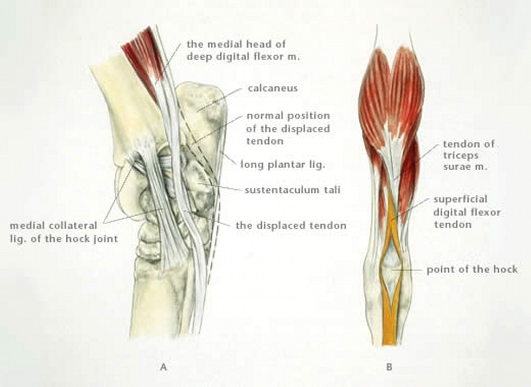
Illustration by Dr. Gheorghe Constantinescu.
In the acute stages, sedation and analgesia are important. If the tendon remains permanently displaced laterally (or medially), treatment involves prolonged stall rest (4–6 mo), possibly with the limb immobilized in a heavily padded full limb bandage or cast. Lameness usually improves, but the horse may be left with a mechanical lameness causing a jerky hindlimb action that may limit its use as a dressage horse, although horses may be able to jump or race. Horses with a persistently medially displaced tendon usually have a greater degree of mechanical lameness and a poorer prognosis for return to athletic function.
In horses in which the tendon is unstable and subluxates on and off the tuber calcanei, endoscopy of the calcaneal bursa reveals disruption of both the medial retinacular/calcaneal insertion of the superficial digital flexor tendon and its associated fibrocartilage, with disruption of the medial wall of the bursa creating or establishing communication with an acquired subcutaneous bursa. Treatment is by radical resection of the disrupted fibrocartilage and division of remaining attachments of the fibrocartilage or retinacular insertions with the unstable main body of the superficial digital flexor tendon, thereby creating a stable lateral subluxation. Although repair or reconstruction techniques have been reported, results have been unreliable. Permanent stable subluxation eliminates anxiety and usually results in a “sound” functional horse.


