


Bacterial Diseases in Ferrets
Most bacterial infections in ferrets are similar to those seen in other carnivores. However, there are some bacterial infections specific to ferrets. The most important ones include Helicobacter mustelae gastritis and Lawsonia intracellularis enteritis. In addition, several different types of mycobacteria have been found to cause atypical disease in ferrets.

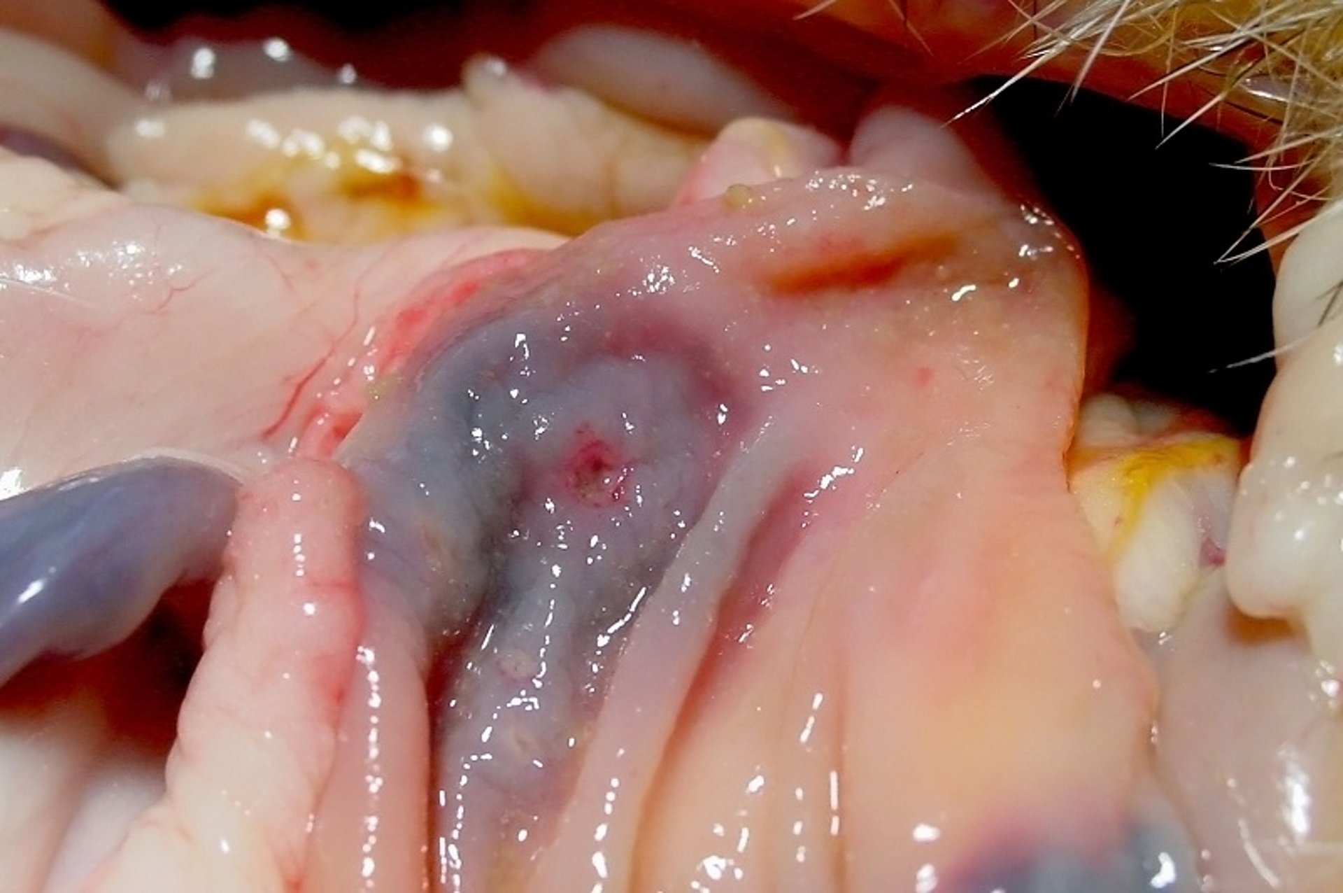
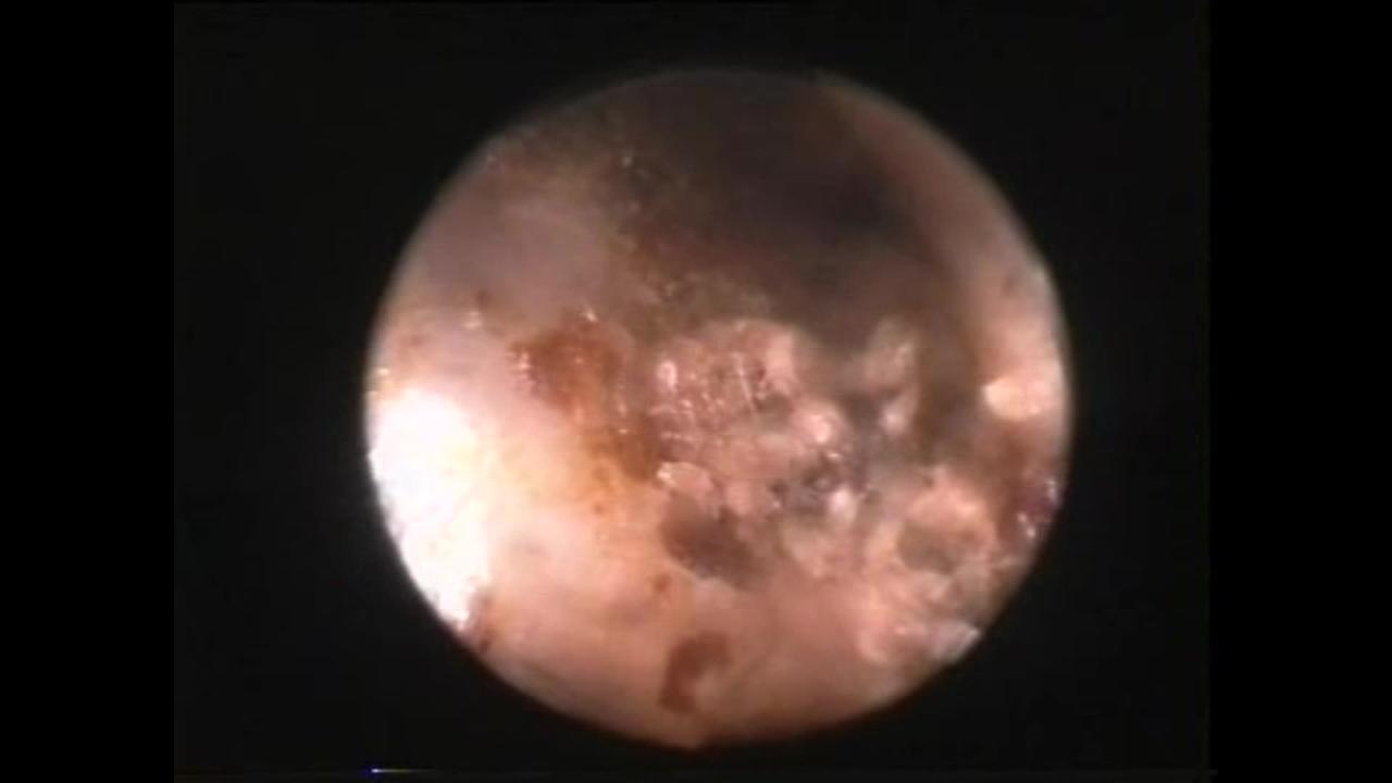
Courtesy of Dr. N. J. Schoemaker.
Helicobacter mustelae is found in the stomach and duodenum of ferrets after weaning. It is an opportunistic pathogen and can induce chronic, persistent gastritis and ulcer formation similar to gastric ulcer disease in humans. Gastric lymphoma may occur in chronic cases. Clinical signs may be absent in infected ferrets but can also include inappetence, vomiting, bruxism, diarrhea, melena, and hypersalivation. Lethargy, weight loss, and dehydration may also occur. When ulcers are present, ferrets may show signs of pain on cranial abdominal palpation. Although Helicobacter may be found during histologic examination of surgical or endoscopic biopsy samples, the presence of bacteria should not be considered diagnostic, unless associated with the presence of ulcers.
Treatment is commonly initiated when clinical signs are evident, prior to confirmation of presence of H mustelae, and usually comprises multidrug treatment, including amoxicillin (20 mg/kg, PO, every 12 hours) or clarithromycin (50 mg/kg, PO, every 24 hours); metronidazole (20–25 mg/kg, PO, every 12 hours), omeprazole (1–4 mg/kg/day, PO) and sucralfate (25–125 mg/kg, PO every 8–12 hours). Treatment is usually for 21 days. Because of the opportunistic nature of this pathogen, it is important to reduce stress and consider the possibility of trichobezoar formation.
The strict intracellular bacterium Lawsonia intracellularis is the causative agent of proliferative bowel disease, most commonly seen in ferrets up to 16 weeks of age, kept in large groups under suboptimal conditions. The bacteria invade the ileum and colon and cause hypertrophy of the mucosa. Clinical signs include diarrhea, weight loss, and rectal prolapse. Treatment with chloramphenicol (25 mg/kg, PO, twice daily) for 14–21 days is reported to be successful, while optimizing housing and diet are apparently of minimal benefit in preventing the disease.
Viral Diseases of Ferrets
The most important viral diseases in ferrets include canine distemper (due to infection with morbillivirus of the family Paramyxoviridae), influenza A and B (caused by anorthomyxovirus infection), coronavirus infections, and Aleutian disease (due to a parvovirus).
Ferrets are susceptible to canine distemper virus; however, clinical signs of disease are currently rare because most ferrets kept as pets are vaccinated. Transmission occurs by via aerosol droplets or through contact with (conjunctival or nasal) exudates, urine, feces, skin, or some combination of these routes. Clinical signs often develop within 7–10 days after infection and include fever and lymphopenia followed by anorexia, erythema of the mucous membranes, and serous to mucopurulent ocular and nasal discharge. Dermatologic signs, including a hyperkeratotic and crustaceous dermatitis of the face (eyelids, chin), abdomen, and inguinal and perineal area, and hyperkeratosis of the footpads, also occur. Respiratory and gastrointestinal signs develop and progress rapidly, complicated by secondary bacterial infections. Neurologic signs resulting from acute encephalomyelitis have been described but are not always present in affected animals. Diagnosis is via history, clinical signs, and results of histopathologic examination of relevant samples. Mortality is close to 100%, and death typically occurs 12–14 days after infection. As a result, vaccination is highly recommended.

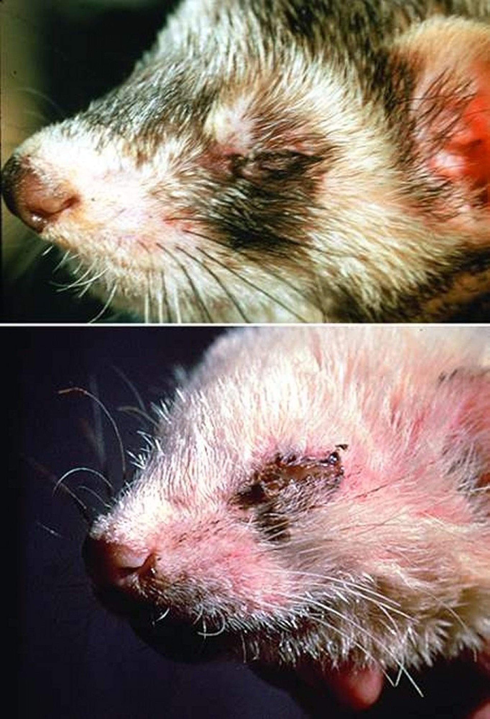
Courtesy of Dr. John Gorham.

Courtesy of Dr. Louise Bauck.
The influenza virus causes fever, lethargy, anorexia, nasal discharge, sneezing, and depression in ferrets. In the early stage, an infection may be easily mistaken for a canine distemper infection (and vice versa). Treatment is supportive and includes antimicrobials for secondary infections, although amantadine (6 mg/kg, nasally, twice a day) has been suggested as an experimental antiviral treatment. Recovery usually occurs within 7–14 days. Because ferrets may be infected by humans and may also infect people, it is important that strict hygienic measures are taken when either veterinary staff or ferrets are suspected of having influenza infections.
At least two coronaviruses cause disease in ferrets. The ferret enteric coronavirus causes epizootic catarrhal enteritis. Ferret enteric coronavirus is highly transmissible and is often brought into a group of ferrets by an asymptomatic juvenile. Clinical signs begin 2–14 days after introduction of the new ferret or after exposure through fomites. After infection, the virus causes blunting of the intestinal villi and consequent maldigestion and malabsorption, leading to clinical signs such as anorexia, vomiting, green or mucoid diarrhea, melena, dehydration, lethargy, and weight loss. In recent years, the development of clinical signs has become rare. Disease, if seen, is most severe in older ferrets, which may need months to fully recover. Treatment is supportive and includes fluids, nutritional support, GI protectants, and broad-spectrum antimicrobials if secondary bacterial infection is suspected. Prevention is by means of quarantine of new ferrets, thorough cleaning of new bedding and toys, and washing hands and changing clothes after handling unaffected ferrets.
A second related coronavirus, ferret systemic coronavirus causes a systemic pyogranulomatous inflammatory disease resembling the dry form of feline infectious peritonitis. The disease is therefore now frequently referred to as ferret infectious peritonitis. This disease is seen in young ferrets (average 11 months) with a progressive course of several weeks to months. Clinical signs include anorexia, weight loss, diarrhea, and enlarged intra-abdominal and, less commonly, peripheral lymph nodes. Hypergammaglobulinemia, anemia, and CNS signs can be seen as the disease progresses. Average survival time is ~2 months. Treatment, if undertaken, is primarily supportive; use of prednisolone has been anecdotally reported to increase survival times to some extent.
Aleutian disease is a parvovirus originally seen in mink, but at least 2 distinct ferret strains of the virus have been identified. The virus causes immune complex deposition in organs, which results in a variety of nonspecific clinical signs such as progressive weight loss, weakness, ataxia, hepatomegaly, and splenomegaly. Severe hypergammaglobulinemia is the most consistent finding on laboratory testing. A presumptive diagnosis is based on clinical signs and the presence of hyperglobulinemia. The most common tests used to diagnose viral infection in the US are PCR assay and serologic testing (through the Infectious Diseases Laboratory of the University of Georgia). In other countries, the counterimmunoelectrophoresis (CIEP) is the most commonly used test to detect parvovirus antibodies. Definitive diagnosis is difficult because apparently normal ferrets in shelters may have positive titers. Moreover, the virus has been found in the urine, feces, and blood of both symptomatic and asymptomatic animals.
Treatment with anti-inflammatories and immunosuppressants such as prednisolone and cyclophosphamide can be considered and may have clinical benefit. There is no vaccine available for this disease in ferrets.
Fungal Diseases of Ferrets

Courtesy of Dr. N. J. Schoemaker.
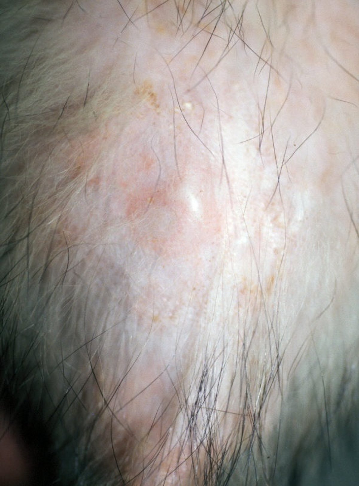
Ferrets are susceptible to Microsporum canis and Trichophyton mentagrophytes infection, although clinical signs of disease are rare. Transmission is by direct contact or via fomites and is often associated with overcrowding and exposure to cats. Infection is more common in kits and young ferrets and is often seasonal and self-limiting. Diagnosis and treatment follow similar guidelines as described for ringworm in dogs and cats. A pyogranulomatous dermatitis and fungal pododermatitis has been associated with Microsporum nanum. Other fungal diseases in ferrets include cryptococcal meningitis and blastomycosis causing granulomatous meningoencephalitis. Fungal pneumonia is uncommon in ferrets but can be caused by Blastomyces dermatitidis and Coccidioides immitis in endemic areas. Cryptococcosis from Cryptococcus bacillisporus and C neoformans var grubii has been diagnosed in ferrets. Signs include pneumonia, pleuritis, rhinitis, and regional lymph node enlargement.
Parasitic Diseases of Ferrets

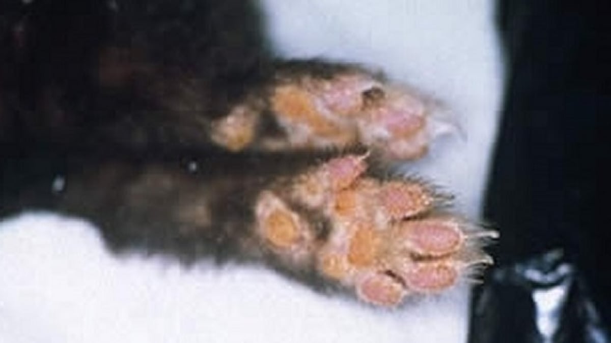
Ear mite infection is the most common ectoparasitic disease in ferrets and is caused by Otodectes cynotis. The same ear mite is found in dogs and cats, and it can be passed between species. Diagnosis is by otoscopy, as for dogs and cats. Treatment is also similar as for dogs and cats, and several of the available drugs are registered for use in ferrets. Fleas are also common in ferrets and can be transmitted between ferrets and other household pets. Diagnosis is by visualization, and treatment is the same as for dogs and cats. Many of the long-acting topical treatments, such as fipronil, last longer in ferrets because of increased sebum concentration in the coat. Mange in ferrets is caused by Sarcoptes scabiei and can manifest as generalized dermatitis or can be limited to the feet (pedal form), specifically affecting the toes and foot pads, unique to ferrets.
Heartworm disease, caused by Dirofilaria immitis, can affect ferrets, especially if given outdoor access in endemic areas. Disease can be caused by even a single worm. Clinical signs include lethargy, coughing, dyspnea, and ascites. Ferrets are typically infected with a very small number of worms (1–20), making diagnosis difficult. Echocardiography is warranted, because the parasites often obstruct blood flow and cause right-side heart failure. Echocardiography may also be helpful in identification of the worms in the right ventricle, pulmonary arteries, and vena cavae. Peripheral microfilaremia is uncommon in ferrets; therefore, antigen testing is more beneficial. Long term treatment with antithrombotic drugs and adulticides can be initiated but may have adverse effects. Selamectin (18 mg/kg, topically) is currently the recommended mode of prevention in endemic areas.
Coccidiosis, caused by Eimeria or Isospora spp, can cause disease in young ferrets, with clinical signs including diarrhea and lethargy. Diagnosis and treatment are similar to that for dogs. Rectal prolapse can also occur with coccidiosis and usually resolves after treatment of the underlying disease.
Giardia is another protozoal disease that may be seen in ferrets. When clinical signs are present, diarrhea and weight loss are most commonly seen. Treatment is with metronidazole (20 mg/kg, PO, twice a day).


