


Osteochondrosis is one of the most important and prevalent developmental orthopedic diseases of horses. Although its specific etiology is not known, it is considered to arise from a focal disturbance in endochondral ossification, with subsequent trauma or physiologic loading resulting in lesion formation. The term osteochondrosis is currently used to describe the clinical manifestation of the disorder; however, the term dyschondroplasia is preferred when referring to early lesions.
Osteochondrosis has a multifactorial etiology that includes rapid growth, high carbohydrate diet, mineral imbalance, and biomechanics (ie, trauma to cartilage). Genetics has been implicated, with some breeds predisposed (eg, Standardbred and Swedish Warmblood). The condition mainly affects articular growth cartilage, but the metaphysis may also be involved. If the physeal metaphyseal cartilage is affected, bone contours and longitudinal growth are disturbed ( see Physitis in Horses). Dyschondroplasia at articular surfaces may progress to formation of cartilage flaps or osteochondral fragments (osteochondrosis). At some sites, subchondral cysts may develop ( see Subchondral Bone Cysts). Axial skeletal involvement includes vertebral articular facets, which may be associated with stenosis of the vertebral canal and, therefore, ataxia and proprioceptive deficits (ie, wobbler syndrome), but the relationship between these conditions is not clear.
Clinical Findings:
The clinical signs of equine osteochondrosis are difficult to characterize specifically because of the wide range of lesions and sites involved. In young horses, many cases have no detectable clinical signs and are identified only on presale radiographs. Furthermore, lesions of dyschondroplasia may not progress to osteochondrosis, and radiographically observed osteochondrosis lesions may resolve over time without producing clinical signs. In severe cases, other signs of developmental orthopedic disease also may be apparent.
The most common presenting sign of osteochondrosis is a nonpainful distention of an affected joint (eg, gonitis, bog spavin). The exceptions to this are joints in which swelling is difficult to detect (eg, shoulder joint, medial femorotibial joint), in which case lameness is more often the first sign observed. Clinical signs may be divided broadly into two categories: those seen in foals < 6 mo old and those seen in older animals. Often the first sign noted in foals is a tendency to spend more time lying down. This is accompanied frequently by joint swelling, stiffness, and difficulty keeping up with other animals in the paddock. An accompanying sign may be the development of upright conformation of the limbs. Fetlock osteochondrosis is particularly seen in younger foals (< 6 mo old).
Lameness is usually absent or mild except for those sites mentioned above for which the earlier sign of joint swelling is difficult to detect. For example, lesions in the shoulder frequently result in moderate to severe lameness, muscle atrophy, and pain on joint flexion. In the stifle, some horses with subchondral bone cysts in the medial femoral condyle present with lameness severe enough that a fracture may be suspected, and swelling may only be detected on careful examination. More severe signs are also observed when osteochondral fragments come loose within the joint. This is often seen in yearlings or older horses that present with stiffness, flexion responses, and varying degrees of lameness. These signs are usually associated with the onset of training.
Diagnosis:

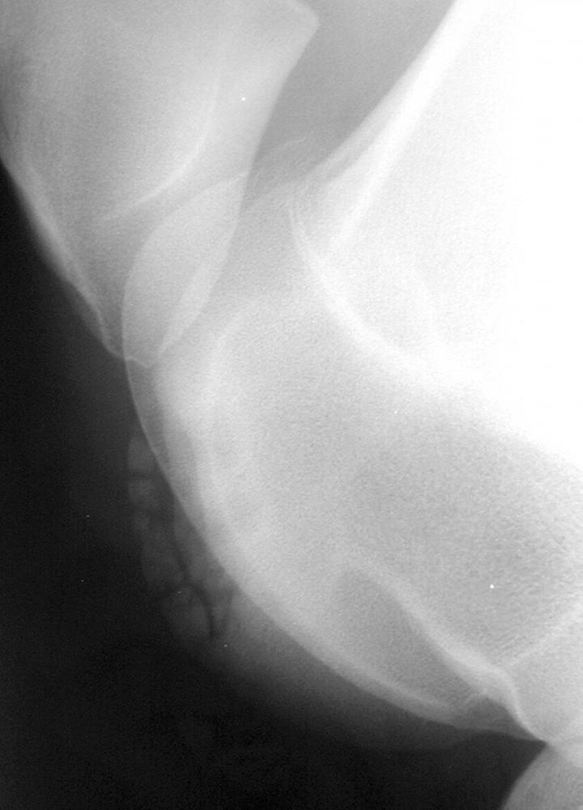
Courtesy of Dr. Chris Whitton.
Clinical diagnosis can often be made on the basis of signalment and signs. More definitive diagnosis requires use of some specific clinical aids. Radiographic examination has been the traditional way to confirm diagnosis; however, early lesions involving cartilage without significant subchondral bone damage may not be visualized. In the distal limb, oblique views may be helpful; in the hock, because the most common site of a lesion is the distal intermediate ridge of the tibia, the best view is a plantarolateral/dorsomedial oblique. Ultrasonographic examination of the swollen joints can help delineate articular damage and synovial inflammation and determine whether osteochondral fragments are intra- or extra-articular. The most accurate way to confirm diagnosis is by arthroscopy, and most of the predilection sites are accessible.
Scintigraphy has limitations in growing horses because of normal high activity in physes and sites of active endochondral ossification. It is a useful technique to detect subchondral cysts and secondary degenerative changes in older horses. MRI is ideal for diagnosis of both early and late lesions but is usually not necessary. Also, sites that are most diagnostically challenging are generally in the proximal limb, where access is difficult. Clinical pathology and the evaluation of synovial fluid is rarely helpful but can be used to eliminate inflammatory causes of swollen joints.
Treatment and Management:
Management of osteochondrosis depends on the site and severity of signs. Mild cases recover spontaneously, and a conservative approach may be appropriate. In young animals (< 12 mo old), this involves restricted exercise for some weeks combined with reduced feed intake to slow the growth rate. Particular care should be taken to ensure appropriate mineral supplementation (eg, in cases of suspected copper deficiency). It is controversial whether correcting the diet, once signs have developed, will actually assist resolution, but it may help limit or prevent further cases on stud farms. Intra-articular medication with hyaluronic acid may be beneficial, but injection of long-acting corticosteroids is not recommended in young, growing horses.
Cases considered for surgery are treated arthroscopically. This technique has been successful in most affected sites, particularly the hock, stifle, and fetlock. Damaged cartilage, osteochondral fragments, and compromised subchondral bone are removed and the joint flushed extensively with sterile fluid. Prognosis after removal of discrete osteochondral fragments is good. In cases with more extensive osteochondral damage, prognosis depends on the extent of the joint surface that must be removed. Prognosis is poor for cases with instability resulting from joint surface loss or in which secondary osteoarthritis (degenerative joint disease) is advanced. This is often the case with shoulder osteochondrosis because of the difficulty in detecting early signs. Cases involving subchondral cysts have a guarded prognosis, because these cysts are often in important weightbearing areas of the joint, and restoration of the joint surface is rarely possible.


