


Thrombosis (clot formation within a blood vessel), embolism (process by which unattached material (emboli) such as a blood clot, fat or cholesterol deposit, gas, tissue, or foreign material travels within the bloodstream and occludes flow within a vessel), and aneurysm (dilation or outpouching of a blood vessel wall) are pathologic abnormalities that can occur within the vasculature. Clinical signs, diagnostic testing, and therapy vary based on the classification, location, severity, chronicity, and associated or concurrent comorbidities. Typically, mainstays of treatment include addressing the underlying disease process and antithrombotic drugs.
A thrombus is an aggregation of platelets and fibrin that may form when certain conditions exist. Historically, these have included some combination of Virchow's triad such as blood stasis (reduced flow), endothelial injury, and/or an existing hypercoagulable state. A thrombus can develop in a cardiac chamber and be attached to the endocardial wall (mural) or less likely free floating (ball), or can originate in situ within a blood vessel where it can cause a partial or complete obstruction. The thrombus can be classified based on its location and the clinical signs it produces (eg, jugular venous thrombosis in large animals associated with prolonged venous catheterization, pulmonary arterial thromboembolism associated with heartworm disease in dogs).
All or part of a thrombus may break off and be carried through the bloodstream as an embolus that lodges distally at a point where the size of the embolus exceeds the vascular diameter. Poor IV injection or catheterization techniques along with inferior catheter material can result in vascular thrombosis. However, clinically significant vascular thrombosis is more commonly seen in animals with underlying diseases that result in a hypercoagulable state, such as systemic inflammation, endotoxemia, neoplasia, endocrinopathies (eg, hyperadrenocorticism), immune-mediated hemolytic anemia in dogs, or antithrombin deficiency (eg, protein-losing nephropathy or enteropathy). If left untreated or uncontrolled, systemic thrombosis can result in hemorrhagic diathesis or disseminated intravascular coagulation (DIC), a life-threatening disorder of hemostasis with deposition of microthrombi and consumption of coagulation factors that results in concurrent hemorrhage.
Thrombus formation can occur in both large and small arteries and veins. Horses and cattle are more likely to develop venous thrombi, whereas in dogs and cats, arterial thrombi appear to be more clinically important. However, aortic-iliac thrombosis has been reported in horses and critically ill calves, and cranial vena caval thrombosis has been reported in dogs. Arterial thrombosis or embolization results in ischemia of the tissues supplied by the infarcted vessel (eg, cats with cardiac disease and subsequent arterial thromboembolism). Emboli from infective conditions such as endocarditis are classified as septic (bacteria contained in the embolus). Septic emboli can result in bacterial dissemination and infection of distal capillary beds. Neoplastic emboli can also occur and may contribute to metastasis. Systemic arterial thromboembolism is more important in cats, whereas dogs and large animals appear to develop in situ arterial thrombosis more commonly. Thrombosis of limb arteries causing lameness and gangrene has been reported in adult horses and foals. This occurs secondary to hypercoagulation and systemic inflammation (eg, septicemia in foals).
An aneurysm is a vascular dilation caused by weakening of the tunica media of blood vessels. The weakness might be primary or caused by degenerative or inflammatory changes progressing from an intimal lesion. False aneurysms (pseudoaneurysms) are caused by damage to all three layers of the arterial wall and result in extravascular accumulation of blood. Disruption of the endothelium associated with a true aneurysm can cause formation of a thrombus with subsequent embolization; thus, aneurysms, thrombi, and emboli may be recognized simultaneously. Aneurysms are rare in domestic animal species, although they have been reported in dogs, cats, horses, primates, and turkeys.
Clinical Findings and Diagnosis of Thrombosis, Embolism, and Aneurysm in Animals
Diagnosis is based on routine laboratory tests (eg, CBC, serum biochemistries), screening for hypercoagulability, and diagnostic imaging
Acute onset of dyspnea is often associated with pulmonary thromboembolism, although some animals may develop hemoptysis; the latter is most associated with pulmonary arterial disease such as that resulting from heartworm infection. Septic cardiac thrombi are associated with endocarditis; nonseptic cardiac thrombi are associated with myocardial disease (most commonly in cats), or rarely with cardiac or pulmonary neoplasia. Infarction within the genitourinary system can present with hematuria, abdominal pain, and splinting. Splanchnic infarction usually results in abdominal pain, with vomiting seen in small animals.
Aneurysms cause no clinical signs unless hemorrhage occurs or an associated thrombus develops. Except for hemorrhagic vasculopathy of turkeys, aortic or sinus of Valsalva rupture in horses with sudden death, hemorrhage associated with guttural pouch mycosis in horses, or pulmonary arterial aneurysm in cattle, spontaneous aneurysmal hemorrhage is rare, and clinical signs usually relate to thrombosis. An aneurysm of the abdominal aorta and its branches in large animals may be palpated rectally as a fixed firm swelling with a rough, irregular surface that pulsates with the heart beat. Fremitus may be present. In excess thrombus formation, the pulse may be delayed distally and have a slow rate of rise in pressure, or it may be absent. Other helpful diagnostic modalities include ultrasonography and angiography.
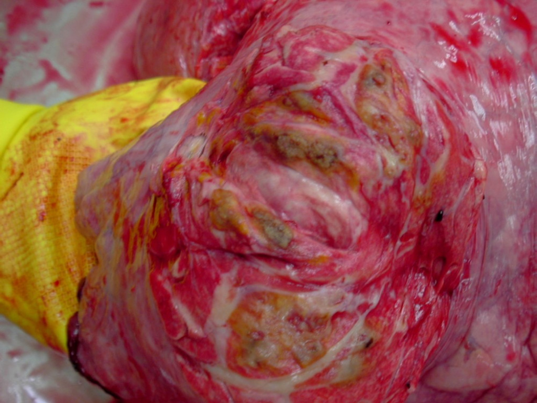
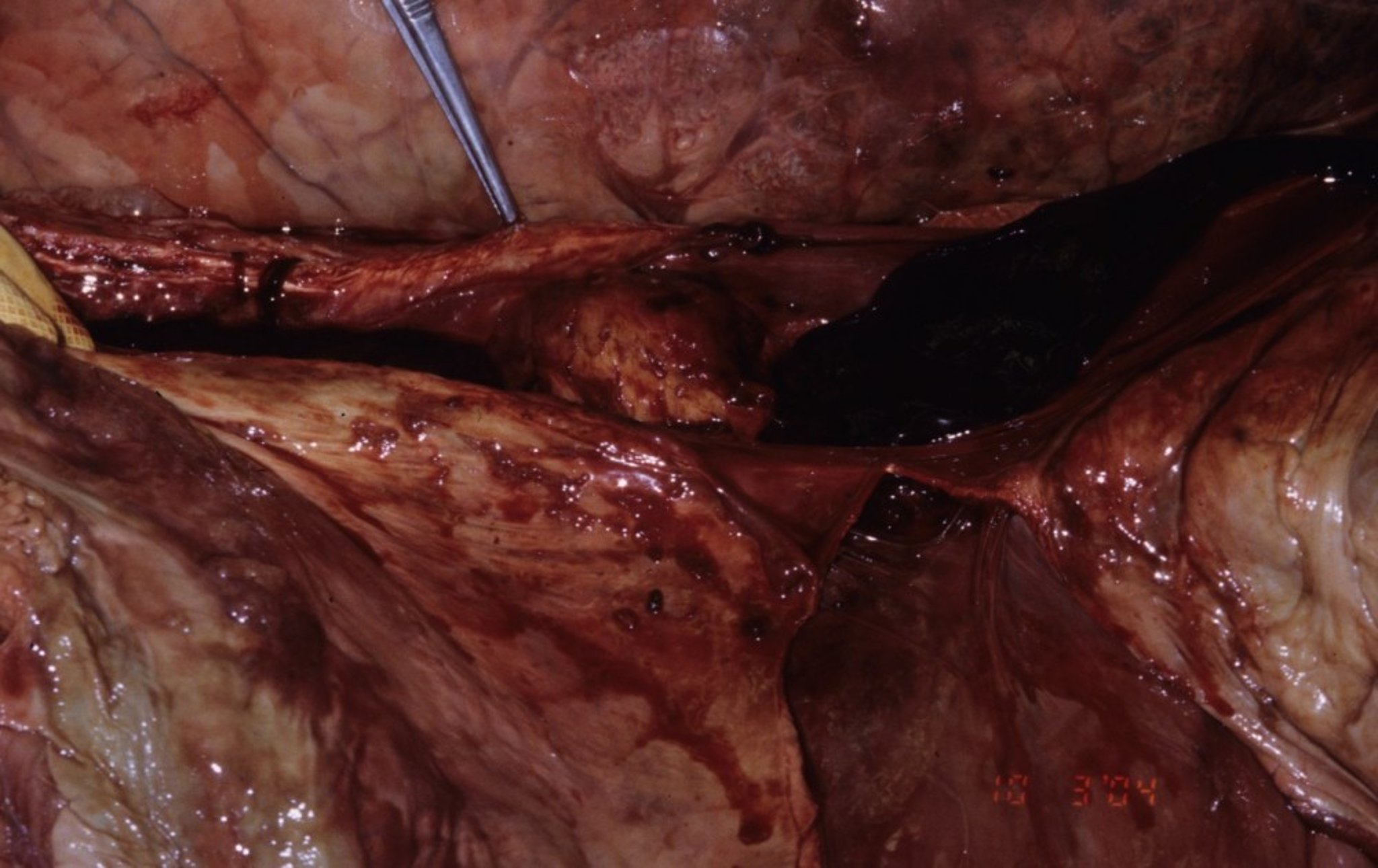
Cattle

Courtesy of Dr. Sameeh M. Abutarbush.

Courtesy of Dr. Sameeh M. Abutarbush.
Thrombosis of the caudal vena cava occurs in association with hepatic abscessation and vascular erosion of the abscess. Most often, the thrombus is visualized within the hepatic portion of the caudal vena cava, although it can also be found in the perirenal, subphrenic, or intrathoracic portion.
Typical clinical signs in cattle with caudal vena caval thrombosis include:
poor general condition
chronic weight loss
intermittent fever
Embolic pneumonia with secondary pulmonary abscessation, thromboembolism, and pulmonary arterial aneurysms are common sequelae. Affected animals with pulmonary involvement may present with coughing, tachypnea, dyspnea, and abnormal lung sounds. Aneurysms in pulmonary arteries that contain septic emboli may rupture and cause intrapulmonary hemorrhage, or pulmonary abscesses may erode into bronchi and result in hemorrhage into the airways. The sequelae to these disorders may include epistaxis, hemoptysis, and death. Clinical pathologic data usually support a diagnosis of vena caval syndrome but are not specific. Increased fibrinogen, anemia, and in cases with an active abscess process, increased liver enzymes may be seen.
Pulmonary arterial embolism and embolic pneumonia are also frequent complications of tricuspid or pulmonic valvular endocarditis in cattle, but aneurysms rarely develop. Intermittent fever and anorexia due to bacteremia at times of embolic showering are often present, and the animal typically has a history of a chronic active infection (eg, foot abscess, reticular abscess) as well as chronic weight loss with poor body condition score. Most cases of right heart endocarditis in cattle are bacterial and are commonly associated with a cardiac murmur, with a point of maximal intensity over the tricuspid valve. Echocardiography and blood cultures help identify right heart vegetative lesions and the causative bacterial agent, respectively.
Thrombosis of the cranial vena cava in cattle produces bilateral jugular engorgement; edema of the head, submandibular area (so-called "bottle jaw"), and brisket; and pronounced oral mucosal hyperemia. However, similar clinical signs are seen with right-side congestive heart failure, which could be a sequela of tricuspid valve endocarditis. Significant lingual, pharyngeal, or laryngeal edema may develop and result in dysphagia and dyspnea. Use of ultrasonography of the vena cava and echocardiography can aid in the diagnosis.
Horses
Cranial vena cava thrombosis may result from extension of a jugular thrombus. Jugular vein thrombosis in horses is often associated with phlebitis following catheterization or extravasation of injected material and will cause swelling, heat, and pain of the affected area. Bilateral jugular vein thrombosis can cause edema and swelling of the head and neck, mimicking cranial caval thrombosis. Ultrasonographic examination of the affected vein can determine the extent of the thrombus and degree of occlusion. Doppler ultrasound is a more sophisticated method to determine blood flow and vessel patency. If a catheter-associated thrombophlebitis is suspected, blood culture and catheter-tip culture can be performed.
Horses with colitis and other GI disorders are at increased risk of developing jugular thrombosis; ruminants are much less prone to jugular thrombosis than horses. Caudal vena cava–like syndrome has been described in a Quarter horse with respiratory signs. Hepatic abscesses, caudal vena cava thrombosis, pulmonary thromboembolism, and embolic pneumonia were identified at necropsy.
Migrating Strongylus vulgaris larvae ( see Strongylus vulgaris–Associated Disease) can cause arteritis with development of thrombi and verminous aneurysms in the aorta, cranial mesenteric, or iliac artery. In some horses, emboli develop and partially or completely occlude terminal branches of the mesenteric arteries. Affected intestinal segments show changes ranging from ischemia to hemorrhagic infarction. Clinical signs are those of colic, constipation, or diarrhea. The colic usually is recurrent, and attacks may be severe and prolonged. Newer anthelmintics and improved therapeutic regimens have resulted in verminous arteritis becoming an uncommon disorder.
Thrombosis with or without aneurysm of the terminal aorta and proximal iliac arteries produces a characteristic syndrome in horses. Although associated with parasitism, other causes are possible. Affected horses appear normal at rest; however, graded exercise results in an increasing severity of weakness of the hindlimbs with unilateral or bilateral lameness, muscle tremor, and sweating. Severely affected animals may show signs of exercise intolerance, weakness, and atypical lameness that resolves after a short rest. Subnormal temperature of the affected limbs may be detectable, along with decreased or absent arterial pulsations and delayed and diminished capillary filling. Rectal palpation may show variation in pulse amplitude of the internal or external iliac arteries (or both) and asymmetric vasculature. In severe cases, the hindquarter muscles atrophy, and lameness may become evident with only mild exercise.
Complete embolic or thrombotic occlusion of the distal aorta may produce acute bilateral hindlimb paralysis and recumbency in horses. Affected animals are anxious, appear painful, and rapidly go into shock. The hindlimbs are cold, and rectal palpation reveals an absence of pulsation in either iliac artery. Transrectal ultrasound can help determine blood flow in the aorta and iliac arteries.
Aneurysm of the aortic root has been reported in horses, commonly noted at the right aortic sinus with or without concurrent endocarditis. Similar to those in people, aneurysms of the sinuses of Valsalva in horses can be congenital or acquired. Rupture of an aortic aneurysm typically leads to sudden death, a scenario most commonly seen in breeding stallions during live cover.
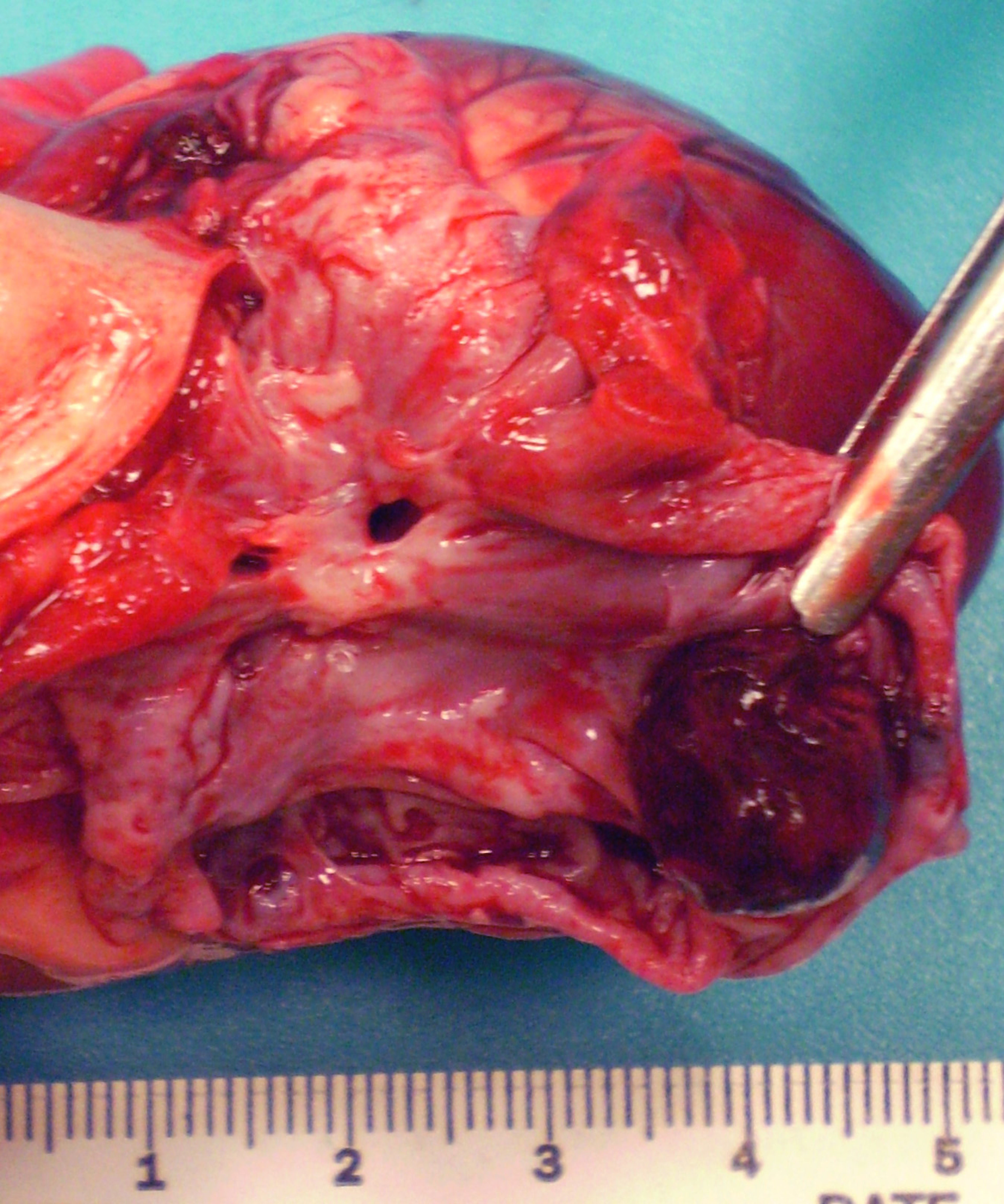
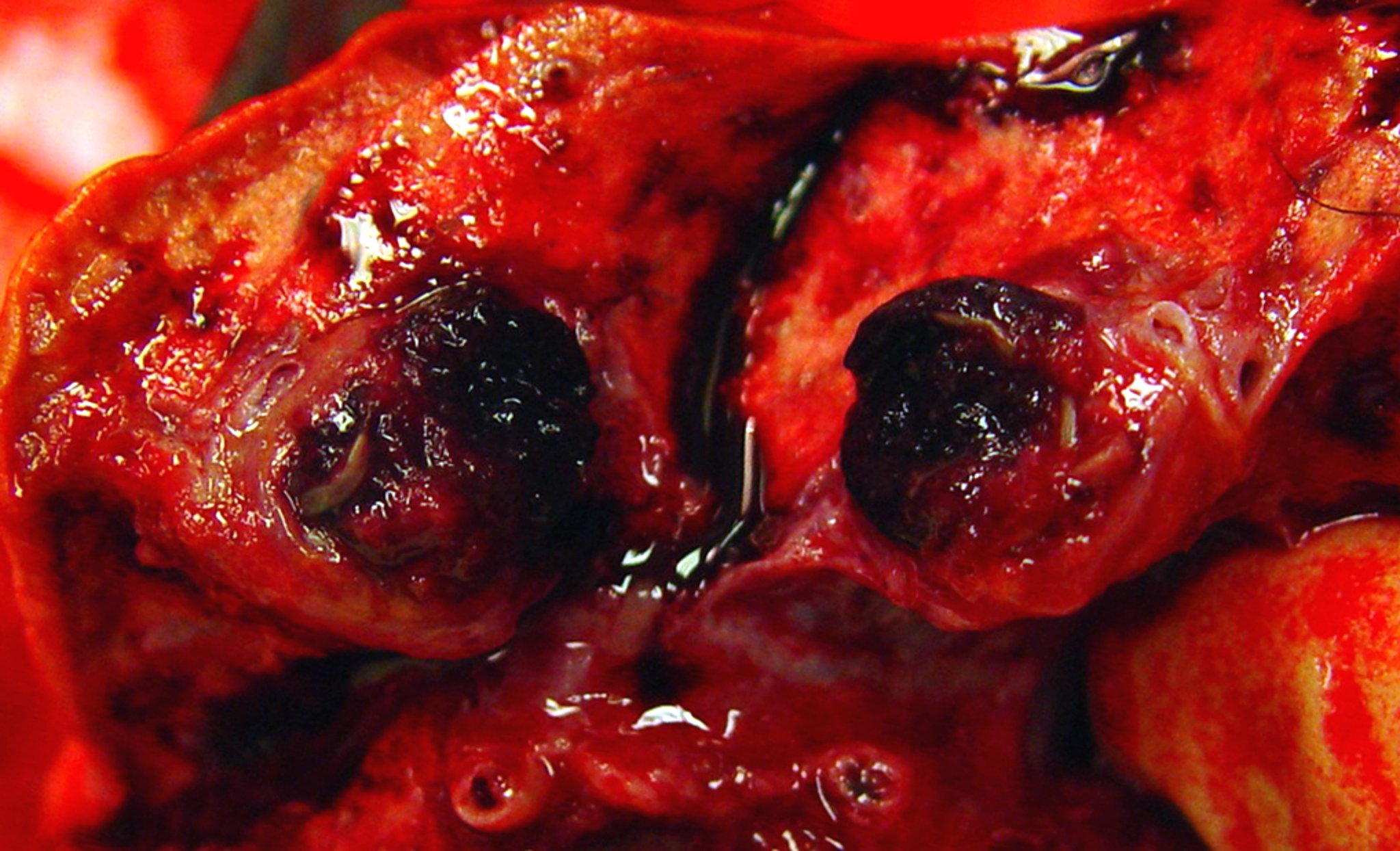
Dogs and Cats

Courtesy of Dr. Suzanne Cunningham.

Courtesy of Dr. Suzanne Cunningham.
In dogs, and less commonly in cats, heartworm disease may lead to pulmonary arterial thromboembolism (PTE) that commonly results in dyspnea, and collapse. Affected animals are often reportedly healthy until sudden onset of coughing, hemoptysis, respiratory distress, or sudden death. Clinical signs most commonly develop in the weeks after treatment with adulticide; however, pulmonary thromboembolism may also develop from spontaneous worm death, or pulmonary thrombosis can form in situ secondary to pulmonary endothelial damage. Chest radiographs in affected animals may be normal or show underperfusion of the affected lung lobe, interstitial to alveolar infiltrates, or pleural effusion. Visualization of tortuosity of the pulmonary arterial branches may be seen on thoracic radiographs in patients with heartworm disease or pulmonary hypertension secondary to PTE. Arterial blood-gas analysis typically demonstrates hypoxemia with a normal or low level of CO2 in the blood. Ventilation/perfusion scanning with radionuclide-labeled macroaggregated albumin and gases or pulmonary CT angiography can confirm the diagnosis.
Other diseases associated with pulmonary thromboembolism in dogs and cats include protein-losing nephropathy or enteropathy, hyperadrenocorticism, immune-mediated hemolytic anemia, and neoplasia. Additional diagnostic tests to evaluate for PTE and underlying etiologies may include echocardiography, thromboelastography, heartworm antigen (antigen/antibody for cats) testing, CBC, biochemical analyses, urinalysis with urinary protein:creatinine or urinary cortisol:creatinine ratio, low-dose dexamethasone suppression test, or ACTH stimulation test.
In cats, cardiogenic embolism (arterial thromboembolism) is a devastating complication of cardiac disease. Although hypertrophic cardiomyopathy is the most common type of cardiac disease in cats, any condition that results in left atrial enlargement, including other cardiomyopathies, hyperthyroidism, or congenital heart disease, can predispose to arterial thromboembolism. Intracavitary thrombi typically form in the dilated left atrium where stagnant flow exists or, less commonly, within abnormal areas in the left ventricle.
Although the condition is poorly understood, these cats may also possess some degree of concurrent hypercoagulability, because all cats with cardiomyopathy do not develop cardiogenic embolism. Portions of these intracavitary thrombi can break off and form emboli that infarct arterial branches, most commonly the aortic trifurcation (saddle emboli). Clinical signs include pain and paresis or lower motor neuron paralysis of the hindlimbs. The arterial pulse (either femoral or pedal) is reduced to absent in the affected limbs, which are cooler than normal and have firm, swollen gastrocnemius muscle bellies. These clinical signs can be unilateral, bilateral, or bilateral but asymmetric.
Emboli may also infarct other arterial beds, including the right forelimb, renal, splanchnic, cerebral, or myocardial circulation. Decompensation of the underlying myocardial disease is not uncommon and may result in congestive heart failure (pulmonary edema or pleural effusion). Ischemia and necrosis of infarcted pelvic limb musculature results in increases in serum CK and AST, and subsequent reperfusion of affected muscles can result in life-threatening hyperkalemia and acidosis. Differential glucose and lactate levels can be performed to help confirm a diagnosis of arterial thromboembolism.
Echocardiography is the imaging modality of choice to assess cardiac structure, function, and presence of an intracardiac thrombus, whereas chest radiographs are used to diagnose left-side congestive heart failure. Nuclear perfusion studies, using the unbound radioisotope 99mTc can give sensitive information regarding the degree of perfusion of the hindlimbs and areas that may require amputation in select cases, although this is rarely done. Infrared thermography may provide additional support in diagnosing arterial thromboembolism in cats and other animals.
Systemic hypertension in cats and patent ductus arteriosis, aortic coarctation, degenerative processes, and infections in dogs have been associated with aortic aneurysms. Obtaining differential blood pressure measurements in the forelimbs and hind limbs may be part of the diagnostic workup.
Cranial vena caval thrombosis and subsequent chylothorax and acute respiratory distress has been reported in dogs and cats with transvenous pacemakers and other indwelling jugular devices/catheters. Cranial vena caval thrombosis has also been associated with caval cannulation for open-heart surgeries performed under cardiopulmonary bypass. Successful treatment of caval thrombosis in affected animals has been described using a combination of anticoagulants, systemic and local infusions of thrombolytics, and balloon venoplasty, with or without placement of an endovascular stent.
Treatment of Thrombosis, Embolism, and Aneurysm in Animals
Treatment of thrombosis or embolism is intended to restore blood flow and perfusion, preventing further clot formation, while addressing the underlying etiology
Common approaches include supportive care, use of pharmacologic therapies (ie, antithrombotic, thrombolytics, antibiotics if sepsis is present ) as well as additional interventions (ie, angioplasty/venoplasty, endovascular stenting, thrombectomy)
Specific therapeutic approaches will vary based on the species, location, severity, chronicity, underlying etiology, and comprehensive clinical picture of the patient
Treatment of endocarditis includes longterm antibiotics (several weeks to months) and in some cases intermittent administration of antipyretic, anti-inflammatory, or antithrombotic drugs. Antibiotic choice should be based on culture and sensitivity results obtained from blood cultures. The prognosis for recovery is poor to guarded at best, and persistent debilitating cardiac disease is common even if the active infection can be controlled.
Treatment of venous thrombosis in horses and cattle is usually limited to supportive care, including hydrotherapy of accessible veins, anti-inflammatory agents, and systemic antimicrobials to control secondary sepsis. Surgical removal of thrombosed jugular veins has been performed successfully in horses, but unless both veins are severely affected, inflammation will resolve with medical treatment, and formation of collaterals will usually result in sufficient venous circulation. Thrombosis of the cranial or caudal vena cava results in more severe clinical signs and requires more aggressive therapy, which could include thrombolytic drugs and/or intravascular/surgical removal followed by aggressive anticoagulation. Response to anticoagulation therapy alone is generally inadequate.
Measures to minimize trauma to, and bacterial contamination of, veins remain the best means to prevent venous thrombosis. Extreme care should be taken when placing catheters or giving IV injections. The effectiveness of antiplatelet therapy in horses (aspirin at 5–10 mg/kg, PO, every 24 or 48 hours; or clopidogrel at 1–3 mg/kg, PO, every 24 hours), anticoagulant therapy (unfractionated heparin at 40–80 IU/kg, SC or IV, every 8–12 hours; or low-molecular-weight heparin [enoxaparin at 0.5 mg/kg, SC, every 24 hours; or dalteparin at 50 IU/kg, SC, every 12–24 hours for adults, or 100 IU/kg, SC, every 24 hours for foals]) to facilitate intrinsic thrombus resolution is unknown but should at least prevent further thrombus formation.
In horses, aneurysms due to Strongylus vulgaris rarely rupture; the chief concern is thromboembolism of intestinal vasculature with subsequent colic. Generally, the arterial wall is sufficiently involved that thrombus removal is impractical. Antibacterial treatment and anthelmintics to kill the migrating larvae are of considerable value. The most rational approach to cranial mesenteric and aortic-iliac thrombosis in horses is prevention and control of strongylosis.
Ultrasound-guided balloon thrombectomy for treatment of aorto-iliac-femoral thrombosis in a horse has been reported.
Acute management of aortic emboli in cats can be approached in several ways. More than 50% of cats that survive a cardioembolic event will regain some function of the hindlimbs over 4–6 weeks with conservative medical therapy. More aggressive therapy aimed at dissolution of the thrombus through thrombolytic drugs or rheolytic intervention may result in improved short-term functional outcome, but survival is no better than that from conservative therapy and, in some cases, may actually be worse.
Conservative therapy usually consists of initial pain management (hydromorphone at 0.1–0.2 mg/kg, SC, IM, or IV, every 4–6 hours; or buprenorphine HCl at 0.01–0.03 mg/kg, SC, IM, IV, or oral transmucosal [buccal], every 6–8 hours) and anticoagulant therapy (enoxaparin at 0.75–1 mg/kg, SC, every 6–12 hours for cats; dalteparin at 75 U/kg, SC, every 6 hours; or heparin at an initial dose of 250–300 U/kg, SC, every 6 hours).
The activated partial thromboplastin time can be used to monitor heparin therapy, with a goal of 1.5–1.7 × the pretreatment value. The use of antiplatelet therapy (clopidogrel at 37.5 mg total for loading dose, PO, once on admission, then 18.75 mg, PO, every 24 hours) should be considered to further reduce the thrombotic potential; in addition, it may have a beneficial effect on collateral circulation. Thrombolytic therapy, although not routinely recommended, could include streptokinase (90,000 IU/cat, IV over 20 minutes, followed by 45,000 IU as a continuous infusion for 2–24 hours), recombinant tissue-type plasminogen activator (tPA, 0.25–1 mg/kg/hr, IV, up to a total dose of 1–10 mg/kg), or urokinase (4,400 IU/kg, IV over 10 minutes, then 4,400 IU/kg/hr for 12 hours).
These drugs promote thrombolysis by converting plasminogen to plasmin, which subsequently breaks down fibrin strands. Streptokinase is considered a nonspecific activator of plasminogen, because it activates circulating fibrin as well as fibrin contained within thrombi/emboli, which can lead to a systemic proteolytic state and bleeding. Although urokinase and tPA are more fibrin-specific than streptokinase, bleeding can also be seen with these agents. Moreover, all of these agents are prohibitively expensive and difficult to obtain.
The use of antiplatelet agents such as clopidogrel has been shown to hasten thrombus dissolution and reduce acute arterial rethrombosis in experimental studies and human clinical trials, respectively. However, an in vitro feline study did not identify a significant difference in thrombolysis rates. It is not known whether these results can be applied to the natural clinical disease. Thrombolytic therapy appears to have the best response in cats with acute onset of clinical signs and incomplete or unilateral infarction. However, these cats may respond similarly well to conservative therapy, without the risk of reperfusion injury or expense of these agents.
Although a severe complete infarction is more likely to develop reperfusion injury with thrombolytic therapy, these cats are also less likely to recover with conservative therapy alone. The reported survival rates for initial aortic infarction events are similar whether conservative (35%–39%) or thrombolytic (33%) therapy is used. Cats with single limb infarction do much better (68%–93%) than cats with bilateral hindlimb infarction (15%–36%) regardless of therapy used. Aspirin (25 mg per cat, PO, every 48–72 hours; or 5 mg/kg, PO, every 48–72 hours) has historically been the most widely used preventive therapy for cardioembolic disease in cats. Although aspirin appears relatively safe in cats (up to 20% GI adverse effects) and is inexpensive unless compounding is done, the antiplatelet efficacy of aspirin in cats has been called into question, and currently there is no evidence that aspirin prevents first-time or recurrent cardiogenic embolism. Clopidogrel may be a more effective antiplatelet drug in this species.
Clopidogrel (18.75 mg/cat/day, PO) inhibits both primary and secondary platelet aggregation. These effects are more potent than those induced by aspirin. Clopidogrel also impairs the platelet release reaction, decreasing the release of pro-aggregating and vasoconstrictive agents. Adverse effects are rare but can include vomiting in up to 10% of cats; this appears to be ameliorated by giving the drug with food.
A combination protocol of aspirin and clopidogrel has been used previously, but has fallen out of favor. Aspirin as the sole antithrombotic in cats at risk for arterial thromboembolism is not recommended (CURATIVE consensus).
A multicenter, randomized, prospective study (FATCAT) revealed that clopidogrel was associated with a significantly prolonged survival time compared with aspirin in cats that presented with cardiogenic arterial thromboembolism. The time to recurrence of arterial thromboembolism or death in the clopidogrel group was >365 days versus 192 days in the aspirin group.
Thus, clopidogrel is currently recommended instead of aspirin for cats at risk of arterial thromboembolism. A double-blind, randomized active control study comparing clopidogrel vs rivaroxaban for prevention of recurrent arterial thromboembolism in cats (SUPERCAT) is currently underway.
Warfarin (0.25–0.5 mg/day/cat, PO) has also been used for prevention of primary or secondary cardiac embolism. Dosing is adjusted to prolong the prothrombin time to 1.5–1.7 × the pretreatment value. Because warfarin decreases the anticoagulant proteins C and S before reduction in factors II, VII, IX, and X, joint treatment with heparin is recommended for the first 5–7 days of warfarin therapy. Problems with warfarin therapy include large inter- and intra-individual variability, difficult dosing because of tablet size, and bleeding, including fatal hemorrhage. Warfarin is no longer recommended to use in cats, because of marked interindividual variation coupled with a narrow therapeutic index.
The low-molecular-weight heparins are smaller in size than unfractionated heparin but maintain the ability to inhibit factor Xa, with a greatly reduced inhibition of IIa. The reduced anti-IIa activity translates into a negligible effect on the activated partial thromboplastin time, but measurement of anti-Xa activity can be used to monitor dosing efficacy. Enoxaparin (1–1.5 mg/kg, SC, 2–3 times daily or 0.75-1 mg/kg SC every 6 hours) and dalteparin (150–170 IU/kg, SC, 2–3 times daily or 75U/kg every 6 hours) have both been used in cats. These drugs have been well tolerated with only rare bleeding reported, but objective clinical studies evaluating their efficacy have not been performed. These agents have been frequently combined with clopidogrel in an attempt to provide a more complete antithrombotic effect. This protocol appears to be well tolerated, although some minor bleeding has been seen.
The use of factor Xa inhibitors such as rivaroxaban (0.5–1 mg/kg, PO, every 24 hours) or apixaban has been reported in cats for thromboprophylaxis. These drugs appear to be well tolerated in combination with clopidogrel.
The reported cumulative risk of arterial thromboembolism in cats with cardiomyopathy at 1, 5, and 10 years is 3.5%, 9.5%, and 11.3%, respectively. Reported recurrence rates for cats receiving some form of antithrombotic prevention are 17%–75%, with a 1-year recurrence rate of 25%–50%. Longterm median survival times after an initial cardioembolic event have ranged from 51–376 days. Although these numbers may seem daunting, many of these cats can do well. If owners are willing to treat, they should be encouraged to give cats 24–72 hours of supportive care before deciding on euthanasia, unless severe infarction, severe CHF, severe azotemia, or reperfusion injury are present.
In the 2019 published consensus on the rational use of antithrombotics in veterinary critical care (CURATIVE) guidelines for small animals, high risk for thrombosis was identified for dogs with immune-mediated hemolytic anemia or protein-losing nephropathy, cats with cardiomyopathy and associated risk factors, and dogs and cats with >1 disease/risk factor for thrombosis.
Arterial thrombosis in dogs is most commonly associated with protein-losing nephropathy and neoplasia, though idiopathic thrombosis is also seen. There is little clinical experience with arterial thromboembolism in dogs, but thrombolytic therapy using streptokinase, urokinase, and tPA have been reported in isolated cases with variable success. There are no clinical trials evaluating the efficacy of antithrombotic therapy for prevention of arterial thromboembolism in dogs, but dosing protocols for aspirin (0.5–15 mg/kg/day, PO), clopidogrel (1.1–3 mg/kg/day, PO), warfarin (0.1–0.22 mg/kg/day, PO), unfractionated heparin (150–300 U/kg, SC, every 6 hours), dalteparin (100–175 U/kg, SC, 2-3 times daily), enoxaparin (1–1.5 mg/kg, SC, 2–3 times daily or 0.8 mg/kg, SC, every 6 hours), and rivaroxaban (0.5–2 mg/kg/day, PO) have been reported.
Treatment recommendations for pulmonary embolism in dogs are similar to those for cardioembolic disease in cats, including a combination of antiplatelet and anticoagulant therapy in addition to treatment of the underlying cause and pulmonary hypertension, if present. Reported antiplatelet medications in dogs includes aspirin, clopidogrel, abciximab, and ticagrelor). Aspirin (0.5 mg/kg/day, PO) has improved survival in dogs with immune-mediated hemolytic anemia when added to standard immunosuppressive therapy. Anticoagulant medication use reported in dogs includes heparin, low-molecular-weight heparin (enoxaparin, dalteparin), and factor Xa inhibitors (rivaroxaban, apixaban). Warfarin is not recommended, because of inconsistent outcomes associated with severe bleeding complications.
Successful treatment of cranial vena caval syndrome in dogs has been described using a combination of anticoagulants (clopidogrel and apixaban), systemic and local infusions of thrombolytics such as tPA, and balloon venoplasty with or without placement of an endovascular stent.
Key Points
Thrombosis, embolism, and aneurysms can occur in any species. Clinical signs, diagnostic testing, and therapy may vary based on the classification, location, severity, chronicity, and underlying etiology.
Treatment can include management of the underlying disease process, administration of antithrombotic or thrombolytic (systemic or local infusion) drugs, and surgical (surgical thrombectomy) or interventional procedures (balloon thrombectomy, balloon angioplasty/venoplasty with or without endovascular stent placement).
For More Information
Also see pet health content regarding blood clot disorders in dogs, in cats, and in horses.
JVECC 2019. Clinical application of the American College of Veterinary Emergency and Critical Care (ACVECC) Consensus on the Rational Use of Antithrombotics in Veterinary Critical Care (CURATIVE) guidelines to small animal cases. Sharp et al. First published: 06 February 2019 https://doi.org/10.1111/vec.12804
JVECC 2019. American College of Veterinary Emergency and Critical Care (ACVECC) Consensus on the Rational Use of Antithrombotics in Veterinary Critical Care (CURATIVE) guidelines: Small animal. Goggs et al. First published: 17 January 2019 https://doi.org/10.1111/vec.12801


