

A true hernia is defined as having a hernia ring, sac, and contents. Hernias of the abdominal wall are common in all domestic species and include umbilical hernias and inguinal or scrotal hernias. Hernias may be direct (through a rent in the body wall) or indirect (through an already existing ring, such as the inguinal ring or umbilical ring). Congenital hernias tend to be indirect.
Umbilical hernias vary in size and may contain only fat or omentum or, in more severe cases, intestinal loops. In dogs, Weimaraners, Pekingese, Basenjis, and Airedale Terriers are overrepresented. In many cases, umbilical hernias occur in dogs with concurrent cryptorchidism. Hereditary etiology is suspected but not proved. In cattle, the Holstein Friesian breed is overrepresented. In Holsteins, the sire and previous umbilical infection have been associated with an increased risk of umbilical hernia. Diagnosis in all animals is based on observation of the hernia sac, palpation, ultrasonographic examination, and possibly radiographic evaluation. Surgical closure of the body wall defect is indicated in most cases to lower the risk of future intestinal incarceration.

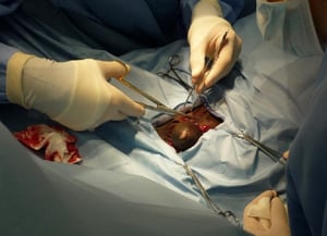
Intraoperative photograph of the surgical approach to a congenital umbilical hernia in a foal, showing a circumferential incision around the hernia sac.
Intraoperative photograph of the surgical approach to a congenital umbilical hernia in a foal, showing a circumferentia
Courtesy of Dr. Lisa Pearson.

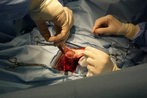
Intraoperative photograph in which the hernia sac of a congenital umbilical hernia in a foal is being opened.
Intraoperative photograph in which the hernia sac of a congenital umbilical hernia in a foal is being opened.
Courtesy of Dr. Lisa Pearson.

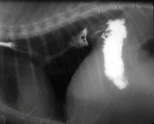
Lateral radiograph with contrast showing a hiatal hernia in a dog. The oral portion of the stomach is present in the thoracic cavity, having passed through the esophageal hiatus of the diaphragm.
Lateral radiograph with contrast showing a hiatal hernia in a dog. The oral portion of the stomach is present in the th
Courtesy of Dr. Ronald Green.

This photograph of a colt shows the gross appearance of a congenital inguinal hernia on the left side (right side of image with patient in dorsal recumbency), which is characterized by swelling and asymmetry of the scrotum.
This photograph of a colt shows the gross appearance of a congenital inguinal hernia on the left side (right side of im
Courtesy of Dr. Lisa Pearson.
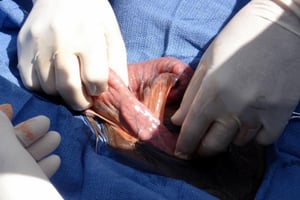
This photograph shows the contents (small intestine) of a congenital inguinal hernia in a colt.
This photograph shows the contents (small intestine) of a congenital inguinal hernia in a colt.
Courtesy of Dr. Lisa Pearson.

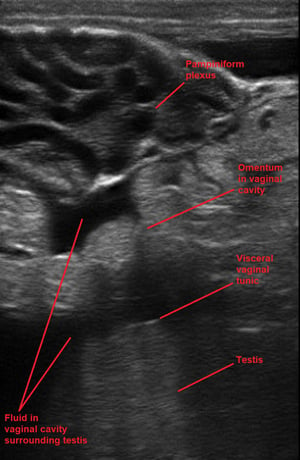
Ultrasonographic image of the pampiniform plexus and dorsal scrotum of a ram with an inguinal hernia. The vessels of the pampiniform plexus are dilated, and fluid and omentum are present within the vaginal cavity surrounding the testis. The top of the image is dorsal and the bottom is ventral. The probe is oriented longitudinally.
Ultrasonographic image of the pampiniform plexus and dorsal scrotum of a ram with an inguinal hernia. The vessels of th
Courtesy of Dr. Jennifer Roberts.

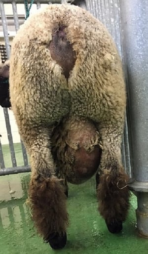
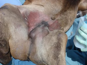
This photograph shows an inguinal hernia in a Hampshire ram, with herniation of abdominal contents and extensive fluid accumulation in the scrotum.
This photograph shows an inguinal hernia in a Hampshire ram, with herniation of abdominal contents and extensive fluid
Courtesy of Dr. Jennifer Roberts.

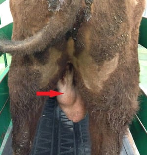
The inguinal hernia of the bull in this photograph is evident as an asymmetrical swelling (arrow) at the proximal aspect of the left side of the scrotum.
The inguinal hernia of the bull in this photograph is evident as an asymmetrical swelling (arrow) at the proximal aspec
Courtesy of Dr. Jennifer Roberts.
Intraoperative photograph of the surgical approach to a congenital umbilical hernia in a foal, showing a circumferential incision around the hernia sac.
Intraoperative photograph of the surgical approach to a congenital umbilical hernia in a foal, showing a circumferentia
Courtesy of Dr. Lisa Pearson.
Intraoperative photograph in which the hernia sac of a congenital umbilical hernia in a foal is being opened.
Intraoperative photograph in which the hernia sac of a congenital umbilical hernia in a foal is being opened.
Courtesy of Dr. Lisa Pearson.
Lateral radiograph with contrast showing a hiatal hernia in a dog. The oral portion of the stomach is present in the thoracic cavity, having passed through the esophageal hiatus of the diaphragm.
Lateral radiograph with contrast showing a hiatal hernia in a dog. The oral portion of the stomach is present in the th
Courtesy of Dr. Ronald Green.
This photograph of a colt shows the gross appearance of a congenital inguinal hernia on the left side (right side of image with patient in dorsal recumbency), which is characterized by swelling and asymmetry of the scrotum.
This photograph of a colt shows the gross appearance of a congenital inguinal hernia on the left side (right side of im
Courtesy of Dr. Lisa Pearson.
This photograph shows the contents (small intestine) of a congenital inguinal hernia in a colt.
This photograph shows the contents (small intestine) of a congenital inguinal hernia in a colt.
Courtesy of Dr. Lisa Pearson.
Ultrasonographic image of the pampiniform plexus and dorsal scrotum of a ram with an inguinal hernia. The vessels of the pampiniform plexus are dilated, and fluid and omentum are present within the vaginal cavity surrounding the testis. The top of the image is dorsal and the bottom is ventral. The probe is oriented longitudinally.
Ultrasonographic image of the pampiniform plexus and dorsal scrotum of a ram with an inguinal hernia. The vessels of th
Courtesy of Dr. Jennifer Roberts.
This photograph shows an inguinal hernia in a Hampshire ram, with herniation of abdominal contents and extensive fluid accumulation in the scrotum.
This photograph shows an inguinal hernia in a Hampshire ram, with herniation of abdominal contents and extensive fluid
Courtesy of Dr. Jennifer Roberts.
The inguinal hernia of the bull in this photograph is evident as an asymmetrical swelling (arrow) at the proximal aspect of the left side of the scrotum.
The inguinal hernia of the bull in this photograph is evident as an asymmetrical swelling (arrow) at the proximal aspec
Courtesy of Dr. Jennifer Roberts.
Inguinal hernias and scrotal hernias are common in pigs, horses (particularly draft breeds and warmbloods), and many breeds of dogs. They are suspected to be hereditary. Inguinal hernias can occur in bitches and may involve the uterus. Clinical signs vary from nonpainful inguinal or scrotal swelling to acute colic in horses or vomiting in dogs, particularly if the small intestine is strangulated.
In horses, rectal palpation can be used to diagnose intestinal loops in the vaginal ring, which can be gently removed to provide relief before transport to a surgical facility. Any devitalized bowel is resected via midline celiotomy. In stallions, testis-sparing laparoscopic closure of the inguinal rings has been performed in both standing and recumbent horses with good outcome and subsequent fertility. In bulls and rams, inguinal hernias may contain loops of intestine or omentum. Increased temperature due to abdominal contents in the scrotum results in abnormal spermatogenesis, spermatozoal defects, and decreased fertility.
Unilateral castration may be indicated if there is irreparable damage to the testis or extensive adhesion formation. In foals and calves, medical management through reduction of the hernia and placement of a figure-eight bandage has been successful in some cases. Hernias that do not spontaneously resolve early in life should be surgically corrected to prevent later complications.
Hernias between the abdominal and thoracic cavities that involve the diaphragm are of several types. Congenital pleuroperitoneal hernias have been described in small animals, horses, and calves. In horses, a specific type of hernia—the retrosternal or Morgagni hernia—has been described in which a hernial sac protrudes into the thorax in the left dorsal tendinous portion of the diaphragm. The sac is characterized by a pleural covering and a peritoneal lining. In described cases, the presenting clinical sign was colic, and diagnosis was made during exploratory celiotomy.
Defects can be surgically repaired with mesh products to decrease the risk of recurrence. The hernial sac is usually left in situ. In cases of direct herniation, clinical signs include dyspnea, exercise intolerance, lethargy, and weight loss. In cattle, herniation of the reticulum into the thorax has been described, with a right-side diaphragmatic defect. Clinical signs include anorexia, scant feces, tympani, and decreased or no rumination. Diagnosis is based on radiographic or ultrasonographic evaluation.
Peritoneopericardial hernias are defined as an embryologic defect that results when the septum transversum fails to fuse during diaphragmatic development, allowing communication between the abdominal cavity and pericardial sac. Weimaraners and domestic long-haired cats may be more commonly affected than other breeds. Clinical signs reflect the contents of the hernia, which may include omentum, liver, gallbladder, or small intestine; these signs include cardiac tamponade, dyspnea, tachypnea, exercise intolerance, coughing, vomiting, and GI obstruction.
In many cases of peritoneopericardial hernias, diagnosis is an incidental finding during imaging or celiotomy for other reasons. Other congenital defects are found in many cases, including umbilical hernia, cryptorchidism, cleft palate, portosystemic shunt, and sternal or vertebral abnormalities. Patients with clinical signs are treated with surgical herniorrhaphy; patients with no clinical signs should be closely monitored.
Hiatal hernias occur through the esophageal hiatus. Type I, the sliding hernia, is the most common in small animals and is characterized by intermittent displacement of the lower esophageal sphincter and gastric fundus into the thoracic cavity. Type II is less common and involves only the displacement of the gastric fundus. Brachycephalic breeds are overrepresented, with a hereditary nature suspected in Shar-Pei.
Clinical signs of hiatal hernias include dysphagia, regurgitation, vomiting, ptyalism, and esophagitis due to decreased function of the lower esophageal sphincter. Diagnosis is based on radiographic or fluoroscopic evaluation; however, the intermittent nature can make diagnosis challenging. Medical treatment of esophagitis is required. Hiatal hernias are surgically corrected by a combination of hiatal plication, esophagopexy, and left-side gastropexy.


