


Common GI Parasites of Cattle
Name | Size | Anatomical Location of Adults | Appearance on Fecal Flotation | Extent of Pathogenicity |
|---|---|---|---|---|
Paramphistomum spp (flukes) | Adults: 5–15 mcm Eggs: 160 mcm | Rumen | Egg | Low |
Haemonchus spp (barber's pole worm, large stomach worm, wire worm) | Adults: 10–30 mm Eggs: 65–100 × 34–50 mcm | Abomasum | Egg | High |
Ostertagia ostertagi (medium or brown stomach worm) | Adults: 6.5–9 mm Eggs: 65–100 × 34–50 mcm | Abomasum | Egg | Medium-high |
Mecistocirrus digitatus | Adults: 40 mm Eggs: 65–100 × 34–50 mcm | Abomasum | Egg | Low |
Trichostrongylus spp (stomach or bankrupt worms) | Adults: 4–7 mm Eggs: 65–100 × 34–50 mcm | Abomasum, small intestine | Egg | Medium-high |
Cooperia spp (cattle bankrupt worm, small intestinal worm) | Adults: 4.5–6 mm Eggs: 65–100 × 34–50 mcm | Small intestine | Egg | Medium-high |
Strongyloides papillosus (intestinal threadworm) | Adults: 3.5–10 mm Eggs: 40–60 × 32–40 mcm | Small intestine | Larvated egg | Low |
Bunostomum phlebotomum (hookworm) | Adults: 12–26 mm Eggs: 79–97 × 47–50 mcm | Small intestine | Egg | Low-medium |
Nematodirus spp (thread-necked intestinal worms) | Adults: 10–23 mm Eggs: 175–260 × 106–110 mcm | Small intestine | Egg | Low |
Toxocara vitulorum (roundworm) | Adults: 200–300 mm (20–30 cm) Eggs: 75–95 × 60–75 mcm | Small intestine | Egg | Low-medium |
Moniezia spp (tapeworms) | Adults: Width 15–25 mm, length variable Eggs: 65–75 mcm in diameter | Small intestine | Egg or segments | Low |
Aonchotheca (formerly Capillaria) bovis | Adults: 8–20 mm Eggs: 45–50 × 22–25 mcm | Small intestine | Egg with bipolar plugs | Low |
Oesphagostomum spp (nodular worms) | Adults: 12–21 mm Eggs: 73–89 × 34–45 mcm | Cecum, colon | Egg | Low-medium |
Chabertia ovina (large-mouth bowel worm) | Adults: 12 mm Eggs: 65–100 × 34–50 mcm | Colon | Egg | Low-medium |
Trichuris spp (whipworms) | Adults: 35–80 mm Eggs: 70–80 × 30–42 mcm | Cecum, colon | Egg with bipolar plugs | Medium |
Common Parasites of the Rumen in Cattle
Paramphistomum spp Rumen Parasites in Cattle
Adult flukes of the species Paramphistomum are occasionally found in ruminal fluid or on necropsy in cattle, and eggs may be found on fecal sedimentation. Adults attached to the rumen papillae are 5–15 mm long and appear light tan-pink. Eggs are the approximate size and shape of Fasciola hepatica eggs (160 mcm); however, they are lighter brown to clear in color. This parasite follows a typical trematode life cycle, passing eggs that hatch in water and liberate miracidia that infect the intermediate host, snails. The parasite develops in the snail, and cercariae are released that encyst on herbage and are consumed by the ruminant host. Disease is rare in the US.
Common Parasites of the Abomasum in Cattle
Nematodes of the abomasum in cattle cause erosion and ulceration of the gastric mucosa and resulting gastritis. Damage to the gastric mucosa decreases the amount of pepsin and hydrochloric acid produced, resulting in a potentially less acidic abomasum. Some abomasal nematodes cause profound anemia and ill thrift.
Haemonchus spp Abomasum Parasites of Cattle

Courtesy of Dr. Grace VanHoy.
Haemonchus placei is the most common Haemonchus species in cattle in temperate North America; however, Haemonchus similis and Haemonchus contortus are occasionally encountered. Haemonchus differs from other trichostrongyles in that it requires a blood meal for survival. The trauma to the mucosa produced by this parasite as it uses its cutting mouthparts to take blood meals also allows blood to seep into the lumen of the abomasum. The amount of blood loss caused by each nematode has been calculated to be 0.05 mL per day. Fecal occult blood tests can detect blood in feces, but they are only moderately sensitive and specific. The pelleted nature of small-ruminant and camelid feces make false negatives common.
In addition to the loss of red blood cells, the loss of plasma proteins through mucosal defects that worms create can be appreciable. Moderate to severe hypoproteinemia with hypoalbuminemia develops during chronic protein loss in the GI tract.
Although there are hyperacute, acute, and chronic forms of haemonchosis, the chronic form is the most common one to affect cattle.
Cattle infected by Haemonchus parasites are anemic, unthrifty, and weak, and they may have bottle jaw or sternal edema if protein loss is severe. Haemonchosis does not generally lead to diarrhea unless the animal is coinfected with other trichostrongyles. Cattle with severe disease often require blood transfusions and intense supportive care, or they may be found dead before clinical signs are apparent.
FAMACHA scoring (observation of the ocular mucous membranes for signs of pallor indicating anemia) can be used to monitor for the development of clinical signs related to Haemonchus infection; however, this scoring system may not be as reliable as in small ruminants and camelids. FAMACHA scoring is discussed in Monitoring.
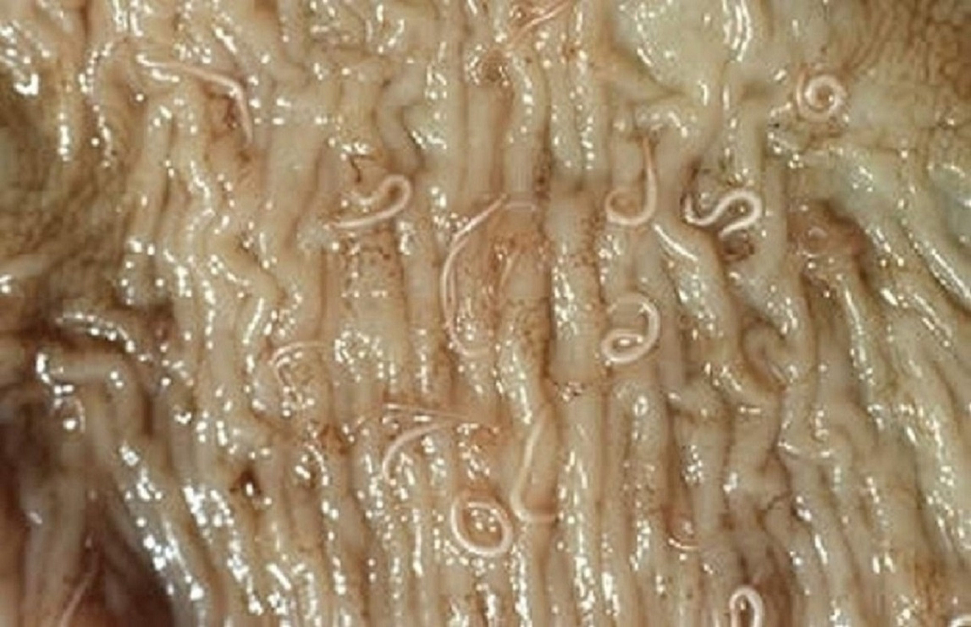
Adult Haemonchus females are up to 30 mm long and have a characteristic barber pole appearance because the reproductive and GI systems of the worm are entwined in a spiral pattern. Adult male worms are up to 18 mm long and white. Haemonchus is a highly fecund species: a single female produces up to 10,000 eggs per day under the right conditions. On a fecal flotation the eggs have the typical appearance of trichostrongyle eggs (see image in Etiology).
The life cycle of Haemonchus is the same as of other trichostrongyles, and the prepatent period is 18–21 days. Larvae on pasture remain infectious for 4–6 days after the eggs are passed in feces. Eggs are more resistant to environmental conditions such as freezing or desiccation than are larvae; however, all stages have some tolerance. After being ingested, infectious L3s can enter an arrested development stage in the abomasum similar to that of Ostertagia ostertagi in type II ostertagiasis (see below).
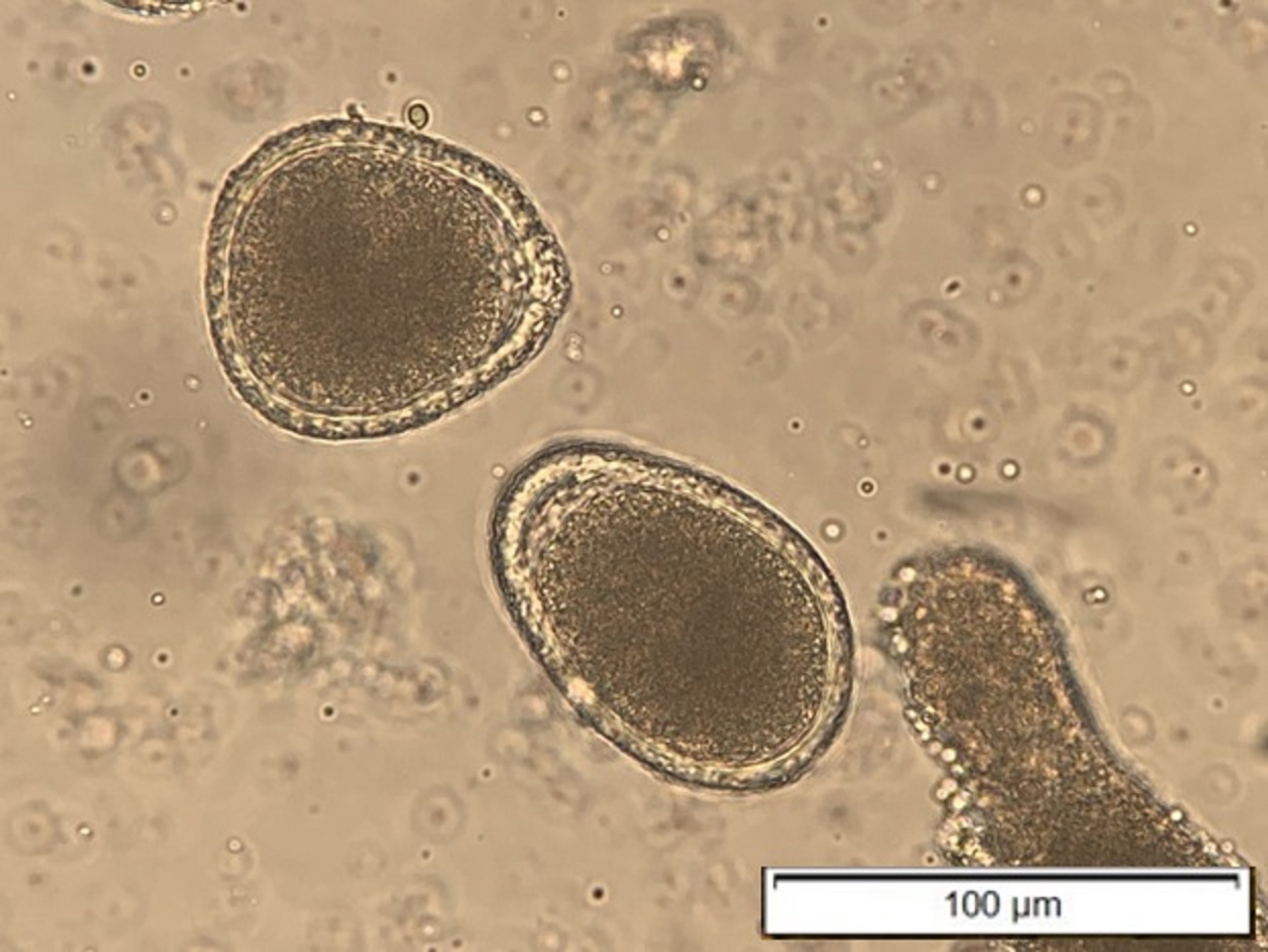
Ostertagia ostertagi Abomasum Parasite of Cattle

Courtesy of Dr. Sameeh M. Abutarbush.
Ostertagia ostertagi is one of the most important species of GI parasites in cattle in North America. Its life cycle and the appearance of its eggs on fecal flotation are characteristic of a trichostrongyle, its prepatent period is 3 weeks, and Ostertagia adults are 6–9 mm long.
Ingested larvae enter the lumen of the abomasal glands and molt by the fourth day; they remain there during the prepatent period, growing and undergoing a final molt before emerging as young adult worms from the gastric glands onto the abomasal mucosa. During this time the specialized cells (pepsinogen-producing zymogen cells, acid-producing parietal cells) lining parasitized glands are lost and replaced by hyperplastic, undifferentiated cuboidal cells, resulting in nodules that may be discrete or confluent.
Around the time of worm emergence, the changes evident in parasitized glands also appear in neighboring nonparasitized glands, rapidly extending the effects of the parasite burden. As a result, in heavy infections abomasal pH may rise from 2 to > 6. From a clinical viewpoint, when pH rises above 4.5, digestion in the abomasum ceases. A protein-losing gastropathy results and, together with anorexia and impaired protein digestion, leads to hypoproteinemia and weight loss. Diarrhea is persistent.
In type I ostertagiasis, which results from recent infection by O ostertagi, most worms present are adults, and the response to anthelmintic treatment is good. Type I disease occurs primarily in calves 7–15 months old, most commonly from the time of weaning and ensuing months in warm temperate regions. In cool temperate regions, it is most common in young cattle during summer and early fall. Cattle may retain their appetite until later in the disease, mimicking the weight loss, hypoproteinemia, diarrhea, and appetite retention observed in Johne disease (which is caused by Mycobacterium avium paratuberculosis).
In type II ostertagiasis, large numbers of larvae that had become dormant or inhibited in development at the early L4 stage emerge from the glands weeks or months later. Type II ostertagiasis occurs primarily in cattle 12–20 months old in the beginning of their second grazing season. In warm temperate regions, inhibition-prone larvae are acquired in spring, and disease may result when large numbers of larvae resume development to the adult stage in late summer or fall. In cold temperate regions, inhibition-prone larvae are acquired during late fall and mature during late winter or early spring. Type II disease is difficult to confirm with fecal examination alone because larvae are not producing eggs yet.
Larval inhibition in O ostertagi and other nematodes has been interpreted as a survival mechanism in which the preparasitic stages on pasture avoid the adverse conditions of winter in cool regions and of hot and dry (or hot and alternately wet and dry) conditions of many warm regions. The factors that lead to and later terminate inhibition are not completely known but are related to ambient temperature and moisture. In warm regions of both the Northern and Southern Hemispheres, inhibition develops principally during spring before the hot and dry conditions of summer. The resumed development or maturation of the parasites is likely to be genetically predetermined and may be influenced by parturition, nutrition, concurrent infection, and host immune response.
Trichostrongylus axei Abomasum Parasite of Cattle
In cattle, Trichostrongylus axei causes gastritis with superficial erosion and hyperemia of the abomasal mucosa, as well as diarrhea. Protein loss from the damaged mucosa and anorexia result in hypoproteinemia and weight loss. Inhibition (hypobiosis) does not occur to the same extent as with Ostertagia (see above). Adult worms are small and hairlike, are 5 mm long, and have low fecundity. Clinical signs occur only with heavy infection, malnourishment, or stress.
Mecistocirrus digitatus Abomasum Parasite of Cattle
Mecistocirrus digitatus is a hematophagous trichostrongyle with a pathology similar to that of Haemonchus; however, M digitatus adults are much larger, ~40 mm long. This parasite is typically present only in tropical climates.
Common Parasites of the Small Intestine in cattle
Infection of the small intestine by nematodes will result in enteritis and varying amounts of protein-losing enteropathy, depending on virulence and worm burden. The small intestine cestodes (tapeworms) rarely cause disease in adult cattle; however, they can cause ill thrift and weight loss in very heavy infections of young animals.
Cooperia spp Small Intestine Parasites of Cattle
Several species of Cooperia are found in the small intestine of cattle; Cooperia punctata, Cooperia oncophora, and Cooperia pectinata are the most common in North America. The red, coiled adults are 5–8 mm long, and the male has a large bursa. Most adults can be found in the first 10–20 ft (3–6 m) of the duodenum. Owing to their small size, they may be difficult to observe grossly without running luminal contents through a screen or making scrapings of the mucosa. The Cooperia life cycle is the same as that of other trichostrongyles, and the prepatent period is 12–15 days.
Cooperia spp are not hematophagous; however, C punctata and C pectinata can cause severe enteritis with anorexia and emaciation. Lesions in the duodenum include marked congestion of the mucosa with petechial hemorrhages. C oncophora produces a milder disease, with poor productivity and weight loss. C oncophora is now the most prevalent parasite causing disease in cow-calf operations in the US, resulting in dramatic decreases in average daily gain.
Trichostrongylus spp Small Intestine Parasites of Cattle
Trichostrongylus spp are also found in the small intestine. Parasite specifics are similar to those of trichostrongyles found in the abomasum (see Common Parasites of the Abomasum in Cattle.
Strongyloides papillosus Small Intestine Parasite of Cattle
Strongyloides papillosus is not a trichostrongyle and has a complex life cycle in which larvae or larvated eggs are passed in the feces and mature on pasture to become free-living adults. The prepatent period is 10 days. Only adult females (3.5–6 mm long) are parasitic and are embedded in the mucosa of the upper small intestine.
Strongyloides infections result from the ingestion of filariform larvae or by transmammary or percutaneous transmission. Strongyloides infections arise most commonly in poorly drained pasture or confined dairy operations with moist conditions and macerated pedal skin. S papillosus generally behaves like a commensal; it causes disease only in severe infections.
Young animals are the most commonly affected; clinical signs, though rare, may include intermittent diarrhea, loss of appetite and weight loss, with occasional blood or mucus in the feces. Large numbers of worms in the small intestine can produce catarrhal enteritis with petechiae and ecchymoses. Sudden death without premonitory clinical signs can occur.
Bunostomum phlebotomum Small Intestine Parasite of Cattle
Bunostomum phlebotomum is hematophagous and has a well-developed buccal capsule into which the mucosa of the small intestine is drawn and abraded by cutting plates during feeding by the worm. Adult females are 25 mm long; adult males, 15 mm.
Infection is by ingestion or skin penetration, which is common in areas with high stocking density and moist conditions. Transcutaneous infection may lead to stamping and uneasiness in cattle. The prepatent period is 60 days, and the appearance of the egg and the life cycle are similar to those of a typical trichostrongyle.
This parasite is generally nonpathogenic; however, heavy infection can produce clinical signs including anemia, intermittent diarrhea, and rapid weight loss with variable hypoproteinemic edema. On necropsy, the small intestine mucosa may appear congested or swollen with petechial hemorrhages where worms were attached. Abrasions, edema, and scabs may develop where larvae penetrate skin.
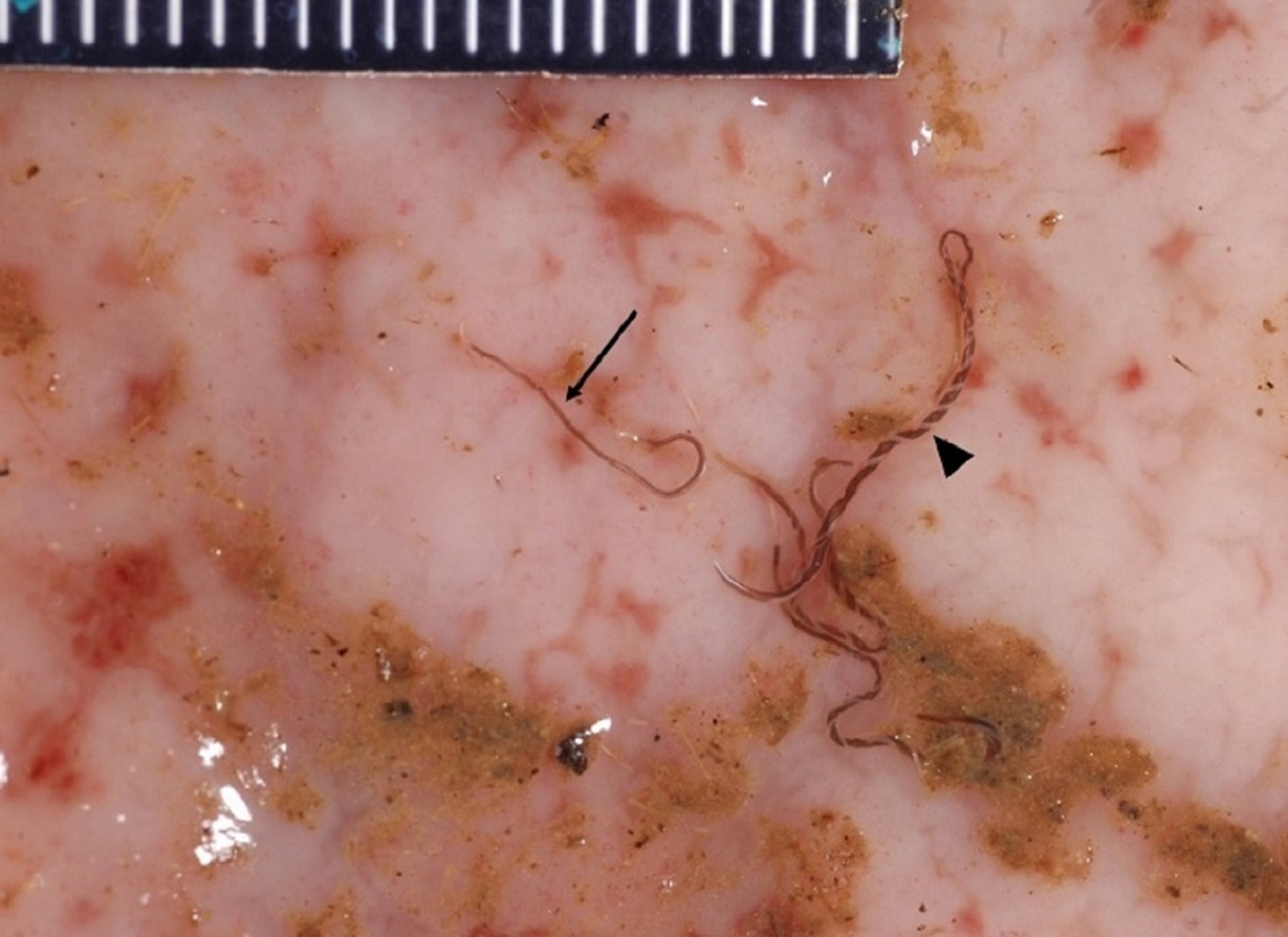
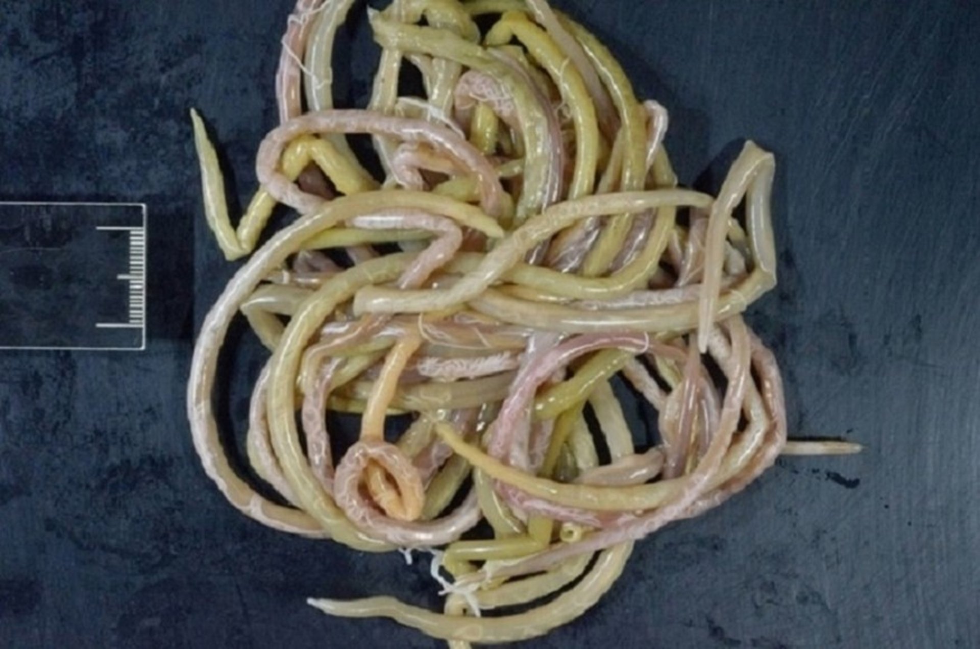
Nematodirus spp Small Intestine Parasites of Cattle

Courtesy of Dr. Grace VanHoy.
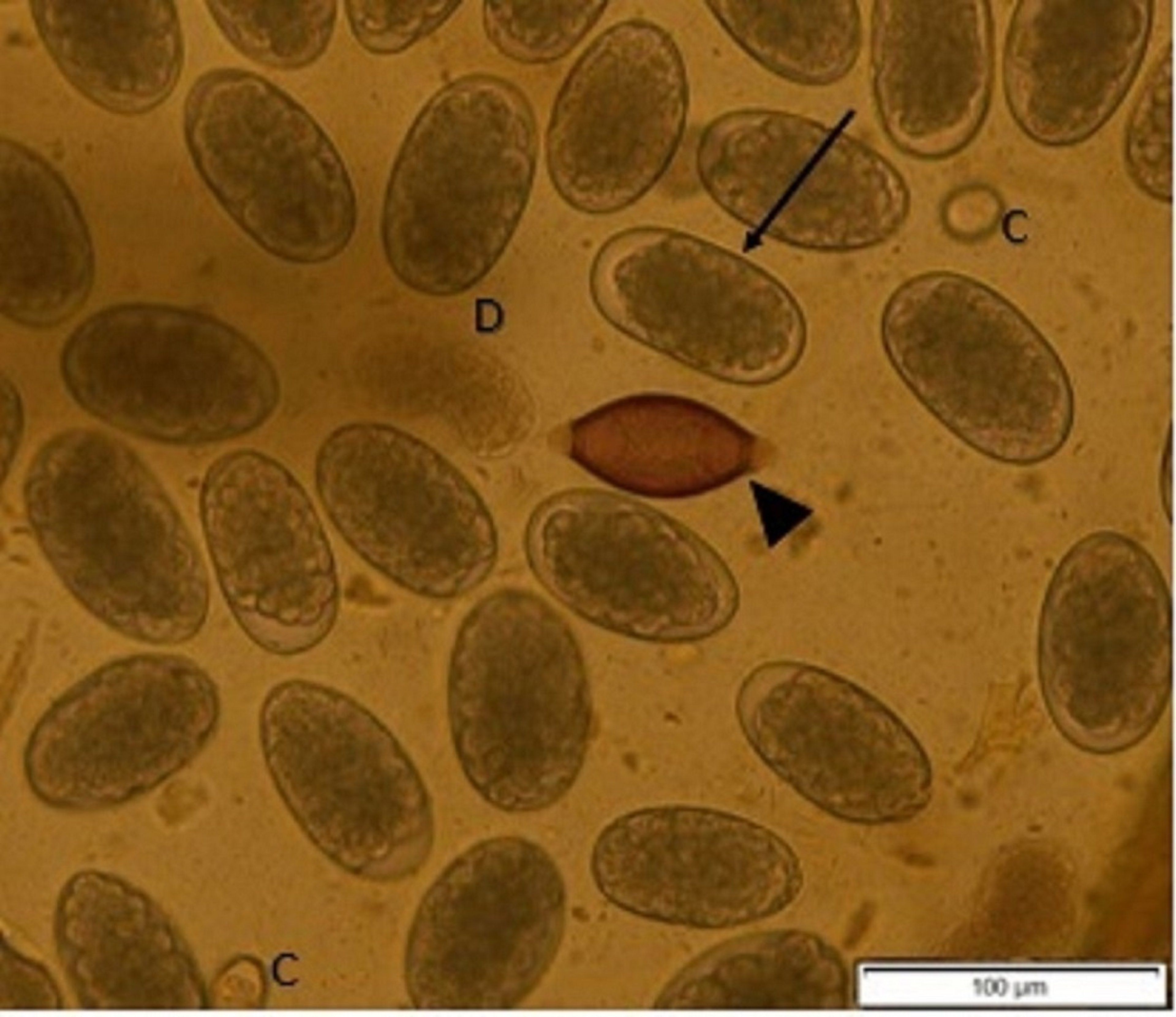
Nematodirus helvetianus is generally recognized as the most common bovine species of Nematodirus in North America; however, Nematodirus spathiger, Nematodirus filicollis, and Nematodirus battus can also infect cattle. Adult females are 18–25 mm long with a spine on the end of the tail and very large eggs in the uterus, and adult males are 12 mm long and are commonly found in the first 10–20 ft (3–6 m) of duodenum. The Nematodirus life cycle is typical of a trichostrongyle, except it is much longer than the life cycles of most other trichostrongyles and requires a period of overwintering for full development.
Eggs are passed in the feces and slowly develop over 2–3 months to larvated eggs. Larvated eggs overwinter in the soil, and low temperatures induce eggs to hatch and release larvae. In the spring, as soil temperatures begin to rise, mobile infectious L3 larvae climb onto vegetation and are ingested. After ingestion, L3s penetrate the intestinal mucosa and molt to L4 and L5 stages. L5s leave the mucosa and become adults in the lumen of the small intestine. The prepatent period after ingestion is 15 days, and fecundity is generally lower than for other trichostrongyles. Adult parasites live in the small intestine for a few weeks before dying.
Because larvated eggs require overwintering, pasture contamination and risk of infection in a given year depend directly on contamination of the pasture the previous year. After larvae hatch, they live for only a month on pasture; therefore, depending on the timing of larval hatching and calving, neonates may be at risk. Nematodirus is the most pathogenic to young ruminants that have no acquired immunity; adult ruminants may harbor some parasites and contribute to pasture contamination, but overt disease is rare.
The low fecundity of Nematodirus means that lower fecal egg counts may still represent a large adult population, and false negatives are more possible. This parasite has a very distinctive, large egg. Necropsy may reveal only thickened, edematous mucosa.
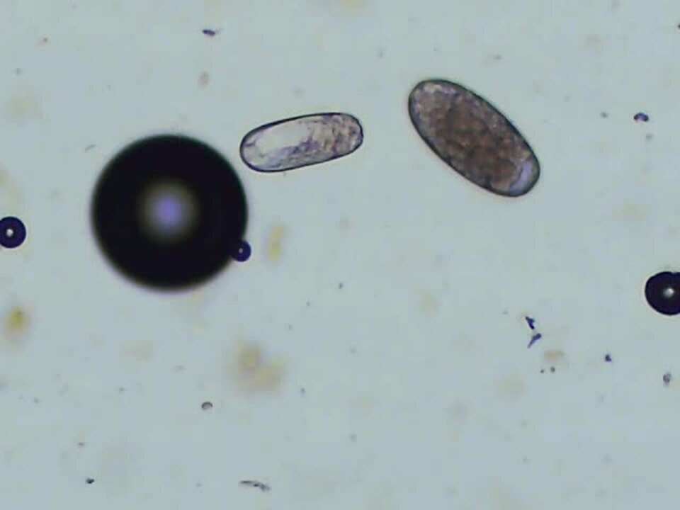
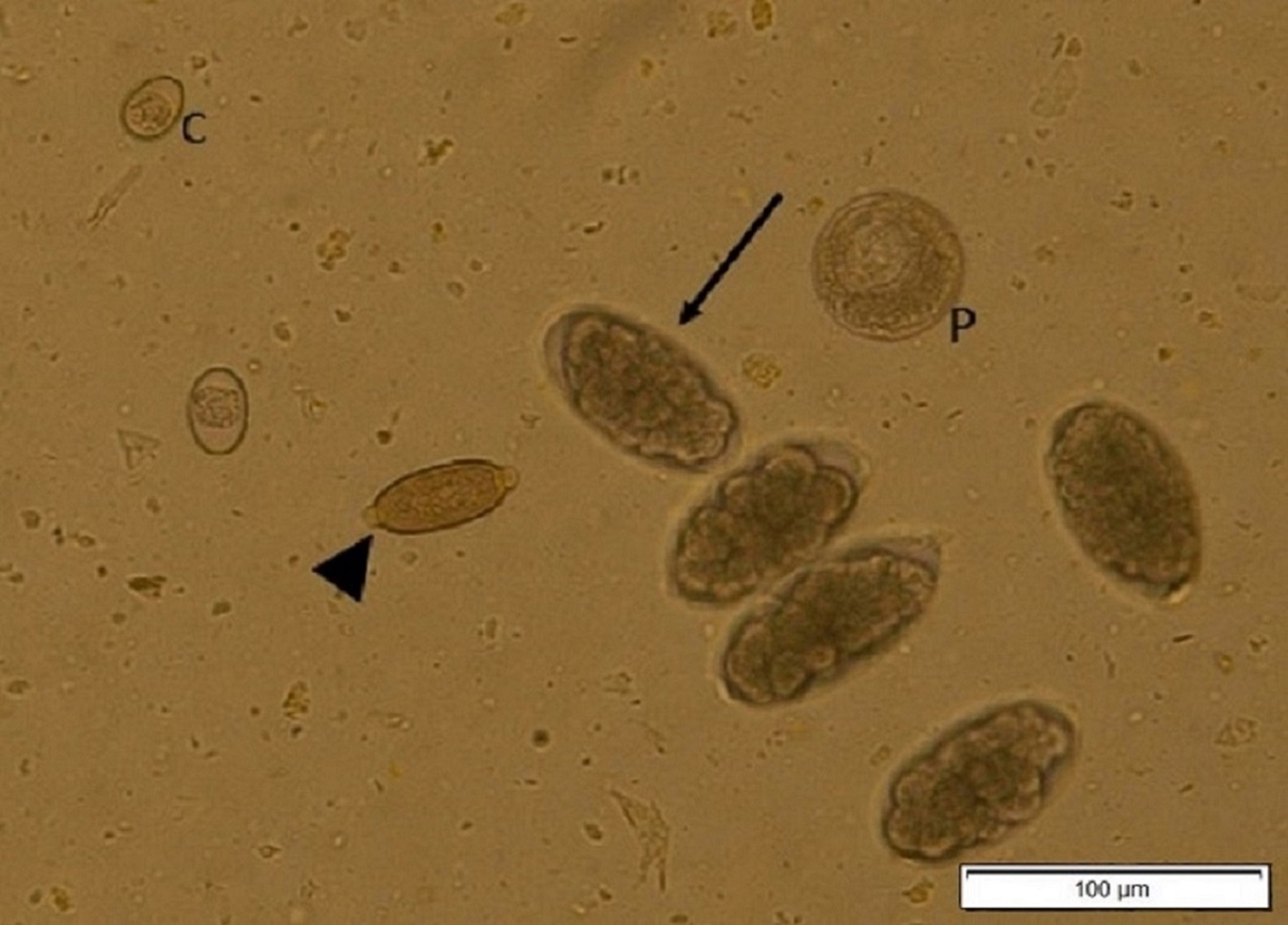
Toxocara vitulorum Small Intestine Parasite of Cattle

Courtesy of Dr. Lauren Camp.

Courtesy of Dr. Kristina Vu.

Courtesy of Dr. Kristina Vu.
The ascarid Toxocara vitulorum is a stout, white worm (females 25–30 cm, males 20–25 cm) found in the small intestine of calves < 6 months old. Older calves are resistant because they have robust acquired immunity. Larvae hatching from ingested eggs pass to the tissues and, in pregnant cows, are mobilized late in pregnancy and passed via the milk to calves. Eggs appear in the feces of calves from 3 weeks old and are easily recognized by their thick, pitted shells and dark brown center.
Light to moderate infection may be tolerated without clinical signs. Heavy infections can produce diarrhea, weight loss, and death.
Moniezia spp Small Intestine Parasites of Cattle
The anoplocephalid tapeworms Moniezia benedeni and Moniezia expansa are found in young cattle. The worms of this group are characterized by the absence of a rostellum and hooks, and by segments that are wider than they are long. The eggs are triangular or rectangular and are ingested by the intermediate host, free-living oribatid mites that live in the soil and grass. After 6–16 weeks, the infective cysticercoid Moniezia larvae are present in the mites. Infection occurs after ingestion of the mites; the prepatent period is 4–5 weeks.
Moniezia are generally considered nonpathogenic in calves, but intestinal stasis has been reported.
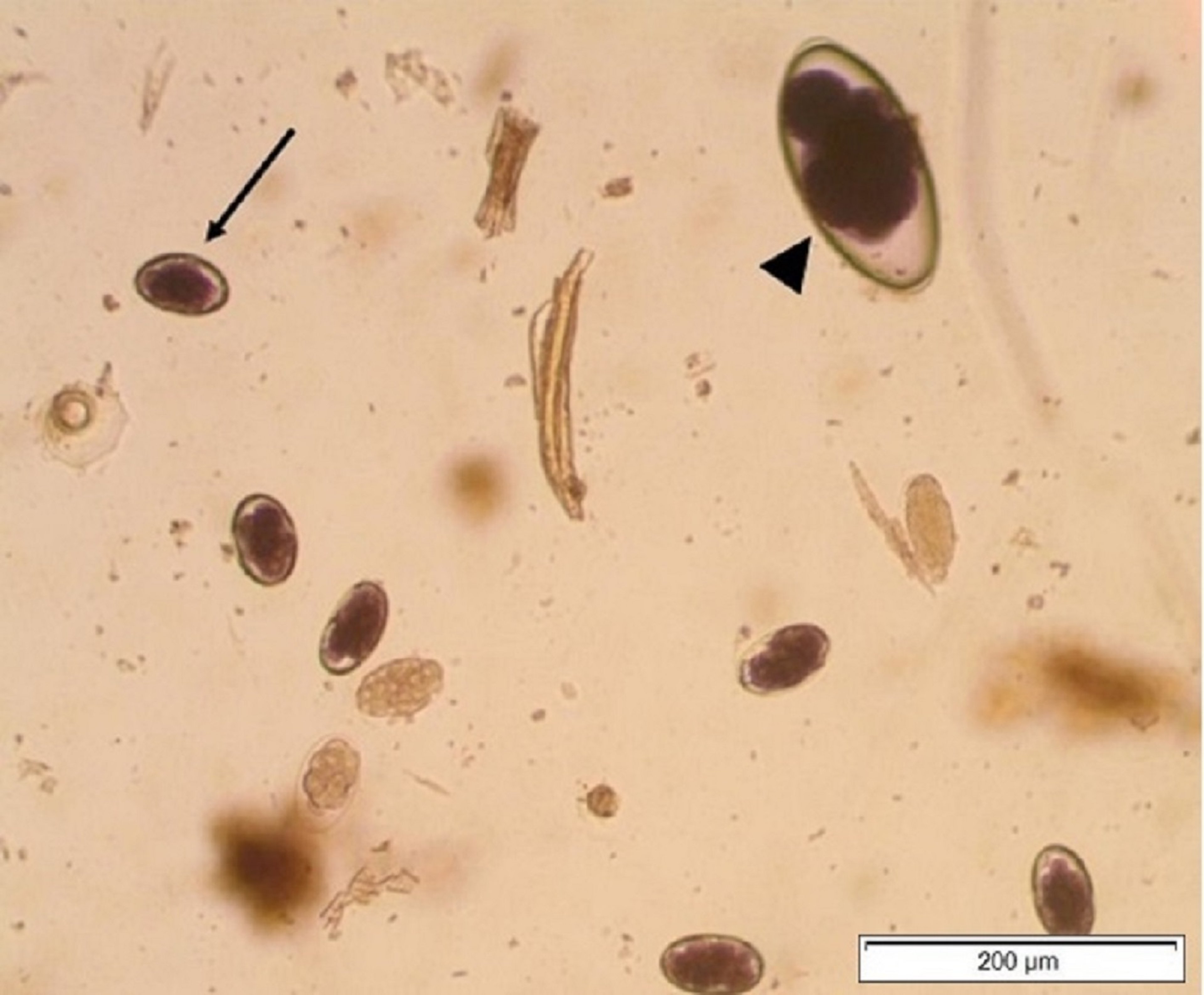
Aonchotheca (Capillaria) bovis Small Intestine Parasite of Cattle

Courtesy of Dr. Grace VanHoy.
Aonchotheca are closely related to Trichuris; however, their eggs are slightly smaller and lighter brown, and the bipolar plugs are less prominent. This parasite has been detected on routine fecal examination, but no specific pathogenicity has been reported in cattle.
Common Parasites of the Large Intestine in Cattle
GI parasites of the large intestine in cattle cause enteritis with petechial hemorrhages and intestinal wall edema, as well as varying amounts of protein-losing enteropathy, depending on the parasite's virulence and the worm burden. Some parasites of the large intestine can cause calcified nodules to form on the serosal surface in chronic infections.
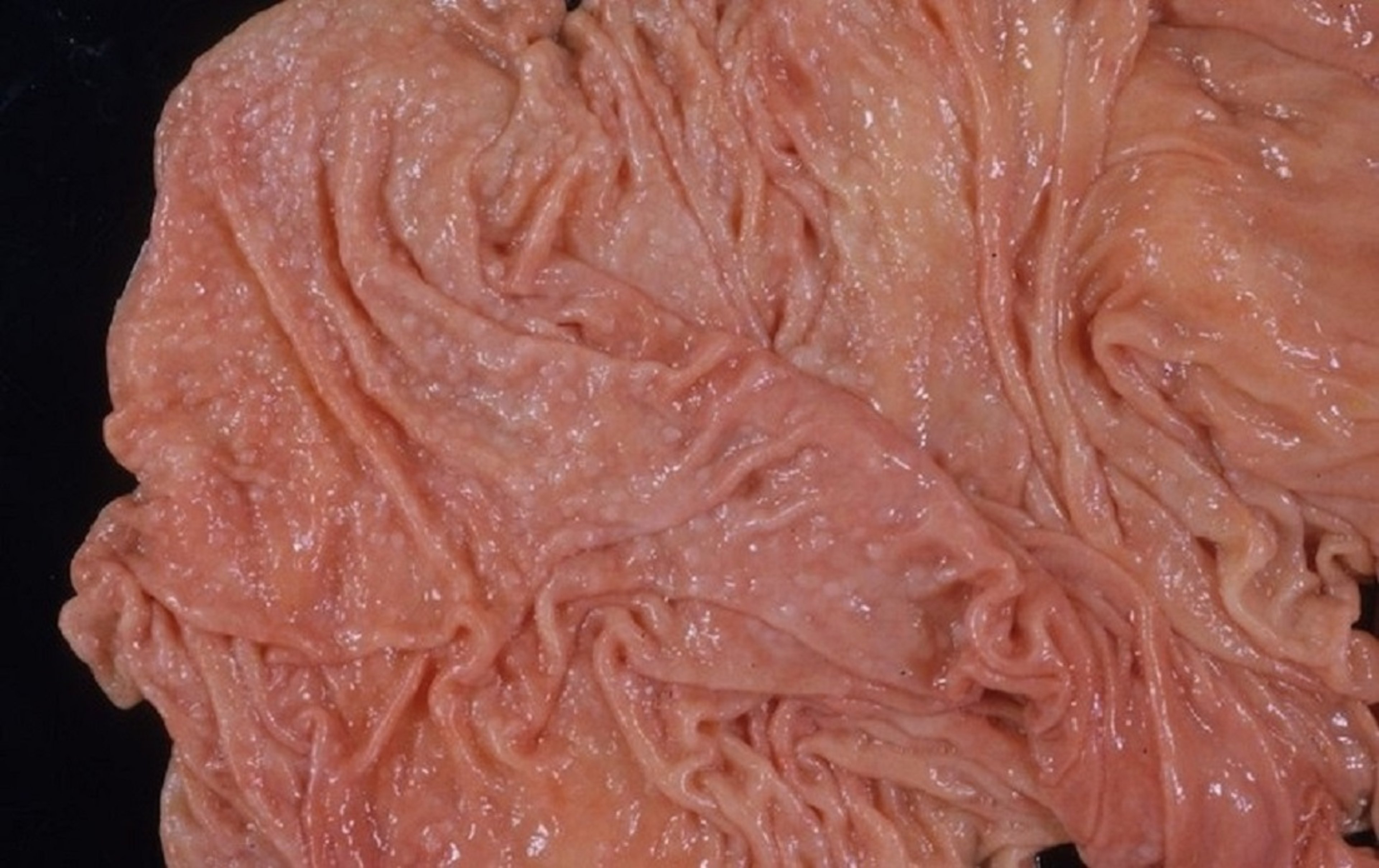
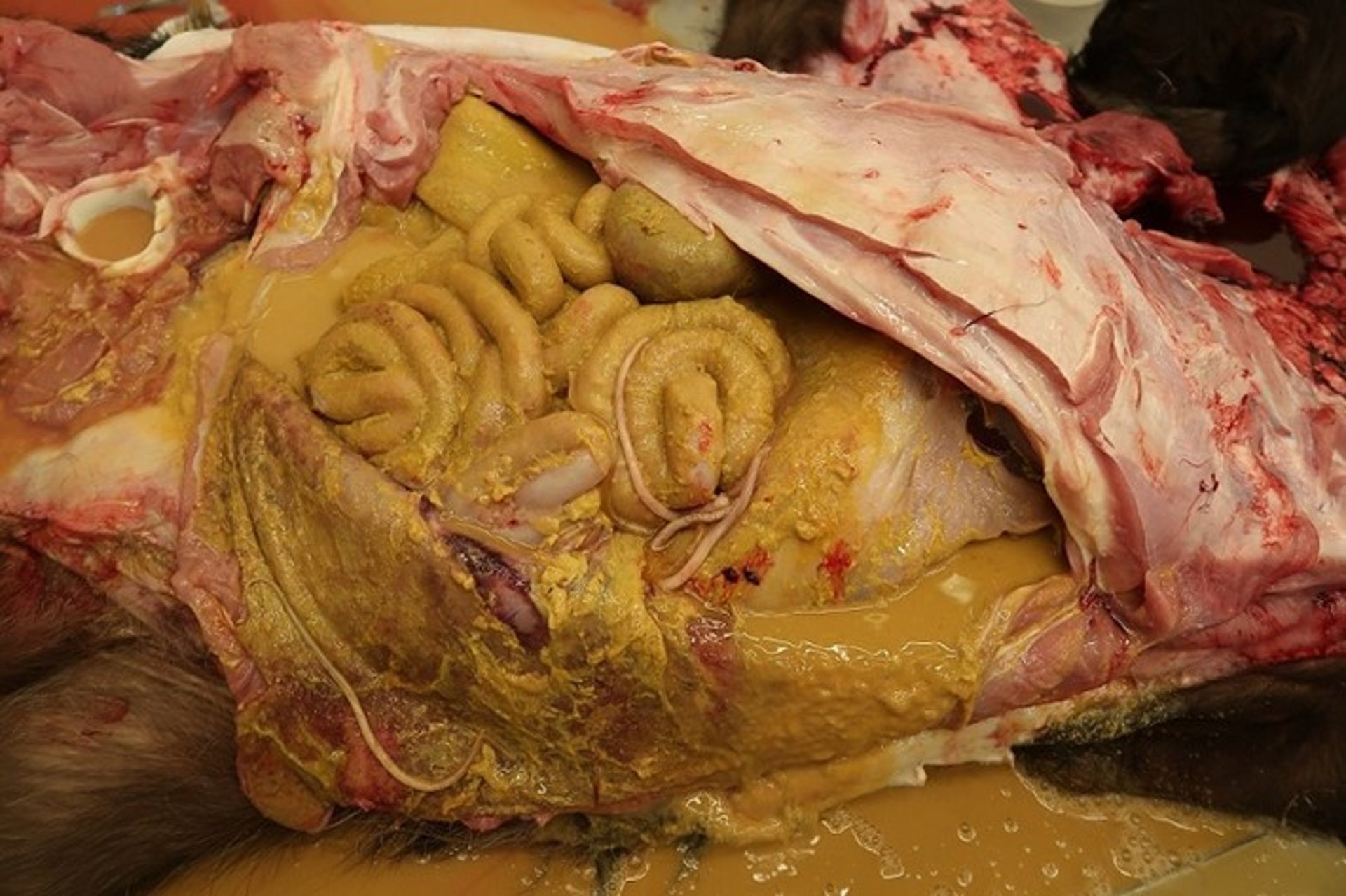
Oesophagostomum spp Large Intestine Parasites of Cattle
Adults of Oesophagostomum radiatum, Oesophagostomum venulosum, and Oesophagostomum columbianum are 12–15 mm long. The life cycle and appearance of eggs on fecal flotation are typical of a trichostrongyle. The larvae can penetrate the distal ileum but are more commonly found in the cecum and colon. Larvae remain embedded in the mucosa for 5–10 days and then return to the lumen as fourth-stage larvae (L4). The prepatent period is 6 weeks.
Upon reinfection and acquisition of partial immunity, larvae can become arrested and may encyst in mucosa or serosa and eventually cause the formation of distinctive, calcified nodules. An exaggerated inflammatory response to the encystment of immature larvae can lead to the clinical signs in acute infection of watery, dark, fetid diarrhea; weight loss; and death. Calcified nodules in chronic infection may disrupt intestinal motility and cause intussusception.
Disease usually results from nonpatent infections, so eggs are not observed on fecal flotation. On necropsy or abdominal exploratory surgery, nodules may be observed on the serosa of cecum and colon. These nodules either are filled with caseated pus and larvae, or are calcified and inactive.
Chabertia ovina Large Intestine Parasite of Cattle
Chabertia ovina adults are 12 mm long and bent ventrally at the anterior end. The life cycle of C ovina and the appearance of its eggs on fecal flotation are typical of a trichostrongyle. The larvae penetrate the mucosa of the small intestine in cattle and later emerge and pass to the colon, where most pathology is found. The prepatent period is 7 weeks. Larvae and adults may cause small hemorrhages with edema in the colon and passage of feces coated with mucus; however, clinical chabertiosis is rare in cattle.
Trichuris spp Large Intestine Parasites of Cattle

Courtesy of Dr. Raffaele Roncalli.

Courtesy of Dr. Grace VanHoy.
Trichuris spp infections are common in young calves and yearlings; however, the worms are seldom very numerous. The adults are found in the cecum and colon and are characterized by a long, slender anterior segment that coils into the intestinal mucosa and a thicker posterior segment. Trichuris spp are not trichostrongyles, but they have a similar direct life cycle. Infective eggs are ingested and hatch to larvae within the ruminant. Larvae then penetrate the wall of the anterior small intestine, where maturation proceeds for 2–10 days, after which the larvae migrate to the cecum and colon to become adults. The prepatent period is 7–9 weeks.
Trichuris eggs are distinctive from those of trichostrongyles and doubly operculated. The eggs are resistant to poor environmental conditions, and infections may persist on certain premises. Clinical signs are rare; in occasional heavy infections, however, dark feces, anemia, hypoproteinemia, and anorexia may be evident.
Diagnosis, Treatment, and Control of Gastrointestinal Parasites in Cattle
For the diagnosis, treatment, and control of GI parasites in cattle, see Overview of Gastrointestinal Parasites of Ruminants.


