

Nutritional Diseases of Amphibians
See also page Environment and Husbandry for Amphibians
Nutritional diseases in amphibians are common because typical food sources lack macro- and micronutrients. With the exception of earthworms, most invertebrates ingested by amphibians have an inverse calcium:phosphorus ratio. During veterinary visits, it is critical to discuss nutritional needs and supplementation with owners. A liquid diet suitable for carnivores may be used for amphibians to provide additional nutritional support when required.
Metabolic bone disease

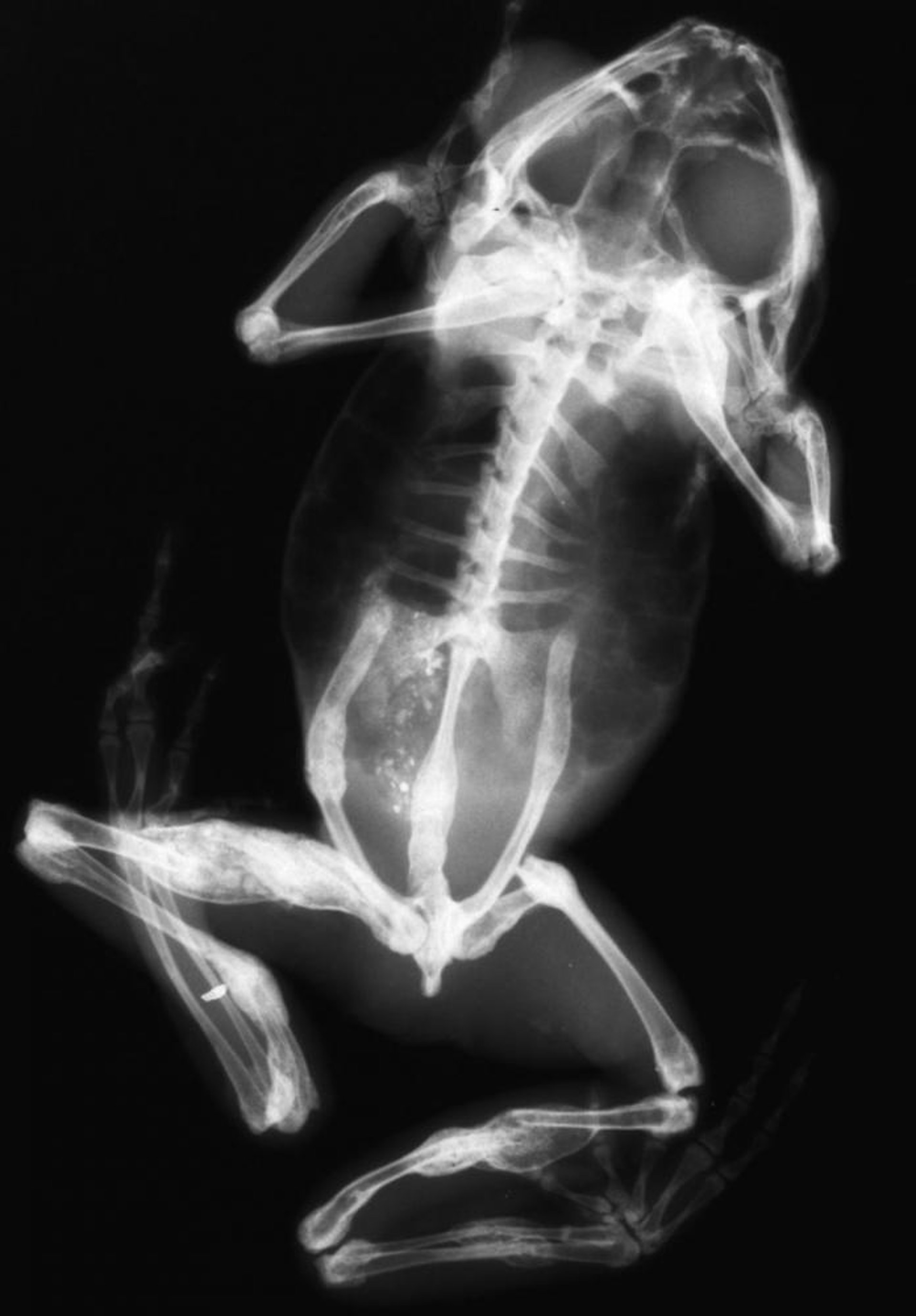
Courtesy of the National Aquarium.

Courtesy of the National Aquarium.
Metabolic bone disease (MBD), or nutritional secondary hyperparathyroidism, is frequently observed in captive amphibians, resulting from a combination of dietary calcium and vitamin D3 deficiency, inappropriate UVB light provision, and water source ratio of calcium:phosphorus. Water sources should also be investigated for fluoride concentration, which can also contribute to MBD-like lesions. Although difficult to diagnose, kidney disease causing secondary hyperparathyroidism must be ruled out.
Inverse ratios of calcium:phosphorus result in mandibular deformity, long bone fracture, scoliosis, and, eventually, tetany and bloating. Diagnosis is made via radiographic evaluation showing thinning cortices of long bones, mandibular and hyoid bone deformities, pathological fractures, and in severe cases, gastrointestinal gas.
Treatment includes correcting the diet and administering calcium glubionate (1 mg/kg, PO, every 24 hours for 30 days). Full-spectrum lighting with biologically active UVB light should be provided. For species more sensitive to prolonged or continuous light cycles, boost UVB application can be used, which consists of high levels of UVB for short periods of 20 minutes weekly until lesions resolve and then every 3–4 weeks thereafter. Starvation, resulting in weight loss, lethargy, and dehydration, must be treated by providing proper nutrition via assisted feeding.
Thiamine deficiency
Thiamine deficiency occurs in amphibians fed frozen fish. The freezing process of fish results in oxidation of the fat-soluble vitamins and vitamins B9 and vitamin C. Clinical signs include tremors, seizures, and opisthotonos. Initial treatment is the administration of thiamine at 25–100 mg/kg, IM or intracoelomically, followed by thiamine (25 mg/kg, PO) with each meal. Thiamine deficiency can be prevented by routinely supplementing diets with 250 mg thiamine per kg of fish fed.
Hypovitaminosis A
The carotenoids, including vitamin A, are not synthesized by amphibians and must be provided via diet. Excessive levels of vitamin A have been hypothesized to interfere with vitamin D metabolism and contribute to metabolic bone disease, whereas deficiency has been associated with lethargy, wasting, and inability to use the tongue to catch prey due to the development of squamous metaplasia of the tongue (short tongue syndrome). Other clinical signs can include brown to black pigmentation on the tongue and facial nodules caused by oronasal fistulae. Diagnosis is typically via dietary review, because confirmation requires hepatic biopsy for retinol assessment, which is not feasible in most amphibian species. Treatment includes providing vitamin A supplementation and force-feeding a proper diet.
Obesity
Obesity is a disease. Overfeeding is the primary cause of obesity; many amphibian species will continue to consume prey as long as it is available and without regard for their energy needs. The oversized fat bodies may be palpated within the coelomic cavity; however, in females, ultrasonography may be necessary to differentiate enlarged fat bodies from egg masses. Treatment for obesity in active species includes enlarging the size of the enclosure to allow increased activity. Maintaining the amphibian at the upper end of its preferred optimal temperature zone (POTZ) will accelerate metabolic rate and increase caloric use. Reducing caloric intake also may be used to control weight. Food sources lower in fat and cholesterol should be considered.
Corneal lipidosis
Corneal lipidosis is a syndrome involving cholesterol and lipid in the cornea. The exact cause of this disorder is unknown, but is likely caused by overfeeding and diets too high in cholesterol content. Temperatures below POTZ also seem to contribute to the syndrome. Treatment is the same as for obesity. If ocular inflammation or pain is observed, topical anti-inflammatory medication can be administered.
Renal oxalosis
Renal oxalosis has been observed in tadpoles fed oxalate-rich plants (ie, spinach). Clinical signs include hydrocoelom and systemic edema. Presumptive confirmation is made at necropsy with histologic findings. Once clinical signs are observed, treatment is rarely successful.
Urolithiasis
Urolithiasis is occasionally observed in captive arboreal anuran species, mainly waxy monkey frogs. Reported bladder stones are made of ammonium urate and formation is likely related to improper diet (high protein), temperature fluctuations, and dehydration. Short-term treatment requires surgical removal, and husbandry changes must be made.
Neoplasia of Amphibians
A variety of neoplastic processes have been reported in amphibians. The most well-known neoplastic disease in amphibians is virally (ranid herpesvirus-1) induced Lucké renal carcinoma that affects populations of northern leopard frogs. Neoplastic processes of the integument are also commonly observed, such as epidermal papillomata in caudatans. Chromatophoromas are neoplastic processes arising from pigmented cells. Depending on the cell of origin, these masses have a variety of morphological presentation. Surgical removal or biopsy and histologic evaluation are required for further evaluation and identification of neoplastic processes.
Trauma of Amphibians
Traumatic injuries are common in captive amphibians and include lacerations, bone fractures, internal bleeding, desiccation, and the loss of digits, limbs, or tail. Rapid assessment followed by supportive care is required for a successful outcome. Desiccation is common in amphibians that escape their enclosure or do not receive proper care. For smaller amphibians (< 30 g), most fractures can be managed conservatively with cage rest. For larger amphibians, the use of external or internal fixation may be beneficial. Pain management must be considered in traumatic cases. The presence of opioid receptors suggests administration of opioids may be beneficial (buprenorphine, 0.02 mg/kg, IM, SC, or PO). Nonsteroidal anti-inflammatory drugs may also be administered (meloxicam, 0.2 mg/kg) and seem to provide pain relief.
Rostral abrasions are common on the rostral maxilla of amphibians, especially in anuran species. These lesions occur from repeated traumatization from striking enclosure glass or screens. Treatment requires enclosure modification, and veterinarians should assess stocking density to determine whether conspecific stress could be resulting in increased movement. Topical antimicrobial administration may be necessary to treat secondary bacterial infections. Chronic abrasions typically have substantial granulation tissue or fibrosis along with abnormal pigmentation of the area.
For More Information
Divers S, Stahl S, eds. Mader's Reptile and Amphibian Medicine and Surgery. 3rd ed. Elsevier, 2017.
Also see pet health content regarding diseases and disorders of amphibians.


