

The identification and characterization of previously undescribed viral diseases in aquaculture is increasing because of recent technologic advances and increasing expertise in the aquatic veterinary field. Viruses may also affect susceptible species in aquaculture settings. Again, management options are limited but vary depending on the type and pathogenicity of the virus, species susceptibility, reportability, and whether the virus is considered endemic or exotic. In general, no approved or effective treatments exist for viral diseases in aquaculture species. Temperature manipulations are problematic, especially because of concerns over latency and potential recrudescence. Vaccine development for economically important viruses is ongoing, and some vaccines exist for some viruses either in the US or internationally. Additional viruses of importance to aquaculture are described below.
Herpesviruses (Alloherpesviruses)
Herpesviruses (alloherpesviruses) are also important pathogens in aquaculture. Herpesviruses have been identified in a number of different aquacultured species, including cyprinids and various cichlid species.
Channel Catfish Virus Disease in Aquaculture

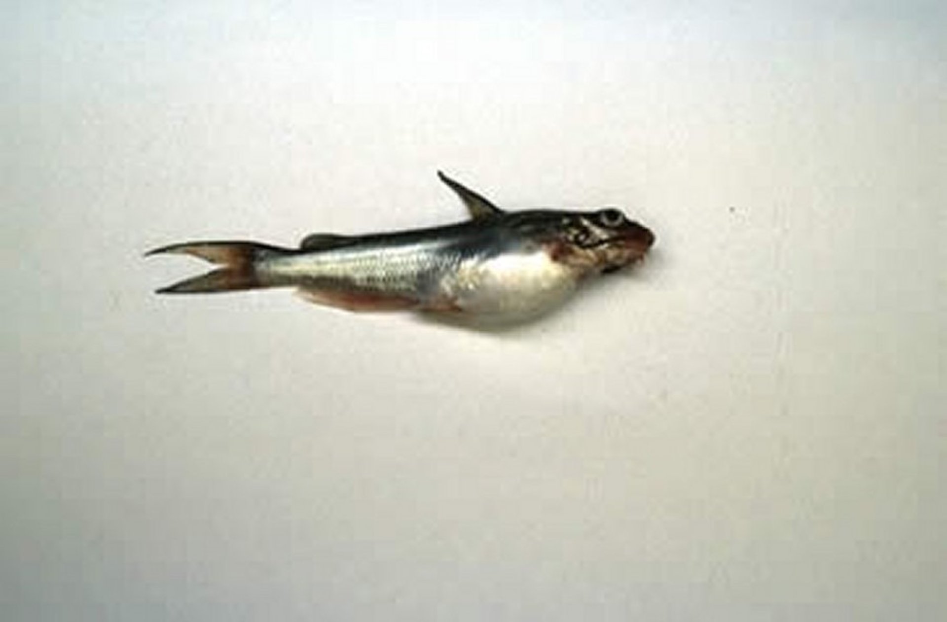
Courtesy of Dr. Ruth Francis-Floyd.
Channel catfish virus (CCV) disease is an acute, virulent herpesvirus infection of fry and fingerling channel catfish that can cause mortality of >80% at water temperatures ≥25°C in small fish (≤5 cm). As fish age, mortality decreases, and clinical infection in fish >1 year old is rare. Acute infection often includes a recent history of a stressful event such as handling or transport, low dissolved oxygen, or chemical treatment. Infected fish show signs of ascites, exophthalmos, and hemorrhages in fins. The cell line of choice for virus isolation is channel catfish ovary, followed by serum neutralization to confirm identification. Typical cytopathic effects include cell fusion, syncytia formation, and intranuclear inclusions. There is evidence for vertical transmission of CCV; consequently, survivors of an epizootic should not be used for broodstock. Although CCV can cause severe mortality when an outbreak is in progress, the annual number of cases of CCV in the catfish industry is relatively low.
Novirhabdoviruses
Infectious Hematopoietic Necrosis in Aquaculture
Caused by a novirhabdovirus in the family Rhabdoviridae, infectious hematopoietic necrosis is listed as an OIE notifiable disease. It is endemic in salmonid (Oncorhynchus spp) populations in the Pacific Northwest and Alaska and has been reported in Atlantic, chum, chinook, sockeye, and kokanee salmon and in cutthroat, steelhead, and rainbow trout. Lake trout and Arctic char, members of the genus Salvelinus, appear resistant. The disease has also been reported in parts of Europe and Asia. Most epizootics have been attributed to importation of infected eggs or fry.
Acute disease in fry < 2 months old may result in high mortality (>90%) with few external signs. Disease usually occurs at water temperatures of 10°–12°C, although outbreaks occasionally occur at temperatures >15°C. Typical signs include lethargy with sporadic hyperexcitability, including whirling. Sick fish may be darkened with distended abdomens, exophthalmia, pale gills, and mucoid fecal casts. Important differential diagnoses include infectious pancreatic necrosis and viral hemorrhagic septicemia. Hematopoietic tissue in the kidney and spleen are most severely affected by necrosis.
Risk factors include age (fish < 2 months old are more susceptible), density, and water temperature. Hauling young fish around dams in trucks may be a significant risk factor because of crowding during transit. Although most disease outbreaks have been reported in freshwater, active disease has occurred in Atlantic salmon housed in sea cages. Diagnosis is by viral isolation (from kidney and spleen of young fish and ovarian fluid of broodstock), with confirmation by serum neutralization. Rapid serologic tests are becoming more available. A nonlethal test involving viral isolation from mucus has been reported. The disease is transmitted horizontally through the water, and vertical transmission is suspected. Asymptomatic carrier fish serve as reservoirs of infection. A vaccine is available in the US for use against this pathogen.
Viral Hemorrhagic Septicemia in Aquaculture
Viral hemorrhagic septicemia (VHS) is caused by a novirhabdovirus in the family Rhabdoviridae and is a highly regulated disease in the US. VHS can be divided into several different strains based on genotype: VHS strain IVa is the predominant strain in US aquaculture; strain IVb (see below) has been identified in wild stocks only in the US and Canada; and strains I, II, and III are found primarily in Europe and Asia. The disease causes marked necrosis of hematopoietic tissue in the kidney, particularly the anterior kidney, but largely spares excretory tubules in the posterior kidney. Rainbow, brook, and lake trout (genus Salvelinus) and Atlantic salmon and brown and golden trout (genus Salmo) are susceptible. The virus also causes disease in a variety of freshwater and marine coldwater fish, including pike, turbot, white fish, and sea bass. VHS also is found in free-ranging marine fish in the Pacific northwest, including anadromous salmon (coho and chinook), as well as haddock and cod in the North Sea.
The disease occurs in three forms: acute, chronic, and nervous. Acute mortalities occur in rainbow trout fry < 3 g and < 30 days old. In these fish, the kidney is swollen and the anterior segment is necrotic and pale. The liver may be pale with hemorrhagic mottling, and systemic hemorrhage may be visible in the eyes, skin, skeletal muscle, and viscera. The most notable lesion is widespread hemorrhage in the liver, adipose tissue, and within skeletal muscle. Moribund fish lie on the bottom of the tank and may exhibit sporadic flashing and corkscrew swimming behavior. As fish age, mortalities drop from 80%–100% to 10%–50%. The chronic form is a persistent infection with low RBC and WBC counts; virus can be isolated from all tissues. Chronically infected fish may exhibit few visible external signs. The nervous form of the disease has been reported primarily in cultured freshwater fish but has also been reported in marine fish.
The optimal temperature for active infection is 9°–12°C; the virus is unable to replicate at temperatures >15°C. The cell line of choice for virus isolation is bluegill fry (BF-2). Viral identification is confirmed by serum neutralization. Newer diagnostic tests include immunofluorescence, ELISA, and PCR assay. VHS is a highly regulated disease, with disease-free geographic regions defined in Europe. No vaccine is commercially available. Veterinarians working with zoologic collections must ensure that susceptible species received from endemic areas are properly tested and certified disease free.
In 2005, a new strain of VHS, VHS IVb, was identified in the Great Lakes and caused widespread morbidity and mortality of only wild stocks. Affected species included muskellunge, Chinook salmon, brown trout, freshwater drum, black crappie, bluntnose minnow, gizzard shad, largemouth bass, smallmouth bass, blue gill, yellow perch, and channel catfish. Signs of VHS IVb can be nonspecific. Some affected fish have no clinical signs, but exophthalmia, ascites, hemorrhages (in the eyes, skin, gills, fin base), and behavioral changes may be seen.
To date, no aquacultured specimens have been found to be infected with this strain, but veterinarians should be aware of it and avoid introducing stocks or water from infected/unprotected systems.
Iridoviruses
Viral Erythrocytic Necrosis in Aquaculture
Viral erythrocytic necrosis has been reported in >20 species of marine and anadromous fish (both cultured and free ranging) and is characterized by erythrocytic degeneration. Affected species include Pacific herring, Atlantic cod, and Pacific salmonids (chum, pink, coho, and Chinook), steelhead trout, and cultured eels in Taiwan. The disease is chronic, and external signs may be subtle or nonexistent. Sick fish are anemic, which may result in pale gills and internal organs. Severity of the disease is related to age and species of fish, with juveniles < 1 g most severely affected.
The characteristic lesion is a single eosinophilic cytoplasmic inclusion body in the circulating erythrocytes of anemic fish. The inclusions are best visualized on Giemsa-stained fresh blood smears. To date, the agent has not been successfully isolated. Histologically, increased hematopoietic activity may be evident in the kidney, and round cytoplasmic inclusions (0.8–4 μm) are found in circulating RBCs. Inclusions stain pink or magenta with Giemsa.
Other degenerative changes may be evident in RBCs, including cytoplasmic vacuolation and margination of nuclear chromatin. Hemolytic anemia with concurrent hemosiderosis and erythroblastosis has been reported in moribund Pacific herring. Multinucleated giant erythroblasts may occasionally be seen in peripheral blood, and macrophages may phagocytize abnormal erythroblasts. A presumptive diagnosis is based on the presence of typical cytoplasmic inclusions in circulating RBCs of anemic fish. Confirmation requires visualization of hexagonal virus particles in cytoplasm of affected RBCs using transmission electron microscopy. A marine reservoir is suspected but has not been identified. Vertical transmission is suspected because of the high prevalence of infection in fry from infected broodstock.
Epizootic Hematopoietic Necrosis in Aquaculture
The ranaviruses are an important group within the family Iridoviridae and, as a group, include viruses that can infect fish, amphibians, and reptiles. One of these causes epizootic hematopoietic necrosis (EHN), which so far has been reported to cause disease only in fish and is listed as a notifiable disease by OIE. First reported in redfin perch in Australia in 1984, it has since been shown to cause disease, albeit less severe, in rainbow trout. Similar viruses have been reported in sheatfish in Germany and black bullhead catfish in France and Italy.
EHN is endotheliotropic, producing necrotic lesions in the endothelium of blood vessels and some visceral lesions. Behavioral signs include lethargy, darkening, and erratic swimming. Mortality occurs after 4–5 days. The most consistent lesion associated with EHN is focal necrosis of hematopoietic tissue in the anterior kidney and liver. Necrotic hematopoietic cells may be visible within blood vessels. Presumptive diagnosis is based on clinical signs and isolation of the suspect agent in cell culture. Bluegill fry (BF-2) is the cell line of choice. Detection may also be accomplished using ELISA, immunofluorescence, or electron microscopy. Epizootics of EHN in redfin perch are most common in the spring and summer and almost exclusively involve juvenile fish. Survivors seem to be resistant to future infection. There is no evidence of vertical transmission of EHN, and redfin perch carriers have not been detected. An unidentified reservoir and carrier host is suspected. Fomite transmission of EHN has been demonstrated, and birds have also been shown to carry infected material.
Largemouth Bass Virus in Aquaculture
Largemouth bass virus, a ranavirus, was isolated from moribund largemouth bass in South Carolina in 1995. It was previously isolated from largemouth bass in several Florida lakes but had not been directly associated with disease. It has been found in largemouth bass in most southeastern and many midwestern states. The disease is not well understood, because the virus is commonly isolated from tissues of clinically normal fish. In the 1995 fish kill, ~1,000 fish died over a 2- to 3-month period in an area that encompassed >66,000 hectares. Lesions were nonspecific and are still poorly described. Fat-head minnow is the cell line of choice for isolation of virus.
Megalocytiviruses in Aquaculture
The megalocytiviruses are another very important and emerging group of viruses within the family Iridoviridae. They have been associated with disease in freshwater angelfish and gourami species, as well as in numerous other aquacultured freshwater and marine food and aquarium fishes, including oscars and other cichlids (family Cichlidae), swordtails, sailfin mollies, and other common live-bearers (family Poeciliidae), and Banggai cardinalfish. Numerous marine and freshwater food and game finfish species are also naturally susceptible to infection by megalocytiviruses, including jacks and pompanos (several species, family Carangidae), mackerels and tuna (several species, family Scombridae), grouper (several species, family Serranidae), cobia (Rachycentron canadum), largemouth bass (Micropterus salmoides), barramundi (Lates calcarifer), redfish (Sciaenops ocellatus), hybrid striped bass (Morone saxatilis × M chrysops), and gray mullet (Mugil cephalus).
Clinical signs are nonspecific but include lethargy, anorexia, darkening, abnormal swimming (including spinning) or position in the water, increased respiration, coelomic distention, ulceration, hemorrhages/petechiae, pale gills/anemia, fin erosion, white feces, and moderate to heavy mortalities. Higher temperatures may be associated with some outbreaks.
Diagnosis is based on clinical signs, history, histopathology, and PCR identification (many of these viruses have not been successfully cultured). Histopathology often includes areas of acute, diffuse, severe coagulative necrosis in some tissues, and/or variable amounts of necrosis and mild inflammation. Large, finely granular basophilic to amphophilic intracytoplasmic inclusions are most commonly identified in spleen and kidney, although in severe infections, liver, heart, oral cavity, thymus, bone, gills, skeletal muscle, submucosa of the GI tract, and gonads are also affected.
There is currently no treatment for megalocytiviruses other than depopulation. A vaccine is available internationally but not in the US.
Other Viruses
Infectious Salmon Anemia in Aquaculture
Infectious salmon anemia is reportable to USDA APHIS and the OIE. The first report was from farmed Atlantic salmon on the west coast of Norway in 1984. Affected fish were lethargic and severely anemic (PCV < 5% in moribund fish). The causative agent is an orthomyxovirus. Acute outbreaks result in high mortality. Initial signs include lethargic fish hanging around the edges of the cage. As the disease progresses, moribund fish lie on the bottom.
Diagnosis is based on clinical signs, with emphasis on anemia (PCV < 10%), the gross appearance of a dark liver, and hepatic necrosis. Confirmation can be by viral isolation using the SHK-1 cell line. Virus may be visualized in endothelial cells of cardiac blood vessels using transmission electron microscopy. The agent is enveloped, slightly pleomorphic, and ~100 nm in size. Suspected cases can also be verified using an immunofluorescent antibody technique on frozen tissue. Transmission is horizontal, and virus is shed in skin, mucus, feces, and urine. Sea lice (Lepeophtheirus salmonis) may be a vector; disease outbreaks seem worse when sea lice are present. There is no evidence of vertical transmission. Sea trout have been proposed as a possible reservoir of infection. Protective immunity has been demonstrated in salmon that survive an outbreak. Vaccines are available internationally but may not confer complete protection. The disease is heavily regulated in Norway and now in the US, where the USDA should be notified immediately of any suspected cases.
Infectious Pancreatic Necrosis in Aquaculture
Infectious pancreatic necrosis is an acute, systemic, contagious disease of salmonid fry and fingerlings caused by a birnavirus. The virus is the archetype of the aquatic birnaviruses, which are further subdivided into two serotypes, A and B, that do not cross-react using serum neutralization. The serotype B group currently consists of only 10 isolates, all European in origin. In contrast, the A serotype contains >200 isolates that have been further subdivided into 9 serotypes, A1–A9. Morbidity and mortality occur only in young animals, usually < 3 g. However, virus can be isolated from survivors for the duration of their lives, indicating a persistent carrier state, although recrudescence of disease in survivors has not been reported. The virus is vertically and horizontally transmitted, widespread, and reported worldwide, except in Iceland and Australia. Rainbow trout are highly susceptible to disease. In the US, striped bass and their hybrids are recognized as potential carriers. Other species affected include freshwater eels (Anguilla spp), yellowtail, turbot, sea bass, and menhaden, as well as aquatic invertebrates including molluscs and crustaceans. Brook trout are believed to be reservoirs of infection in the US.
Clinical infection is nonspecific. Diseased fish may be anorectic, ataxic, and display a corkscrew swimming pattern. Externally, fish are darkened; exophthalmia and external petechiation may be evident. Internally, petechiae may be visible on viscera; the gut is typically empty and may contain a yellow exudate. Fecal pseudocysts may be evident in the water column. Histologically, focal areas of coagulative necrosis involve acinar and islet cells of the pancreas and hematopoietic cells of the kidney. Intracytoplasmic viral inclusions may be visible in pancreatic acinar cells. Infection should be confirmed with viral isolation followed by serum neutralization. Most fish cell lines are susceptible. The virus can also be identified using fluorescent antibody, complement fixation, and ELISA techniques. There is no treatment for infected fish, but avoidance can be accomplished by purchase of SPF stocks, quarantine, and disinfection of eggs with iodophores (20–50 mg/L). Infectious pancreatic necrosis is not regulated by the USDA, but state regulations exist in various parts of the country.
Salmonid Alphaviruses in Aquaculture
Salmonid alphaviruses are the cause of pancreas disease and sleeping disease in Atlantic salmon, rainbow trout, and brown trout. To date, reports have been from Norway and parts of the UK, although farmed fish in other countries in Europe have also been affected. This suite of diseases and the etiologic agent are reportable to USDA APHIS and the OIE, and this disease is considered a foreign disease to the US. As the disease names suggest, pathology involves necrosis and loss of exocrine pancreatic tissue, as well as changes in heart and skeletal muscle. Mortality rates vary from minimal to >50% in severe cases. As a sequela, a percentage of surviving fish may become slender and long runts. Survivors may also become carriers; however, vertical transmission is still under debate, and horizontal transmission is considered the main route of infection. A vaccine is available in affected countries.
Viral Nervous Necrosis (Betanodaviruses) in Aquaculture
The betanodaviruses are an emerging group of viruses infecting >40 different species of primarily marine fish worldwide. Susceptible species include red drum, cobia, barramundi, tuna, groupers, flatfishes, surgeonfishes, lemonpeel angelfish, the orbicularis batfish, and tilapia. The resulting disease is also known as viral nervous necrosis (VNN) and viral encephalopathy and retinopathy. A few freshwater species have also been reported as susceptible. Betanodaviruses can infect tropical, subtropical, or cold-temperate species, with species susceptibilities and optimal temperature ranges varying depending on the strain of the virus. Four different genotypes are currently recognized. As a group, betanodaviruses can infect fish at temperatures ranging from approximately 15°–30°C.
Younger life stages (larvae, fry, fingerlings) are more frequently affected, although older, market-size fish may become affected. Losses can range from 15%–100%. Betanodaviruses are somewhat unique in that, as the disease name suggests, their target tissue is the CNS. Consequently, clinical signs include abnormal swimming or spinning, vertical position in the water, flexing of the body, muscular tremors, and darkening or lightening of the skin. Hyperinflation of the swim bladder is also often seen, with affected fish positively buoyant and found near the surface. Traumatic lesions resulting from abnormal swimming may be secondary sequelae. Betanodaviruses can be spread horizontally and vertically, and live or frozen fish fed to broodstock or growout stages may be a potential source of infection. Other sources of infection include wild invertebrates and contaminated water. Egg disinfection may help reduce impact of the disease. A commercial vaccine may be available internationally, but none is currently available in the US.
Histopathologically, in properly fixed samples of affected CNS tissue (brain, spinal cord, retina of the eye), the presence of severe vacuolation is a hallmark of the disease. Disease and pathogen confirmation are based on clinical signs, species, histopathologic findings, immunofluorescent antibody techniques, and PCR assay.


