

Sporotrichosis is a sporadic, chronic, granulomatous disease of humans and various domestic and laboratory animals due to Sporothrix spp. Infection usually results from direct inoculation of the organism into skin wounds via contact with plants, soil, or penetrating foreign bodies.
The Sporothrix organism is dimorphic and forms mycelia on vegetation and in Sabouraud dextrose agar at 25°C–30°C (77°F–86°F) but is yeastlike in tissue and media at 37°C (98.6°F). It is ubiquitous in soil, vegetation, and timber; distributed worldwide; and in the US, most commonly found in coastal regions and river valleys. Disseminated disease due to inhalation of spores is rare.
Sporotrichosis has been reported in dogs, cats, horses, cows, camels, dolphins, goats, mules, birds, pigs, rats, armadillos, and humans. Zoonotic infection can occur from cats to humans and has been reported without evidence of trauma. In contrast, transmission from other infected species appears to require inoculation of previously traumatized skin. The large number of organisms found in/on feline wounds, claws, and feces is believed to be responsible for the increased zoonotic potential of feline sporotrichosis.
Epidemics of sporotrichosis have been reported in Brazil associated with the species S brasiliensis. Data from these studies support the importance of cats in the zoonotic transmission of the organism. Caretakers of infected cats were four times more likely than others living in the same household to become infected.
Clinical Findings and Lesions of Sporotrichosis in Animals
Sporotrichosis may be grouped into three forms: lymphocutaneous, cutaneous, and disseminated. The lymphocutaneous form is the most common. Small, firm dermal to subcutaneous nodules, 1–3 cm in diameter, develop at the site of inoculation. As infection ascends along the lymphatic vessels, cording and new nodules develop. Lesions ulcerate and discharge a serohemorrhagic exudate.
In cats, lesions are most often present on the head, especially on the bridge of the nose and pinnae. Although systemic illness is not evident initially, chronic illness may result in fever, listlessness, and depression. Respiratory signs may be apparent.
The cutaneous form tends to remain localized to the site of inoculation, although lesions may be multicentric. Disseminated sporotrichosis is rare but potentially fatal and may develop with neglect of cutaneous and lymphocutaneous forms or if the patient is inappropriately treated with corticosteroids. Infection develops via hematogenous or tissue spread from the initial site of inoculation to the bone, lungs, liver, spleen, testes, GI tract, or CNS.
Diagnosis of Sporotrichosis in Animals
Cytology, especially in cats
Yeasts can be confused with other fungal organisms such as Histoplasma

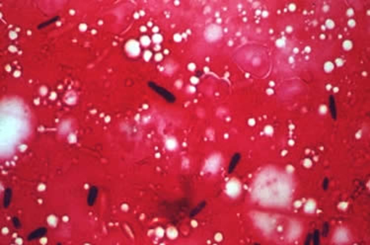
Courtesy of Dr. John Prescott.
Diagnosis of sporotrichosis can be made by culture (samples obtained from unopened lesions) or microscopic examination of the exudate or biopsy specimens. In tissues and exudate, the organism is present as few to numerous, round to cigar-shaped, single cells within macrophages. The fungal cells are pleomorphic and small (2–10 × 1–3 mcm); buds may be present and give the appearance of a table tennis paddle. A nonstaining cell wall surrounds the yeasts when using Wright or Romanowsky-type stains.
Sporothrix yeasts closely resemble Histoplasma yeasts, but Histoplasma does not demonstrate the cigar-shaped morphology common to Sporothrix. A fluorescent antibody technique has been used to identify the yeastlike cells in tissues. In species other than cats, Sporothrix organisms are often sparse in exudate and infected tissue, so diagnosis usually requires culture and/or immunohistochemical staining.
In cultures, a true mycelium is produced, with fine, branching, septate hyphae bearing pear-shaped conidia on slender conidiophores. Culture and PCR assay may be useful to determine the species of Sporothrix involved, because this can affect treatment and prognosis. Antibody ELISAs may be available and are sensitive and specific in cats; other species have not been evaluated.
Treatment of Sporotrichosis in Animals
Itraconazole for sporotrichosis; iodides may be added
Cats often require > 12 months of treatment; failures may occur
Itraconazole (10 mg/kg every 24 hours) is considered the treatment of choice for sporotrichosis. Treatment should be continued at least a month beyond apparent clinical cure. Terbinafine (30 mg/kg, PO, daily) has also been used successfully. Alternatively, potassium iodide, administered PO, has been used with some success alone or in combination with azoles.
During treatment, the patient should be monitored for clinical signs of iodide toxicity: anorexia, vomiting, depression, muscle twitching, hypothermia, cardiomyopathy, cardiovascular collapse, and death can occur. Cats are especially sensitive to iodides and the development of iodism. Surgical resection, cryotherapy, and/or localized hyperthermia has shown some success in limited numbers of cases.
Zoonotic Risk of Sporotrichosis in Animals
Sporotrichosis is an important zoonosis, with feline-to-human transmission well documented. Strict hygiene must be observed when handling patients (especially cats) with suspected or diagnosed sporotrichosis. Humans in contact with infected animals should be informed of the infectious nature of the disease when therapeutic options are discussed. Good biosecurity must be practiced, including personal protective equipment (PPE) and premises disinfection.
Key Points
Sporotrichosis is a chronic fungal disease that manifests with nonhealing wounds and draining tracts.
Cats are a reservoir and a zoonotic risk.
Itraconazole beyond clinical cure is the treatment of choice.


