


Protothecosis is an infection caused by members of the genus Prototheca (achlorophyllous algae found ubiquitously in the environment). The main clinical forms of protothecosis include mammary infections in cattle, cutaneous lesions in cats, hemorrhagic enteritis and systemic signs in dogs, and cutaneous, articular, and disseminated infections in humans. Cases of protothecosis are often refractory to treatment.
Protothecosis is an infection caused by members of the genus Prototheca (achlorophyllous algae found ubiquitously in the environment). Prototheca organisms are opportunistic and infect humans, some domestic animals, and wildlife.
Etiology of Protothecosis
The genus Prototheca (family Chlorellaceae) comprises unicellular, colorless, achlorophyllous, yeast-like microalgae that are closely related to the green algae of the genus Chlorella. Published reviews have substantially improved the understanding of Prototheca taxonomy and epidemiology (1, 2).
Currently, a molecular marker–based approach using sequencing of the mitochondrial cytochrome b (cytb) gene provides the highest resolution for species delimitation. Of the 18 Prototheca species described, 6 have been implicated as etiological agents of opportunistic infections.
Pathogenic species include P bovis (formerly P zopfii biovar II or genotype 2), P blaschkeae, P ciferrii (formerly P zopfii biovar I or genotype 1), P cutis, P miyajii, and P wickerhamii. Of these, P wickerhamii and P bovis account for the majority of infections, predominantly affecting humans and cattle, respectively. P bovis and P wickerhamii have also been reported in both dogs and cats, whereas P ciferrii has been reported in dogs and P cutis in cats (3, 4). P miyajii is a rare human pathogen (5).
P paracutis, P cerasi, P cookei, P pringsheimii, and P zopfii are considered nonpathogenic. Historically, P zopfii genotypes 1 and 2 were reported to cause infections in both animals and humans (6). These genotypes have since been reclassified as P ciferrii and P bovis, respectively. Although P zopfii remains a recognized species, it is not among the pathogenic species identified in the 2019 cytb-based taxonomic revision. The remaining species include P moriformis, P stagnora, P tumulicola, P xanthoriae, P vistulensis, P lentecrescens, and P fontanea.
Among the typical dairy cattle–associated species, P ciferrii has been found in the feces of cattle and the environment of dairy farms, P blaschkeae has been identified in the breeding environment of swine and as a sporadic agent of bovine mastitis, and P bovis has been described as a major primary cause of clinical protothecal mastitis in cattle herds globally (7, 8).
Prototheca reproduce asexually. Sporangia (mother cells) form cytoplasmic sporangiospores (endospores or daughter cells) with spherical to oblong (oval) or wedge-shaped forms ranging from approximately 3 to 30 mcm in diameter, depending on the species. P wickerhamii (3–10 mcm) has wedge-shaped sporangiospores radially arranged; P bovis (7–30 mcm) has oval or spherical sporangiospores, usually larger than those of P wickerhamii.
Pathogenesis of Protothecosis
Because they lack chlorophyll, Prototheca do not perform photosynthesis; they require external sources of carbon and nitrogen (obligate heterotrophs), resulting in a saprophytic lifestyle and opportunistic pathogen behavior in infections in animals and humans.
Prototheca algae are intracellular pathogens that reproduce asexually. During cell maturation (growth), the cytoplasm undergoes cleavage or internal septation, leading to multiple and irregular divisions of the cell, forming 2–20 sporangiospores (endospores). The pressure of the enlarging sporangiospores breaks the cell wall, and sporangiospores are released to begin a new reproduction cycle. In adequate nutrient, temperature, pH, and humidity conditions, this process repeats every 5–6 hours.
The enlargement of internal sporangiospores promotes the destruction of infected cells and tissues. Prototheca-induced infections cause pyogranulomatous inflammation of the tissues. Any cell or tissue can be infected by these algae, including mammary glands, brain, skin, eyes, intestine, skeletal muscles, liver, lymph nodes, kidney tissues, and phagocytes, demonstrating the high infective potential of Prototheca.
The mechanisms that allow Prototheca to infect hosts and cause disease remain unknown. The rigid cell wall structure, suppression of cell-mediated immune response, evasion of phagocytic mechanisms of macrophages and neutrophils, and biofilm-producing ability of the algae probably contribute to the establishment, multiplication, and persistence of the organism in inflammatory foci.
Virulent strains of Prototheca possess intracellular persistence that, in turn, induces pyogranulomatous inflammation that destroys cells and tissues, including mammary parenchyma, and leads to limited immune response and resolution of infection. Among isolates from cattle with mammary infections, biofilm production has been shown to be species dependent, with P bovis being a strong biofilm producer (9).
Epidemiology of Protothecosis
Prototheca spp occupy numerous ecological niches, which contributes to their varied transmission patterns and host associations. Their strong resistance to adverse environmental conditions allows them to persist for long periods in natural aquatic ecosystems and contributes to contamination of urban and agricultural water systems.
Prototheca are commonly found in immediate dairy farm environments, such as drinking troughs, feeders, bedding, and milking equipment, and in water sources including streams, rivers, lakes, and artificial water reservoirs, and they are occasionally detected in the intestinal tract of animals. In environments with high levels of organic pollution, Prototheca can establish densely populated microniches that support localized survival and long-term environmental persistence (1, 6, 10).
Cattle, dogs, and cats are the main domestic animals in which Prototheca have been isolated. However, sporadic cases have been reported in goats, horses, and nondomesticated animals (11, 12).
Traumatic percutaneous lesions, contamination of mucous membranes, and entry through the teat orifice from contaminated milking equipment or environmental sources are likely the main routes of transmission of Prototheca to domestic animals. Calves can become infected by ingesting milk from cows with protothecal bovine mastitis.
Contamination of wounds and traumatic cutaneous-subcutaneous lesions appear to be the main routes of protothecal infection among companion animals, in addition to oral transmission of Prototheca to dogs by ingestion of contaminated water or food.
Systemic or disseminated protothecosis in dogs and cats was once thought to be linked to debilitating conditions or immunosuppressive viral coinfections. However, this association has not been conclusively demonstrated in animals (6).
A substantial increase in the occurrence of clinical mammary protothecosis has been reported worldwide, particularly on farms with adequate control of other infectious bovine mastitis agents, such as staphylococci (principally Staphylococcus aureus), corynebacteria, and Streptococcus agalactiae (8, 13). Outbreaks have been recorded in South America (Brazil), North America (US, Canada, and Mexico), Asia (Japan and China), Oceania (New Zealand), and some countries of Europe (Denmark, England, and Italy) (8).
Infection of endemic herds has economic impacts, including veterinary care expenses for treatment and services, decreased milk production, and premature culling of affected animals.
A large-scale study of the molecular characterization of 342 Prototheca isolates obtained from bovine mammary infections in different countries revealed a major frequency of P bovis (90.6%), followed by minor frequencies of P blaschkeae (8.8%) and P ciferrii (0.6%), confirming the predominance of P bovis as a primary cause of cattle mastitis (14).
Globally, Prototheca spp have been isolated in < 10% of mastitis cases and bulk tank milk samples subjected to microbiological culture:
In a study conducted on 50 dairy farms in the Republic of Korea from 2015 to 2017, P bovis was isolated from 187 of 2,508 (7.5%) quarter milk samples (15).
An overall incidence of bovine mastitis by P bovis of 4.6% has been reported in dairy farms located in southeastern Poland, sampled from 2016 to 2017 (16).
A large-scale investigation of clinical bovine mastitis etiology among 10 Brazilian dairy herds from 2017 to 2019 identified Prototheca spp as the primary agent in 113 of 4,273 (2.6%) clinical cases (17).
Nonetheless, outbreaks of protothecal mastitis might affect approximately 30% of cattle herds and have been associated with contamination of intramammary therapy procedures and with poor milking and environmental hygiene conditions (18, 19).
On endemic farms, pathogenic Prototheca has been recovered from environmental sources with high humidity or wet habitats that contain abundant organic matter in the soil where animals are driven or rest. These farm sources include streams or stagnant ponds; mud and feces around milking areas, outdoor runs, paths, or resting areas; feed; drinking water; and feces of cattle and calves. They are also found on milking machine surfaces, the floor of the milking area, and the material of the compost barn and in bedding materials.
Improper milking hygiene, deficiencies in premilking measures, and excessive amounts of organic material in the milking environment increase the risk of protothecal mammary infections; protothecal mammary infections can also be transmitted from infected to healthy cows during milking.
Pearls & Pitfalls
|
Flies are potential vectors of Prototheca in the farm environment. Poor teat hygiene practices and improper hygiene in intramammary infusions are strong herd-level risk factors for protothecal infections; especially risky are procedures such as the use of dry cow teat sealant and infusions with nonantimicrobial formulations.
P bovis can persistently infect the epithelia of mammary glands during lactation and persist through the dry period into the next lactation, acting as a source of infection to other cows. A 2018 study identified P bovis among fecal samples of cattle and calves from the same herds with a history of mammary protothecosis, suggesting infection of calves by milk and a connection between the occurrence of protothecal mastitis and the fecal cycle of Prototheca between the environment, cattle, and calves (20).
Clinical Findings of Protothecosis
A variety of clinical forms of protothecosis, acute and chronic, have been described in domestic animal species, particularly chronic mammary infections in cattle, hemorrhagic enteritis and systemic infections in dogs, and cutaneous lesions in cats.
Bovine Mastitis
Mammary infections are the major clinical form of protothecosis among domestic animals. Protothecal species are considered environmental agents in the etiology of bovine mastitis.
P bovis is a major species involved in mammary infections of cattle; P blaschkeae and P wickerhamii are less frequently implicated. Most affected cows develop clinical signs of mammary infection; however, some animals can have a silent course of infection (subclinical mastitis).
Prototheca infections can occur during lactation or dry periods. Watery, white to yellow milk with suppurative exudate or flakes, along with edema and indurative mastitis, have been observed in clinical forms. Mammary protothecosis substantially decreases milk production and increases somatic cell count. Supramammary lymph nodes might be enlarged.
Occasionally, Prototheca can spread from mammary glands and infect other organs. Prototheca can be eliminated intermittently in milk—a fact that can make microbiological diagnosis in herds difficult. Protothecal mastitis tends to lead to long-term infections that are commonly refractory to intramammary or systemic therapy.
Canine Protothecosis
Canine protothecal infections are sporadic; however, cases have been reported in companion animals. Dogs are infected predominantly by P bovis and P wickerhamii.Chronic, intermittent, hemorrhagic diarrhea (presence of mucus, hematochezia) and progressive weight loss appear to be the main clinical signs, and they indicate a distal GI tract infection. The oral route is a common cause of canine disease. Other organs affected include the skin, eyes, CNS, liver, spleen, kidneys, and lymph nodes.
A review of canine protothecosis cases reported infections in 21 countries across every continent except Antarctica, with most cases occurring in North America (38.4%) and Europe (29.6%). The disease primarily affected adult, female (51.2%), purebred, medium‑ to large‑breed dogs, especially Boxers, Labrador Retrievers, and Rough Collies, with a median age of four years (6).
Most canine infections presented as systemic disease (67.2%) involving multiple organs; however, enteric (10.4%), ocular (8.8%), cutaneous (7.2%), neurological (4.8%), and myocardial forms (1.6%) also occur. GI signs are common and often begin as mild large‑bowel dysfunction. Most dogs had no underlying comorbidities (88.8%), and transmission was thought to occur through environmental exposure, particularly contaminated water or soil, with fecal–oral transmission considered most likely (6).
Systemic cases frequently involved the eyes, kidneys, heart, liver, colon, and brain. Cutaneous disease has usually been linked to P wickerhamii, whereas systemic infections are more often associated with P bovis, P ciferrii, or P zopfii (which probably would now be identified as either P bovis or P ciferrii) (6).
Treatment typically required multiple antimicrobial and antifungal drugs, yet outcomes were poor, with nearly 90% of affected dogs failing to respond and ultimately dying or being euthanized (6).
Other multisystemic clinical signs of canine protothecosis include fever, urinary incontinence, polyuria, polydipsia, osteomyelitis, myocarditis, lymphadenomegaly, and cutaneous forms (crusts on footpads, ulcerated nodules).
A variety of ophthalmological signs have been described, commonly bilateral, including progressive loss of vision, uveitis, retinitis, synechiae, and glaucoma.
Molecular diagnosis enabled P bovis detection in a dog from Brazil with chronic hemorrhagic diarrhea and weight loss, along with a history of contact with the environment of a dairy herd, suggesting that cows or their environment were the sources of transmission to the dog (21).
Feline Protothecosis
Feline protothecosis is rare. Clinical patterns vary by Prototheca species.
P wickerhamii infection most commonly manifests as cutaneous nodules after presumed traumatic inoculation. Affected cats develop firm, nonulcerated, nonpruritic papules, nodules, or masses within the cutaneous and subcutaneous tissues (22). Lesions can appear singly or in multiples and can range from scabbed to ulcerated. They occur predominantly on the forehead, nose, pinnae, base of the tail, and distal limbs. Regional lymphadenomegaly is typically absent in cats with cutaneous disease.
P bovis and P cutis are more often associated with systemic, intestinal, nasal, or neurological involvement. Infection likely occurs through trauma, mucosal contact, or environmental exposure, and immunosuppression can facilitate systemic dissemination in some cases (4, 23).
Miscellaneous Protothecosis
P wickerhamii infection in a goat with respiratory distress and ulcerated nodules in the muzzle and pinna, as well as pyogranulomatous rhinitis in a mare caused by coinfection of Prototheca spp and Pithomyces chartarum, have been reported. Systemic protothecosis has also been recorded among nondomesticated animals, including fruit bats, deer, beavers, snakes, rats, and fish (11, 12).
Diagnosis of Protothecosis
Microbiological culture
Hematological imaging
Ophthalmological examination
Serological, electron microscopy, and molecular techniques
Pathological findings
Routine identification of Prototheca has been on the basis of microbiological culture and phenotypic aspects of colonies, cytological and histological examination, and micromorphology and biochemical activity (carbohydrate and alcohol assimilation) of the algae. Molecular methods have allowed the determination of species, genotyping, taxonomic reclassification, and characterization of novel species of Prototheca.
Milk, cutaneous lesions, urine, vitreous humor, feces, rectal scrapings, tracheobronchial washing, fragmenting of organs, milking-machine surfaces, and environment material from farms have been used to isolate the algae.
Prototheca spp can be isolated on conventional media, such as blood agar and Sabouraud agar, in aerobic conditions. Growth of the algae is optimized between 25° and 37°C (77° and 98.6°F).
Irregular to mucoid, wet to dry, white to gray or yellow, nonhemolytic yeast-like colonies, 1–2 mm in diameter, are isolated between 2 and 5 days in aerobic conditions on sheep blood agar and Sabouraud agar, depending on the species of Prototheca (see ).

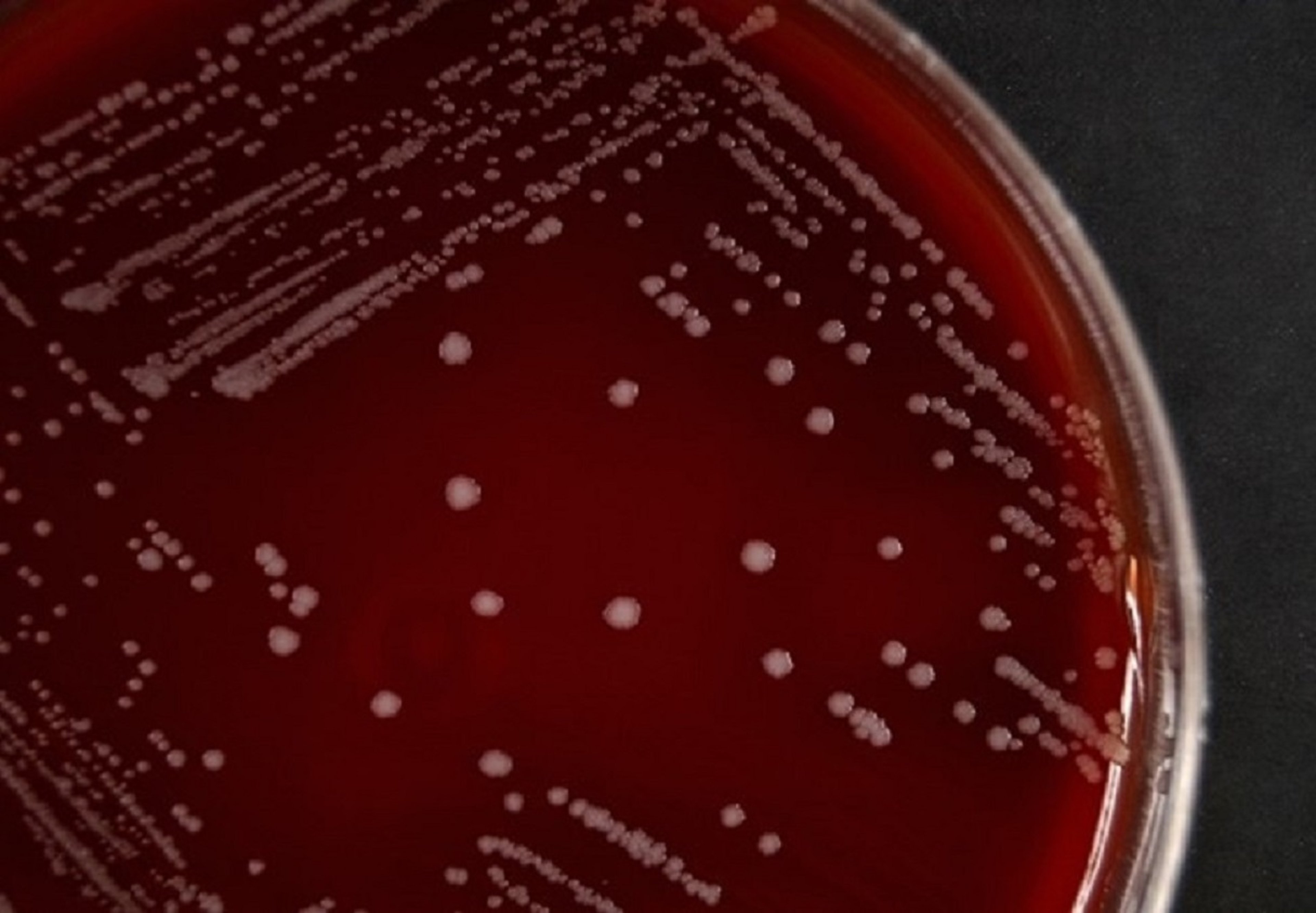
Photograph showing irregular to serrated, white to gray, granular, nonhemolytic, 1–2 mm diameter, yeast-like colonies of Prototheca bovis isolated from a case of clinical bovine mastitis on sheep blood agar after 48 hours of incubation.
Courtesy of Dr. Márcio Garcia Ribeiro.
Prototheca isolation media (PIMs) have enabled selective isolation of the algae, especially from contaminated specimens or environmental material. Gram and fungal staining techniques (eg, lactophenol cotton blue) enable microscopic visualization of Prototheca on the basis of isolated colonies (see ). Spherical to oval or wedge-shaped gram-positive organisms (sporangia) are observed using Gram staining, along with pink structures that constitute broken cell walls of the algae.

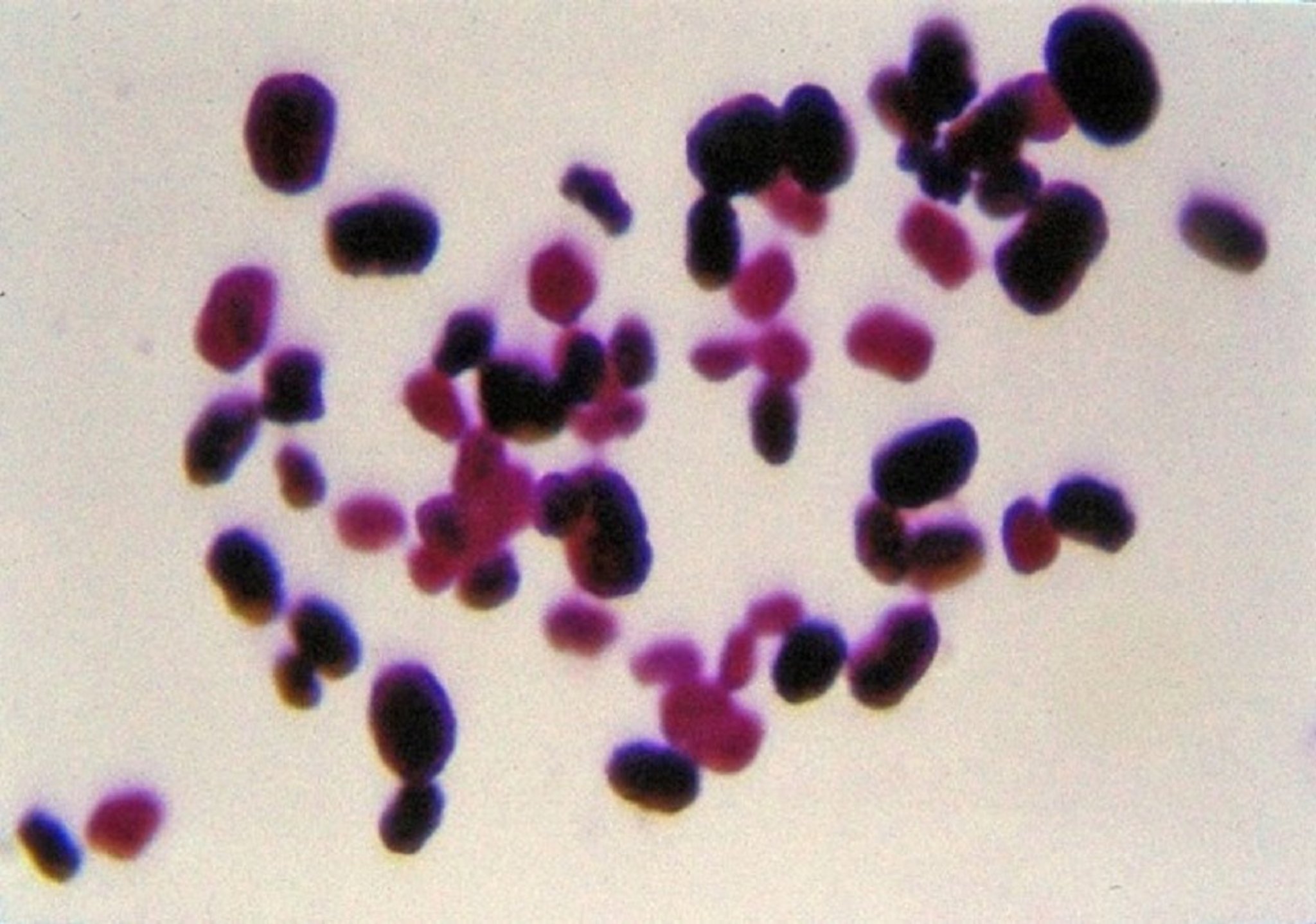
Photomicrograph showing spherical to oval gram-positive organisms (sporangia), compatible with Prototheca spp. Gram stain; original magnification 1,000X.
Courtesy of Dr. Márcio Garcia Ribeiro.
Staining with lactophenol cotton blue and Romanowsky variants enables the observation of sporangiospores. A panel of the assimilation of carbohydrates, alcohol, and other substrates (eg, glucose, galactose, n-propranolol, ethanol, glycerol, trehalose, sucrose, maltose, fructose, arginine, lactose) has been used to determine the species of Prototheca; however, molecular confirmation is recommended.
Hematological and serum biochemical examinations often reveal neutrophilic leukocytosis (nonspecific inflammation) and hyperglobulinemia along with typical renal and hepatic abnormalities when these organs are affected in disseminated canine protothecosis. Marked increases of leukocytes and protein concentrations can occur in dogs with CNS signs.
A rigorous ophthalmological examination should be considered in dogs with clinical signs compatible with disseminated protothecosis.
Imaging findings are commonly nonspecific.
Indirect ELISA (identifying IgG and IgA isotypes) has been used to diagnose Prototheca in serum and whey. Molecular methods such as conventional and multiplex PCR assay, PCR-restriction enzyme analysis (PCR-REA) assay (partial cytb gene), and sequencing (18S rRNA gene) have enabled species identification, genotyping, and reclassification of Prototheca. Matrix-assisted laser desorption/ionization time-of-flight mass spectrometry (MALDI-TOF MS) has enabled the identification of P bovis and P ciferrii recovered from domestic animals.
Transmission and scanning electron microscopy are alternative methods used for confirmation of diagnosis, species, and identification of ultrastructures from Prototheca, including organelles, sporangia, and sporangiospores (see ).

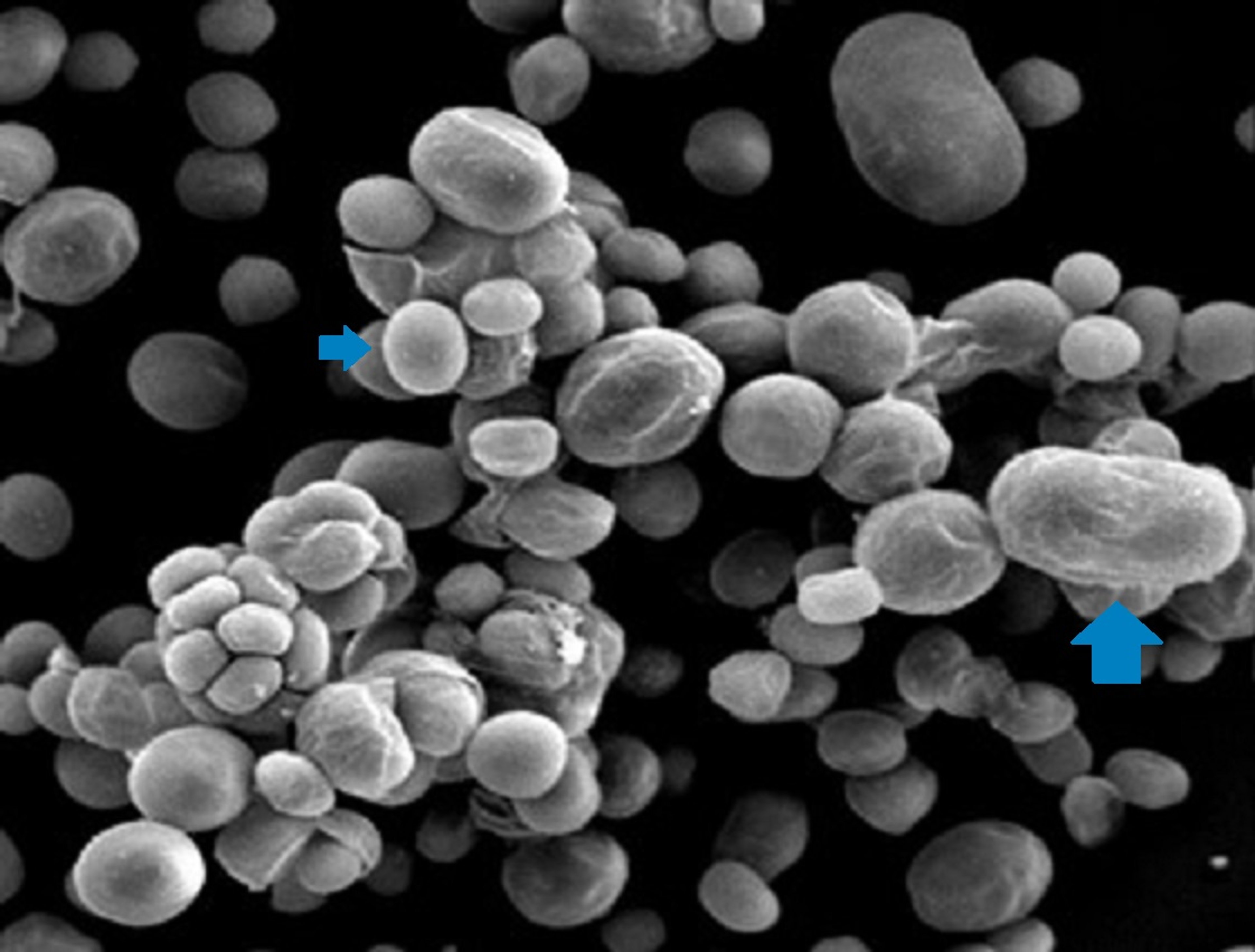
Scanning electron micrograph showing numerous sporangia (mother cells; large arrow) and sporangiospores (endospores; small arrow) representing different developmental stages of Prototheca bovis identified in an outbreak of clinical bovine mastitis. Scanning electron microscopy; original magnification 2,600X.
Courtesy of Dr. Márcio Garcia Ribeiro.
Main gross lesions generally include white to yellow, granular or nodule-like plaques on the serosal surface of organs of domestic animals. Ulcers and cutaneous-subcutaneous nodules occur in canine and feline protothecosis. Canine enteric infections involve erosions, nodules, mucus, and congestion of mucosa, along with mesenteric lymphadenomegaly and hemorrhagic fecal content. Enlargement of supramammary lymph nodes, congestion, nodules, and destruction of mammary parenchyma can occur in protothecal mastitis.
Prototheca spp can be diagnosed by cytological and histological examination of tissue specimens (impression smears, biopsy or fine-needle aspirates, rectal scrapings), and staining by modified Gram stain, Romanowsky variants, Periodic Acid-Schiff (PAS), Grocott-Gomori methenamine Silver (GMS), and others.
Prototheca exhibit typical sporangia (morulae or mother cells) with a thick wall, basophilic cytoplasm, internal septations, and variable forms (oval, wedge-shaped) as well as many sporangiospores (ranging from 3 to 30 mcm in diameter), enabling the identification of species by micromorphology.
Histologically, a chronic pyo- to granulomatous reaction is observed in tissues, with the presence of sporangia containing sporangiospores surrounded by epithelioid macrophages, lymphocytes, neutrophils, plasma cells, occasionally multinucleated giant cells, and a variable contingent of other cells, along with foci of necrosis. Cutaneous lesions exhibit hyperkeratosis, atrophy, ulceration, and follicular loss. Ophthalmic lesions include neuritis and inflammatory cell infiltration of the choroid and retina with a variable number of intralesional algae.
Differential diagnoses of hemorrhagic enteritis caused by P bovis in dogs include Salmonella spp, parvovirus, coronavirus, and intestinal parasites (such as Ancylostoma, Toxocara, Giardia, Isospora, and Cryptosporidium).
Cutaneous-subcutaneous protothecosis of companion animals should be distinguished from infections caused by bacteria (mycobacteria, Nocardia spp, Rhodococcus equi), parasites (Leishmania spp), and fungi and yeasts (Cryptococcus neoformans, Histoplasma capsulatum, Blastomyces spp). Agents that cause chronic bovine mammary infections that are refractory to conventional therapy (eg, Nocardia spp, Trueperella pyogenes, fungal organisms) can induce a similar clinical appearance to protothecal mastitis.
Treatment of Protothecosis
Antimicrobial and antifungal therapy
Surgical excision
Prototheca spp are resistant to a wide spectrum of antimicrobial agents, with azoles and amphotericin B currently being the most promising options.
In vitro susceptibility testing places efinaconazole with the lowest median minimum inhibitory concentration (MIC) and median minimum algicidal concentration (MAC) (0.125 mg/L for both) against canine isolates (P bovis, P ciferrii, and P wickerhamii) followed by ravuconazole (0.5 mg/L for both), amphotericin B (1 and 1.5 mg/L), ketoconazole (16 mg/L for both), itraconazole (32 mg/L for both), and fluconazole (48 and 64 mg/mL) (24). However, in vitro susceptibility testing for Prototheca is not well standardized. The activity of individual drugs differs considerably between Prototheca species and even between strains of the same species, and there is often poor correlation between in vitro drug susceptibility testing results and clinical efficacy (25).
Definitive treatment guidelines for protothecosis have not been formulated, with current treatments being largely empiric and having poor predictability and often unsuccessful outcomes. Only a few documented treatment attempts have resulted in full recovery, and treatment usually focuses on prolonging survival time rather than achieving cure (26).
In dogs, treatment options usually consist of concurrent use of three or more medications, including antimicrobials (most frequently enrofloxacin, followed by amphotericin B and doxycycline), azoles (with itraconazole being used most frequently, followed by ketoconazole, metronidazole, fenbendazole, clotrimazole, fluconazole, posaconazole, thiabendazole, and voriconazole), and other supportive treatment, including corticosteroids, NSAIDs, or crystalloid fluids. However, up to 90% of dogs do not respond to treatment and die or require euthanasia (6). Importantly, treatment outcome is more favorable for cutaneous than systemic protothecosis (6).
Long-term treatment of protothecosis is often required, usually for several months, and treatment cessation can induce relapses. Adverse drug events associated with long-term treatment with these drugs should be considered and hepatic and renal function assessed regularly. New potential drugs and drug-delivery systems, including nanoparticles, cochleated amphotericin B, iodinated carbamates, guanidine, or 3-bromopyruvate, have not been trialed in veterinary patients.
In one reported case, a dog with localized cutaneous protothecosis caused by P wickerhamii underwent two months of pulsed itraconazole administration, which was ineffective. Subsequent posaconazole treatment resulted in clinical improvement; however, lesions recurred when treatment was discontinued because of financial constraints. Intralesional administration of amphotericin B resulted in clinical resolution of the lesions for at least 3 months. (27).
In cases of protothecal mastitis, some antimicrobials, antifungals, and antiseptics or disinfectants that have shown in vitro algicidal effects are irritant or caustic to the mammary glands of cattle, and residues can cause adverse organic reactions in humans if ingested through milk or milk products.
Wide surgical excision has been indicated for protothecal cutaneous lesions of dogs and cats. However, a substantial number of animals subjected to excisional procedure or biopsy of solitary nodules developed systemic disease after the surgical approach (6).
Control and Prevention of Protothecosis
No specific measures are recommended to prevent or control protothecal infections in companion animals, except to avoid contact of wounds with possible environmental sources of the algae.
Given the environment-borne nature of Prototheca that cause bovine mastitis, prevention and control measures applied to environmental mastitis can be applied to mammary protothecosis.
Routine clinical and microbiological diagnosis of mastitis in cattle herds and cows recently acquired, adequate management and milking hygiene, the proper time for teat stimulation, gloves for milkers’ hands, individual towels for drying teats, and use of pre- (especially) and postmilking teat dip are general measures recommended to prevent environmental mastitis in dairy herds.
Studies focused on in vitro action of iodine, sodium hypochlorite, and chlorhexidine against Prototheca spp isolated from milk have revealed algicidal effects in low concentrations, indicating that these sanitizing agents can be used as teat dip solutions to prevent and control protothecal infections (28, 29). In addition, providing feed immediately after milking (so that cows remain standing while the teat canals close), providing clean and dry housing and pre- and postmilking areas (with an emphasis on removing feces and organic material), regularly changing bedding, and chlorinating the water used in milking procedures are measures that should be considered for preventing environmental pathogens, including Prototheca spp.
On endemic dairy farms, early diagnosis, segregation of infected animals into a distinct group during milking, drying of the teat (only one teat), or culling of chronically infected animals or animals with multiple affected teats are the main procedures for controlling protothecal mastitis. Besides an apparent higher virulence and severity of P bovis in mammary infections, prevention and control measures are similar, regardless of algal species or genotypes.
Zoonotic Risk of Protothecosis
P wickerhamii is the most common Prototheca species isolated from human cases of protothecosis. Protothecosis is considered an opportunistic, rare infection in humans. However, systemic cases have been noted among patients with compromised host immunity, particularly those subjected to immunosuppressive therapy, organ transplant recipients, and individuals with HIV (30).
Protothecosis has been associated with certain occupational activities (animal handling such as farming or veterinary work or contact with contaminated water or soil). Infections in humans can occur through percutaneous inoculation of the algae and through contamination of wounds or mucous membranes with environmental reservoirs of the algae. Direct transmission through secondary contact of humans with diseased animals remains unclear.
Ingestion of contaminated bovine milk and milk products (cheese) has been suggested as a source of transmission of Prototheca from cattle to humans, and it poses a public health issue because Prototheca spp can resist the temperatures applied to pasteurize milk and milk derivatives.
Cutaneous articular disorders (olecranon bursitis) and systemic or disseminated infections (peritonitis, hepatitis, encephalitis) are the main clinical signs of human protothecosis. Azoles and amphotericin B are the most common drugs administered to treat protothecosis in humans. However, there is no consistency in the clinical response, particularly among patients with underlying conditions.
Key Points
Prototheca spp are opportunistic microalgae associated with clinical infections in animals and humans.
The algae are ubiquitous in the environment, especially under conditions of high humidity and abundant organic matter.
Chronic mammary infections in cattle, hemorrhagic enteritis and systemic infections in dogs, and cutaneous lesions in cats are the main clinical signs in domestic species.
Besides in vitro susceptibility to some drugs with algicide action, the pathogen commonly is refractory to therapy.
For More Information
Sykes JE. Chapter 90: Protothecosis and chlorellosis. In: Sykes JE, ed. Greene's Infectious Diseases of the Dog and Cat. 5th ed. Saunders; 2021:1126-1134.
Jagielski T, Proskurnicka A, Iskra M, Wronka S. Protothecosis in dogs: a narrative review. J Vet Intern Med. 2025;39(2):jvim70025.
References
Jagielski T, Iskra M, Bakuła Z, et al. Occurrence of Prototheca microalgae in aquatic ecosystems with a description of three new species, Prototheca fontanea, Prototheca lentecrescens, and Prototheca vistulensis. Appl Environ Microbiol. 2022;88(22):e0109222. doi:10.1128/aem.01092-22
Shave CD, Haider MJA, Onyishi CU, et al. Phylogenetic analysis of pathogenic algae reveals lineage-dependent patterns of phagocytosis. mBio. 2025;16(6): e0049825. doi:10.1128/mbio.00498-25
Falcaro C, Furlanello T, Binanti D, et al. Molecular characterization of Prototheca in 11 symptomatic dogs. J Vet Diagn Invest. 2021;33(1):156-161. doi:10.1177/1040638720976423
Maboni G, Elbert JA, Stilwell JM, Sanchez S. Genomic and pathologic findings for Prototheca cutis infection in cat. Emerg Infect Dis. 2021;27(3):979-982. doi:10.3201/eid2703.202941
Kano R, Satoh K, Yaguchi T, Masuda M, Makimura K, de Hoog, GS. Phenotypic characteristics of Prototheca species occurring in humans and animals. Med Mycol J. 2022;63(1):17-20. doi:10.3314/mmj.21-00018
Jagielski T, Proskurnicka A, Iskra M, Wronka S. Protothecosis in dogs: a narrative review. J Vet Intern Med. 2025;39(2):jvim70025. doi:10.1111/jvim.70025
Jagielski T, Krukowski H, Bochniarz M, et al. Prevalence of Prototheca spp on dairy farms in Poland—a cross‐country study. Microb Biotechnol. 2019;12(3):556-566. doi:10.1111/1751-7915.13394
Nardoni S, Mancianti F. Prototheca spp in bovine infections. Encyclopedia. 2023;3(3):1121-1132. doi:10.3390/encyclopedia3030081
Tashakkori N, Rahmani HK, Khoramian B. Genotypic and phenotypic diversity of Prototheca spp. recovered from bovine mastitis in terms of antimicrobial resistance and biofilm formation ability. BMC Vet Res. 2022;18(1):452. doi:10.1186/s12917-022-03546-4
Iskra M, Romaszuk M, Dylag M, Krukowski H, Bielecki J, Jagielski T. Recovery and survival of Prototheca microalgae during wastewater treatment process. Microb Biotechnol. 2026;19(4):e70325. doi:10.1111/1751-7915.70325
Macedo JTSA, Riet-Correa F, Dantas AFM, Simões SVD. Cutaneous and nasal protothecosis in a goat. Vet Pathol. 2008;45(3):352-354. doi:10.1 354/vp.45-3-352
Schöniger S, Roschanski N, Rösler U, et al. Prototheca species and Pithomyces chartarum as causative agents of rhinitis and/or sinusitis in horses. J Comp Pathol. 2016;155(2-3):121-125. doi:10.1016/j.jcpa.2016.06.004
Ely VL, Espindola JP, Barasuol BM, Sangioni LA, Pereira DB, Botton SA. Protothecosis in veterinary medicine: a minireview. Lett Appl Microbiol. 2023;76(6):ovad066. doi:10.1093/lambio/ovad066.
Ahrholdt J, Murugaiyan J, Straubinger RK, Jagielski T. Epidemiological analysis of worldwide bovine, canine and human clinical Prototheca isolates by PCR genotyping and MALDI-TOF mass spectrometry proteomic phenotyping. Med Mycol. 2012;50(3):234-243. doi:10.3109/13693786.2011.597445
Park H-S, Moon DC, Hyun B-H, Lim S-K. Short communication: Occurrence and persistence of Prototheca zopfii in dairy herds of Korea. J Dairy Sci. 2019;102(3):2539-2543. doi: 10.3168/jds.2018-14979
Jagielski T, Roeske K, Bakuła Z, et al. A survey on the incidence of Prototheca mastitis in dairy herds in Lublin province, Poland. J Dariy Sci. 2019;102(1):619-628. doi:10.3168/jds.2018-15495
Lucheis SB, Salina A, Joaquim SF, et al. Milk microbiota of clinical mastitic cows: an etiological approach. Ciência Rural. 2024; 55(02):e20230555. doi:10.20944/preprints202304.0997.v1
Costa EO, Melville PA, Ribeiro AR, Watanabe ET, Parolari MC. Epidemiologic study of environmental sources in a Prototheca zopfii outbreak of bovine mastitis. Mycopathologia. 1997;137(1):33-36. doi:10.1023/a:1006871213521
Pieper L, Godkin A, Roesler U, et al. Herd characteristics and cow-level factors associated with Prototheca mastitis on dairy farms in Ontario, Canada. J Dairy Sci. 2012;95(10):5635-5644. doi:10.3168/jds.2011-5106
Kurumisawa T, Kano R, Nakamura Y, et al. Is bovine protothecal mastitis related to persistent infection in intestine?J Vet Med Sci. 2018;80(6):950-952. doi:10.1292/jvms.17-0710
Ribeiro MG, de Farias MR, Roesler U, et al. Phenotypic and genotypic characterization of Prototheca zopfii in a dog with enteric signs. Res Vet Sci. 2009;87(3):479-481. doi:10.1016/j.rvsc.2009.04.015
Dillberger JE, Homer B, Daubert D, Altman NH. Protothecosis in two cats. J Am Vet Med Assoc. 1988;192(11):1557-1559. doi:10.2460/javma.1988.192.11.1557
Ishikawa K, Chambers JK, Nakashima K, Sakai T, Uchida K. Granulomatous colitis associated with Prototheca bovis infection in a young cat. JFMS Open Rep. 2025;11(2):20551169251348548. doi:10.1177/20551169251348548
Proskurnicka A, Iskra M, Wronka S, et al. Genotyping and drug susceptibility profiling of Prototheca sp. strains isolated from cases of protothecosis in dogs. J Vet Intern Med. 2025;39(1):e17173. doi:10.1111/jvim.17173
Proskurnicka A, Żupnik K, Bakuła Z, Iskra M, Rösler U, Jagielski T. Drug susceptibility profiling of Prototheca species isolated from cases of human protothecosis. Antimicrob Agents Chemother. 2023;67(4):e0162722. doi:10.1128/aac.01627-22
Gmyterco VC, Jagielski T, Baldasso G, Bacher LH, Ribeiro MG, de Farias MR. Cutaneous protothecosis in a dog successfully treated with oral itraconazole in pulse dosing. Acta Vet Scand. 2023;65(1):7. doi:10.1186/s13028-022-00662-x
Sahebjam F, Courtman N, Luk K, Malik R. Successful treatment of the first reported case of Prototheca wickerhamii infection in a dog living in Tasmania, Australia. Aust Vet J. 2026;1-7. doi:10.1111/avj.70107
Hsieh J-C, Hsieh Y-F, Chuang S-T. Prototheca from bovine milk and associated minimal algaecide concentration of chlorhexidine and povidone-iodine in Taiwan. Tierarztl Prax Ausg G Grosstiere Nutztiere. 2020;48(6):380-385. doi:10.1055/a-1274-9023
Salerno T, Ribeiro MG, Langoni H, et al. In vitro algaecide effect of sodium hypochlorite and iodine based antiseptics on Prototheca zopfii strains isolated from bovine milk. Res Vet Sci. 2010;88(2):211-213. doi:10.1016/j.rvsc.2009.08.001.
Shave CD, Millyard L, May RC. Now for something completely different: Prototheca, pathogenic algae. PLoS Pathog. 2021;17(4):e1009362. doi:10.1371/journal.ppat.1009362



