

Developmental orthopedic diseases of horses are an important group of conditions that occur in growing horses. Examples include osteochondrosis, physeal dysplasia, acquired angular limb deformities, flexor tendon deformities, and cuboidal bone malformations.
Osteochondrosis (Osteochondritis Dissecans)
Osteochondrosis (osteochondritis dissecans) is one of the more common developmental orthopedic diseases of horses. The condition mainly affects joint cartilage and underlying bone. However, if the cartilage within growth plates is affected, bone shape and length can be disturbed. Cartilage flaps or bone fragments develop due to abnormal cartilage growth. The disorder can also lead to cysts, an abnormal narrowing of the vertebral canal, and, ultimately, an inability to coordinate muscle movements.
There are many causes of osteochondrosis, such as rapid growth, high carbohydrate diets, mineral imbalance, and biomechanical problems (for example, trauma to cartilage). Inherited conditions have been noticed in some breeds, such as Standardbreds and Swedish Warmbloods.
The signs of osteochondrosis are varied due to the wide range of causes and sites involved. Many young foals show no signs. The most common sign is a nonpainful joint swelling (for example, gonitis and bog spavin). Horses affected by osteochondrosis do not typically become lame, except in cases of damage to particular sites (such as the shoulder or stifle). In severe cases, signs typical of other developmental orthopedic diseases also may be present. In cases involving trauma, joint damage may alter the performance of the horse and cause pain and lameness.
In foals younger than 6 months of age, the first sign noted is often a tendency to spend more time lying down. This is accompanied frequently by joint swelling, stiffness, and difficulty keeping up with other animals in the paddock. An additional sign may be the development of upright conformation of the limbs, presumably as a result of rapid growth. Osteochondrosis of the fetlock is particularly seen in younger foals (less than 6 months old).
The main signs in yearlings or older horses are stiffness of joints, pain when the joint is bent, and varying degrees of lameness. These signs are usually associated with the onset of training, suggesting a preexisting biomechanical problem that the training aggravates.
A diagnosis can often be made on the basis of a detailed physical examination. More definitive diagnosis may require the use of x-rays, ultrasonography, arthroscopy (exploratory surgery using an endoscope), scintigraphy (in older horses), or magnetic resonance imaging (MRI).
Treatment and Outlook
Management of osteochondrosis depends on the location and severity of signs. Mild cases recover spontaneously, and a conservative approach may be appropriate. In young animals (less than 12 months old) this involves several weeks of restricted exercise and a reduced diet to slow the growth rate. Particular care should be taken to ensure appropriate mineral supplementation. (Copper deficiency can be a problem.) Veterinarians debate whether correcting the diet, once signs have developed, actually assists in resolving the condition, but it may limit further cases on stud farms. Medicating the joint with hyaluronic acid may also help.
When surgery is necessary, it is usually performed using an endoscope. This technique has been successful in most affected sites, particularly the hock, stifle, and fetlock. Damaged cartilage and loose pieces of bone below the cartilage (known as joint mice) are removed, and the joint is flushed extensively. The outlook for recovery should be good except in cases of severe joint disruption or degenerative joint disease.
Shoulders are often more problematic to treat surgically because endoscopic access is more difficult, and there is usually more extensive bone damage below the cartilage, often with formation of many cysts. Therefore, the outlook for recovery is guarded.
Physitis
Physitis involves swelling around the growth plates of certain long bones in young horses. It can occur along with osteochondrosis. Suggested causes include nutritional imbalances, defects in conformation, excessive exercise, obesity, toxicosis, and compression of the growth plate. Physitis is frequently seen in fast-growing, heavy-topped foals (often 4 to 8 months of age) during the summer when the ground is dry and hard. It can also be seen in young horses (18 to 24 months of age) that have begun training. Foals affected may be fed high-grain, high-protein, or imbalanced diets.
The condition is characterized by swelling at the level of the growth plate, giving a “boxy” appearance to the affected joints when seen on x-rays. The bones most often affected include the radius, tibia, third metacarpal or metatarsal (cannon) bone, and the first phalanx (long pastern bone). The amount of lameness varies.
Treatment consists of reducing food intake to reduce body weight or at least growth rate; confining exercise to a yard or a large, well-ventilated loose box with a soft surface (for example, peat moss, deep straw, shavings, or sand); ensuring that the feet are carefully and frequently trimmed; and correcting any imbalances in the diet. Your veterinarian can make appropriate recommendations for dietary changes and supplements.
As a preventive measure, the older foal or yearling that is fat or heavy-topped should be watched carefully for signs of physitis, especially when the ground is hard and dry. When these conditions exist, feed rations and exercise should be restricted.
Contracted Flexor Tendons (Club Foot, Knuckling, Flexural Deformities)
Contracted flexor tendons may be congenital (present at birth) or acquired. They are associated with postural and foot changes, lameness, and a lack of strength and energy. A foal that is malpositioned within the uterus, genetic defects, and toxic substances that the mare was exposed to may be causes of contracted limbs in newborn foals. In horses with acquired deformities, contracted tendons are most often a response to longterm pain. The pain may arise from physitis (see above), osteochondrosis (see above), osteoarthritis, coffin bone fractures, or soft-tissue wounds and infection. Pain may cause the horse to withdraw the limb, walking on its toes or knuckles in the fetlocks. This withdrawn position causes the tendon to contract. Nutritional imbalances that are known to cause problems with bone growth (as seen in osteochondrosis and physitis) are also associated with the syndrome and must be addressed.
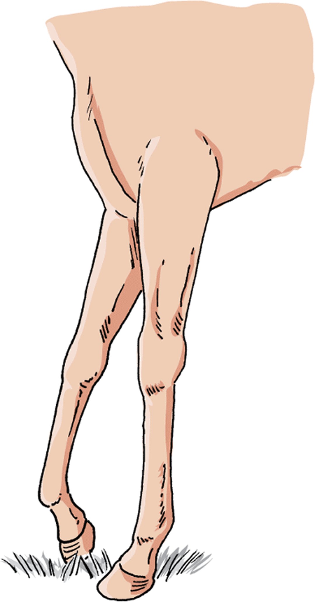
Contracted flexor tendons, foal

Signs vary widely in newborn foals. Some cannot stand, some attempt to walk on the upper part of their fetlocks, and others can stand but knuckle in the fetlocks or carpi (knees). One foal may improve spontaneously, while another, seemingly healthy at birth, may become progressively worse. The onset of signs may be rapid in foals 3 to 12 months old; such animals may walk on their toes with their heels off the ground. A slower onset may produce a “boxy” hoof with an elongated heel and toe that curves inward. Physitis may also occur in these horses. Usually both forelimbs are involved, although one or the other tends to be worse. Infected sores on the toes are a frequent complication that adds to the pain and deformity.
Slightly older horses (1 to 2 years old) commonly knuckle in the fetlock joints. Yearlings usually are more severely affected and more difficult to treat than younger animals. A complete examination by a veterinarian is necessary to determine the specific tendons involved. Any underlying bone or joint diseases or nutrition problems must be identified and corrected.
Mild cases in newborn foals often require no treatment. More severe cases may require supportive care (such as supplemental feeding), splints or casts, and medications. Older foals and yearlings can be managed conservatively with nutritional correction, proper hoof trimming, and treatment to control pain. If the condition persists after one week, however, more extensive treatment is necessary. Surgically cutting the accessory ligament of the deep digital flexor tendon is the most successful and commonly used procedure and does not interfere with future performance. Other types of surgery, including surgical cutting of the tendon and tendon lengthening, may also be recommended. In longterm cases, complications such as abnormal tightening of the joint membrane, malformation of accompanying ligaments, and bone involvement may prevent full recovery. Nutritional correction, proper foot trimming, and treatment to relieve pain are essential to proper healing, even when surgery is recommended. The outlook for recovery is fair to good for horses diagnosed early and managed properly.
Scapulohumeral Dysplasia
The abnormal development of the shoulder joint in miniature horses is called scapulohumeral dysplasia. It causes joint instability and arthritis of the shoulder. Although it is caused by improper development of the shoulder, an affected horse may not show signs until it is an adult. A severe lameness may also appear suddenly. There is often significant muscle wasting of the upper limb. The diagnosis is made based on history, physical examination, and x-rays. There is no simple treatment. Most horses have advanced disease when they are diagnosed, and their condition and pain cannot be controlled. Surgical treatment may be possible but is rarely performed. Euthanasia is often necessary.
For More Information
Also see professional content regarding developmental orthopedic disease in horses.


