


Disorders of the tarsus (hock) include the conditions known as bog spavin, bone spavin, and curb. The tarsus can also be affected by displacement of the tendon from the hock, fracture of the tarsus, hindlimb tendon and muscle ruptures, stringhalt, and thoroughpin.
Bog Spavin (Inflammation of the Hock Joint)
Bog spavin is a term for inflammation of the synovial membrane that results in distention of the joint capsule surrounding the tarsal (hock) joint. This may occur due to a congenital defect, degenerative joint disease, trauma, poor joint conformation, infection, or for an unknown reason (idiopathic). Lameness may or may not be present, depending on the cause.
The joint swelling is observable mainly on the middle back surface of the hock, with smaller swellings on each side of the joint near the back. Idiopathic bog spavin rarely interferes with the usefulness of the horse but may be considered an unsightly blemish. It should be evaluated by a veterinarian, including x-rays to make sure there are no structural changes to the joint. Hock swelling may appear and disappear on its own in weanlings and yearlings.
Excess fluid within the joint membrane may be removed by a veterinarian, to check for infection or bleeding into the joint, using a needle and syringe. Corticosteroid injections to the joint provide variable and short-term relief. Surgery using an endoscope may be necessary when bone and cartilage involvement is known or suspected. Bog spavin tends to recur, especially in cases where poor conformation is involved.
Bone Spavin
Bone spavin is osteoarthritis (bone arthritis), a degenerative condition of bone and joint cartilage in joints that leads to a gradual loss of cartilage and to pain, causing lameness. There are several joints in the hock, and one or more may be affected. Although bone spavin usually causes lameness, this may not always be obvious, especially early on. The cause is not always clear, but may be related to improper conformation of the hock, excessive jarring injury, or mineral imbalance. All breeds can be affected.
In cases of lameness, the horse tends to drag the toe. The forward movement of the hoof is shortened, and hock action is decreased. If the surfaces of the joint have been affected, lameness can be continuous. The heel may become elongated. Racing, jumping, and other sport horses may develop soreness in the gluteal muscles—known as trochanteric bursitis—as a result of the spavin. When standing, the horse may rest the toe on the ground with the heel slightly raised. The lameness often disappears with exercise and returns after rest. The bones of the affected joints may fuse spontaneously, leading to a return to soundness.
Diagnosis of bone spavin is based on the horse’s history, a physical examination, and x-rays to look for joint degeneration and abnormal bony growth. Local anesthesia of the individual tarsal joints and nerves to that part of the leg may be necessary to isolate the exact site of the pain responsible for the lameness.
In the early stages, injection of corticosteroids to the joint may help. Nonsteroidal anti-inflammatory drugs (for example, phenylbutazone) may minimize or eliminate signs. The joints most often affected do not have a large range of motion, and they may fuse naturally or with veterinary intervention. This relieves the pain, and the horse may be more comfortable and able to perform after joint fusion. Corrective shoeing by raising the heels and rolling the toe may help in milder cases but is unlikely to get rid of lameness on its own.
Curb
Curb is a thickening or bowing of the plantar tarsal ligament that runs down the back of the hock. The cause is typically strain. This ligament may become inflamed and thickened after falling, slipping, jumping, or pulling. The condition is most common in Standardbreds, in which poor conformation of the hock makes the horse more prone to develop the condition. An enlargement over the bone about 4 inches (10 centimeters) below the point of the hock may be seen when observing the horse from the side. A recently formed curb typically produces noticeable inflammation and lameness. The horse stands and favors the limb with the heel elevated. In cases of chronic curb, there is rarely any lameness or pain.
Cold packs and rest are recommended for acute cases. Little can be done to overcome curb that results from poor conformation of the hock. Fortunately, the problem seems to be self-limiting, without lasting effects on performance.
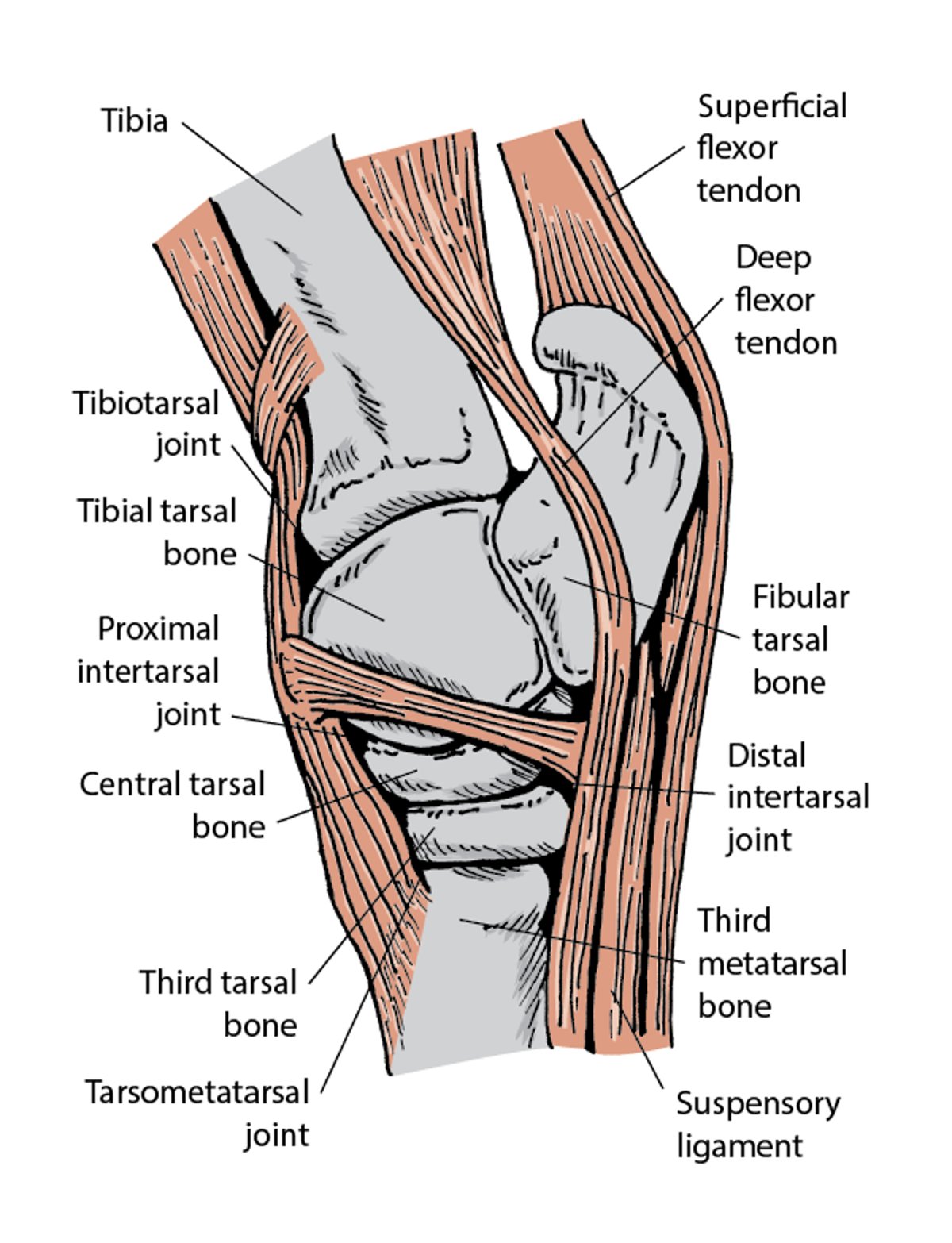
Lateral view of hock joint

Displacement of the Superficial Flexor Tendon from the Point of the Hock
The superficial flexor tendon can be dislocated by damage to its attachment to the point of the hock. A sudden bending of the hock typically causes the injury, after which the tendon may slip to the outside (more commonly) or inside of the hock. The limb may initially become suddenly and severely lame, with heat and swelling at the point of injury. Treatment involves rest for up to 3 months, possibly with a cast. The horse may be left with a permanently displaced flexor tendon and a rather jerky hock action. This usually causes no difficulty during fast exercise or jumping, but dressage movements may be affected. Surgery has been reported in a limited number of cases, but the results have not been very successful, particularly in larger horses.
Fracture of the Tarsus
Fractures of the tarsus or hock are usually caused by trauma or the complications of degenerative joint disease. Because the hock is a complex joint made up of 8 bones, a wide range of fractures can occur. Specific diagnosis depends on careful x-ray examination.
Chip fractures are among the more common fractures of the tarsus. Slab fractures are also seen, particularly in racehorses. Because these often are quite small and may not cause lameness, your veterinarian may need to use local anesthesia to positively identify the site of lameness. In many instances, a rest period of 3 to 6 months is all that is required for full recovery, although with large chip fragments surgical removal may be better. The condition responds well to surgery with an endoscope. Slab fractures may require the placement of bone screws.
Hindlimb Tendon and Muscle Ruptures
Horses rarely tear an entire Achilles tendon (involving both the calf muscle and superficial flexor tendon), but in such cases the outlook for recovery is grave. The hock drops towards the ground and is unable to bear weight.
Gastrocnemius (calf muscle) rupture is more common and can result from excess stress applied to the hock, such as when the horse has to stop suddenly. It can occur in both hindlimbs and weight can be borne, but the excess bending of the hock makes walking difficult. No satisfactory treatment exists. Splinting the limb and slinging the horse have been attempted but are usually unsuccessful.
Wounds to the front of the hock often injure the extensor tendons and digital extensors. When only one tendon is involved, the outlook for recovery is usually good. If both extensor tendons are severed, the horse’s performance gait may be lost, although the horse may remain useful for slower work or for breeding. Conservative treatment will heal the wound, but surgical repair and casting should be considered if both tendons are completely severed or if a return to performance status is desired.
Racing injuries sometimes rupture the superficial and deep flexor tendons. The tendons may also rupture when other tendons are torn. These are serious injuries with obvious lameness and varying degrees of overextension of the fetlock and pastern. Treatment involves surgical repair with splinting and casting the limb, but the outlook for recovery is poor for future performance.
Injury to the fibularis (peroneus) tertius muscle affects the hindlimb and disrupts the action of the stifle and hock joints. The most characteristic identifying feature of this condition is the ability to extend the hock and flex the stifle at the same time. The horse is lame but usually is able to bear weight on the limb. The affected hindlimb exhibits a jerking motion as it is brought forward. Diagnosis is based on clinical signs and can be confirmed with ultrasonography. Treatment consisting of prolonged rest (usually 4 months) is recommended, with a gradual return to exercise, and the outlook for recovery is favorable.
Stringhalt
Stringhalt is the involuntary contraction (spasm) of the muscles of one or both hindlimbs and is seen as exaggerated upward flexion of the joints observed at the walk. Any degree of overflexion in the joint may be seen; in mild cases the horse may spasmodically lift and lower the foot, whereas in extreme cases the foot is drawn sharply up until it touches the belly and then is struck violently on the ground. Mild stringhalt may come and go. Signs may diminish or even disappear during warmer weather. The signs are most obvious when the horse is sharply turned or backed. In some cases, the condition is seen only during the first few steps the horse takes out of its stall. Stringhalt may not materially hinder the horse’s ability to work, except in severe cases when the constant jarring injuries give rise to secondary complications. The condition may also make the horse unsuitable for equestrian sports (for example, dressage).
Its cause is unknown, but it is possible that an abnormality of nerves in the hindlimbs may be involved in some cases. There may be atrophy of the muscles of the legs noted. Cases due to nerve disorders usually occur on one side and may resolve spontaneously. In longterm cases, surgical cutting of a digital extensor, including removal of a portion of the muscle, may yield good results. Improvement may not be evident until 2 to 3 weeks after surgery, however. The outlook for recovery after surgery is considered guarded; not all cases respond.
Severe forms have been attributed to grazing on toxic plants in the United States, Australia, and New Zealand. Both hindlimbs are affected, and the condition may resolve with removal from pastures with the offending plants. The condition is usually seen in later summer or fall.
Diagnosis is based on a veterinarian’s examination but can be confirmed by electromyography, which measures the electrical activity of the muscles and associated nerves. False stringhalt sometimes appears as a result of some temporary irritation to the lower pastern area or even a painful sore in the foot. A stringhalt-like gait may occasionally be seen in a horse with momentary “locking” of the patella (kneecap) in an extended position.
Thoroughpin
Thoroughpin is a swelling of the covering of the deep digital flexor tendon just above the hock. It is characterized by fluid-filled swellings on both sides above the tarsal joint (distinguishing it from bog spavin). It usually affects only one limb and varies in size. It is usually not associated with lameness and may resolve on its own. Essentially a blemish, thoroughpin is chiefly important in show horses. It is treated by withdrawing the fluid and injecting hyaluronic acid or a long-acting corticosteroid. This procedure may need to be repeated until the swelling does not come back. Radiation therapy also helps reduce the fluid buildup from the tendon sheath.
Thoroughpin can also develop as a result of direct trauma, leading to inflammation and bleeding into the tendon sheath. Treatment for this may involve rest, systemic or local anti-inflammatory drugs, and cold therapy. Depending on the cause of trauma, infections are possible and require antibiotic therapy as well.
For More Information
Also see professional content regarding disorders of the tarsus in horses.


