

The paranasal sinuses are tissue-lined cavities in the skull, located behind the nose and eyes. They are divided into several compartments that are interconnected to the nasal passages and to each other. Diseases that originate in one sinus cavity may extend to and involve others.
Most diseases of the paranasal sinuses cause pus-containing or bloody discharge from one nostril. Swelling on one side of the face, excessive tears from one eye, and noise when the horse inhales are common signs of disorders of the sinuses.
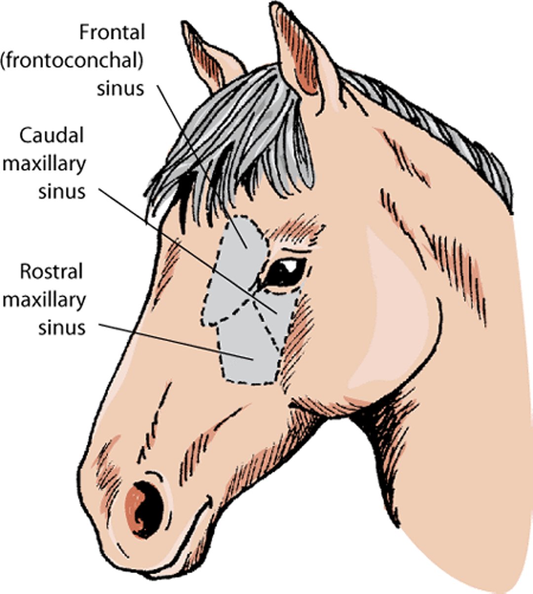
Paranasal sinuses, horse

Using an endoscope, the veterinarian can observe pus-containing material, a mass, or blood in the nasal passage originating from higher up in the head. X-rays of the skull and computed tomography (CT) can also be useful. With sedation and local anesthesia, the veterinarian can obtain a sample from the sinus of a standing horse. Fluid can be sampled from the maxillary or frontal sinuses for bacterial culture, drug sensitivity testing, and microscopic examination.
Sinusitis
Primary sinusitis, or inflammation of the sinuses, occurs after an upper respiratory tract infection that has involved the paranasal sinuses. It usually involves all sinus cavities but can be confined to the lower, deeper sinuses that are difficult to detect on x‑rays and also difficult to access surgically. Secondary sinusitis can result from tooth root infection, fracture, or sinus cyst. The first molar, fourth premolar, and third premolar (from more to less likely) are the most likely to develop tooth root abscesses. Signs of secondary sinusitis closely resemble those of primary sinusitis, including 1-sided nasal discharge and facial deformity. Tooth root abscesses typically produce a foul-smelling nasal discharge. Treatment of primary sinusitis involves rinsing (lavage) of the sinus cavity and antibiotic therapy based the results of bacterial culture and drug sensitivity testing. Secondary sinusitis requires removal of affected cheek teeth or cysts.
Ethmoid Hematoma
Progressive ethmoid hematoma is a destructive mass in the nasal passages and sinuses. The cause is unknown. These masses originate in the sinus and extend into the nasal passage. An expanding hematoma can cause damage to the surrounding bone but rarely causes facial distortion. It is primarily observed in horses older than 6 years. Periodic bleeding from one nostril is the most common sign. Horses with extensive masses may have reduced airflow through the affected nasal passage and bad breath. In longterm cases, the mass may protrude from the nostril. In most instances, the veterinarian can see the lesion extending into the nasal passages on endoscopic examination, and the extent of the mass can be determined with x-rays. Your veterinarian can shrink the mass by injecting it with a chemical. This works rapidly, but recurrence is common. Surgical removal can be performed if needed.
Sinus Cysts
Sinus cysts are fluid-filled cavities. They are typically found in horses less than 1 year old, but can also be seen in those greater than 9 years old. The primary signs are facial deformity, nasal discharge, and partial airway obstruction. X‑rays are more likely to identify a sinus cyst than endoscopic examination. Treatment involves surgical removal of the cyst and associated lining of the sinus. The outlook for complete recovery is good, and the recurrence is low. Some horses may have a permanent, mild discharge of mucus after surgery.
For More Information
Also see professional content regarding disorders of the paranasal sinuses in horses.


