


Lumpy skin disease is a viral infection of cattle. Originally found in Africa, it has also spread to countries in the Middle East, Asia, and eastern Europe. Clinical signs include fever, lacrimation, hypersalivation, and characteristic skin eruptions. Diagnosis is by histopathology, virus isolation, or PCR. Attenuated vaccines may help control outbreaks.
Lumpy skin disease is an infectious, eruptive, occasionally fatal disease of cattle characterized by nodules on the skin and other parts of the body. Secondary bacterial infection often aggravates the condition. Traditionally, lumpy skin disease is found in southern and eastern Africa, but in the 1970s it extended northwest through the continent into subSaharan west Africa. Since 2000, it has spread to several countries of the Middle East and in 2013 extended west into Turkey and several countries in the Balkans. More recently, outbreaks of lumpy skin disease were reported for the first time in Georgia, Russia, Bangladesh, and the People's Republic of China. The recent geographic spread of lumpy skin disease has caused international concern. The disease has not been recorded in the Western hemisphere or in Australia or New Zealand.
Etiology and Epidemiology of Lumpy Skin Disease in Cattle
The causal virus is related to that of sheeppox. Lumpy skin disease appears epidemically or sporadically. Frequently, new foci of infection appear in areas far removed from the initial outbreak. Its incidence is highest in wet summer weather, but it may occur in winter. It is most prevalent along water courses and on low ground. Because quarantine restrictions designed to limit the spread of infection often fail, biting insects have been suspected as mechanical vectors; however, outbreaks have occurred under conditions in which insects practically could be excluded. Experimentally, three species of hard ticks found in Africa have been shown to biologically transmit the virus. Because the disease can be experimentally transmitted by infected saliva, contact infection is another potential route of infection. African buffalo are suspected as maintenance hosts in Africa, but other wildlife species may also be involved.
Clinical Findings of Lumpy Skin Disease in Cattle
Infected cattle develop fever, lacrimation, nasal discharge, and hypersalivation, followed by the characteristic eruptions on the skin and other parts of the body in ~50% of susceptible cattle. The incubation period is 4–14 days.
The nodules are well circumscribed, round, slightly raised, firm, and painful and involve the entire cutis and the mucosa of the GI, respiratory, and genital tracts. Nodules may develop on the muzzle and within the nasal and buccal mucous membranes. The skin nodules contain a firm, creamy-gray or yellow mass of tissue. Regional lymph nodes are swollen, and edema develops in the udder, brisket, and legs. Secondary infection sometimes occurs and causes extensive suppuration and sloughing; as a result, the animal may become extremely emaciated, and euthanasia may be warranted. In time, the nodules either regress, or necrosis of the skin results in hard, raised areas (“sit-fasts”) clearly separated from the surrounding skin. These areas slough to leave ulcers, which heal and scar.

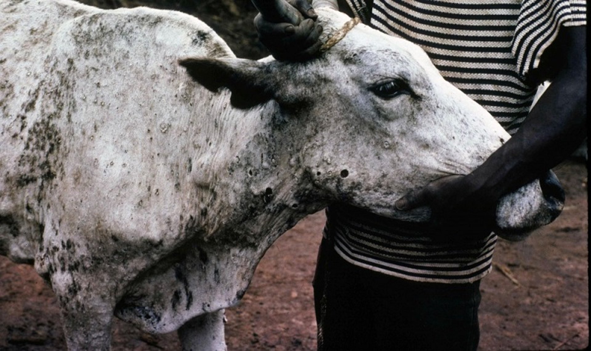
Courtesy of Dr. Max Bonniwell, Oban, Scotland.
Morbidity is 5%–50%; mortality is usually low. The greatest loss is due to reduced milk yield, loss of condition, and rejection or reduced value of the hide.
Diagnosis of Lumpy Skin Disease in Cattle
Histopathology, virus isolation, or PCR
The disease may be confused with the less clinically important pseudo-lumpy skin disease, which is caused by a herpesvirus (bovine herpesvirus 2). These diseases can be similar clinically, although in some parts of the world the herpesvirus lesions seem confined to the teats and udder of cows, and the disease is called bovine herpes mammillitis.
Pseudo-lumpy skin disease is a milder disease than true lumpy skin disease, but differentiation depends essentially on isolation and/or identification of the causal virus. The pox virus of lumpy skin disease can be demonstrated by electron microscopy in the early skin lesions. The two diseases can be distinguished by PCR.
Dermatophilus congolensis also causes skin nodules in cattle.
Treatment and Prevention of Lumpy Skin Disease in Cattle
Attenuated virus vaccines may help control spread
The spread of lumpy skin disease in recent years beyond its ancestral home of Africa is alarming. Quarantine restrictions have proved to be of limited use. Vaccination with attenuated virus offers the most promising method of control and was effective in halting the spread of the disease in the Balkans.
Administration of antibiotics to control secondary infection and good nursing care are recommended, but the large number of affected animals within a herd may preclude treatment.
Key Points
Lumpy skin disease was originally diagnosed in southern and eastern Africa but now also occurs in several countries in the Middle East, Asia, and eastern Europe.
The most distinctive clinical sign is widespread, firm, painful nodules of the skin and mucosal surfaces.
Attenuated virus vaccines may be used to help control the spread of infection.


