


A complete breeding soundness examination (BSE) in dogs consists of a history, physical examination, semen evaluation, and testing for Brucella canis. If infertility is suggested from the history, it should be established that adequate breeding management was practiced and that bitches had normal fertility when bred to other dogs. The time sequence of litters sired and bitches that did not conceive should be recorded, as should any recent illness during or before the time the bitches were bred. These should be assessed to determine whether the infertility may be transient (eg, fever can adversely affect semen production for > 60 days after the initial insult).
A general physical examination should be performed. Dogs with abnormalities such as severe joint disease or spinal problems may not be able to mount. Endocrinopathies such as hyperadrenocorticism or hypothyroidism may decrease fertility; these may be associated with abnormalities in weight or hair coat. The penis and prepuce should be examined; problems such as persistent frenulum, growths, or swelling due to balanoposthitis may prevent normal intromission. Abrasions or lacerations on the penis may bleed during breeding, and blood may be seen in the semen.
The prostate should be digitally palpated per rectum. The most common prostatic problem in mature (> 5 years old) intact dogs is benign prostatic hyperplasia (BPH); the prostate appears uniformly enlarged and is not painful on palpation per rectum. Dogs with BPH might be asymptomatic or have a history of hematuria and/or hemospermia or rectal tenesmus. In dogs with clinical signs, the treatment of choice is castration, although breeding dogs can be treated medically with 5-alpha-reductase inhibitors, which prevent the conversion of testosterone to dihydrotestosterone. Treatment is important, because affected dogs are predisposed to developing prostatitis.
Ultrasonography has become a common adjunctive tool for prostate evaluation in dogs, allowing for accurate measurements and assessment of the gland's echotexture for identification of potential pathologies, in which case a fine needle or biopsy instrument can be guided to sample a lesion.
The scrotum, testes, and epididymides should be palpated. Small, soft testes are usually associated with poor semen quality; greatly enlarged testes suggest orchitis or epididymitis. Lumps suggesting neoplasia may also be palpable. Scrotal abnormalities such as dermatitis may adversely affect semen quality by decreasing scrotal thermoregulation. Length, width, and height of testes should be measured with blunt calipers; these measurements are often of value for future comparison in cases of suspected testicular degeneration. Additionally, total scrotal width is highly correlated with body wt and an estimate of the dog’s sperm production potential.
Ultrasonographic examination of the scrotal contents is valuable not only to obtain more accurate measurements but also to evaluate for the presence of testicular or epididymal masses, which may not always be readily palpable in the early stages of a pathologic process.
Semen collection is performed with the dog on good footing (eg, a rug) rather than on a slippery surface or table. Care should be taken not to intimidate the dog; thus, any general examination procedures are best performed after semen collection. Semen may be collected in the absence of a bitch (although sperm numbers may be lower); however, the presence of a bitch is preferable, especially for inexperienced dogs. The pheromone methyl-paraben may be helpful for collection in the absence of a bitch; some veterinarians freeze swabs of estrous bitch urine or vaginal secretions for this purpose; however, the reaction of male dogs is variable.

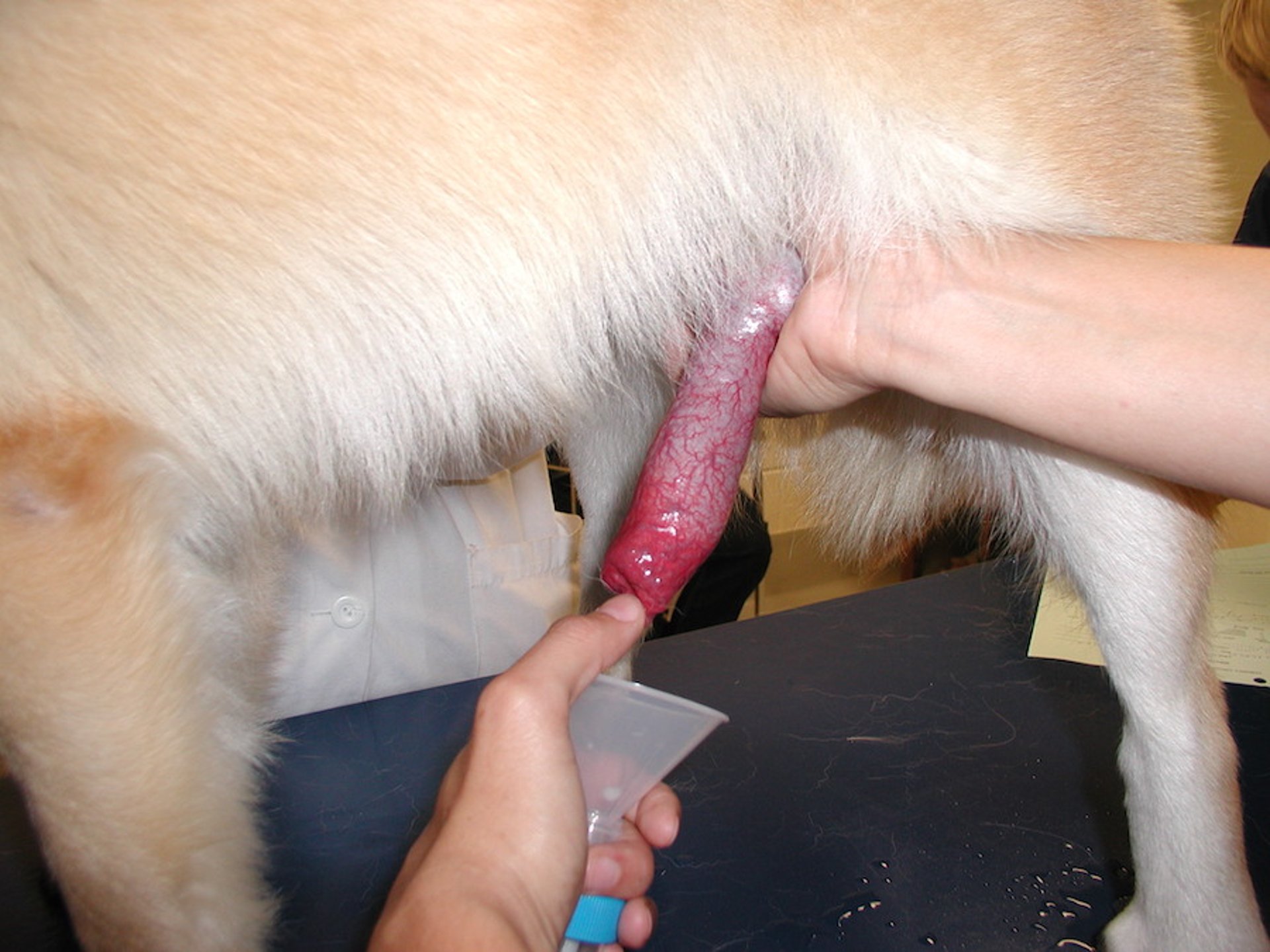
Courtesy of Dr. Sylvia Bedford-Guaus.
A collecting cone such as the liner of a bull artificial vagina, lubricated with sterile nonspermicidal lubricant or petroleum jelly and attached to a test tube, can be used. The penile sheath is gently pulled back, and the cone is slipped over the penis. As soon as the bulbus glandis is exteriorized from the sheath and is within the cone, the penis is grasped through the cone, immediately caudal to the bulbus. Constant pressure is maintained caudal to the bulbus; erection, and eventually ejaculation, should be achieved. Contact of the penis with the lubricated cone typically stimulates the dog to thrust into the cone, and the penis is compressed through the cone caudal to the bulbus as described above.
Semen may also be collected with the gloved hand technique, by stimulating the dog’s penis within the prepuce until a partial erection is achieved; the prepuce is then slid caudally, behind the bulbus glandis, and steady pressure is applied as with the cone technique until the dog ejaculates. A cup or tube fitted with a funnel is held over the tip of the penis to collect the ejaculate. Some breeders simply use a plastic bag held over the penis to collect semen.
The first (prostatic, clear) fraction and the second (sperm-rich, cloudy) fraction should be collected. After these fractions are ejaculated, close inspection of the collection tube should demonstrate that clear (prostatic) fluid is starting to layer on the cloudy second fraction; at this point, the collection may be stopped. The dog may continue to ejaculate prostatic fluid for up to 10 minutes before the erection subsides. The sheath should be examined after the penis is retracted to ensure that the penis is situated normally within the sheath and that no hair is caught within the sheath. Residual protrusion may occur if the sheath rolls inward as the penis retracts.
Semen evaluation consists of determination of appearance, volume, concentration, motility, and percent morphologically normal sperm. Yellow, brown, or red samples may indicate the presence of blood or urine in the ejaculate. The volume is variable, depending on how much prostatic fluid was collected and the size of the dog; it ranges from < 2 to > 20 mL but is typically ~5 mL.
Sperm motility should be evaluated immediately using warmed equipment; this should produce > 70% progressively motile spermatozoa. Sperm morphology is determined as for bulls. At least 80% of the sperm should be morphologically normal. The concentration is determined using a hemocytometer. To do this, the sperm is diluted at 1:100, and the number of sperm in the large central square (made up of 25 smaller squares) on the hemocytometer is counted. The number of sperm counted × 106 is the concentration of spermatozoa per mL. The total number of sperm in the ejaculate is calculated as volume × concentration. Total sperm number in the ejaculate ranges from 400 × 106 to > 1,000 × 106 and is correlated with body wt; as a general rule, a dog should produce ~10 × 106 sperm/lb body wt.
Every dog investigated for infertility should be screened for Brucella canis.
Sperm quality may be normal or abnormal, or no sperm may be seen in the ejaculate. Infertility is rare in dogs with a normal sperm evaluation and, in these cases, the history should be reviewed for mismanagement or bitch infertility. The presence of WBCs or RBCs in the ejaculate suggests inflammation of the tract, most commonly prostatitis; culture of prostatic fluid and appropriate treatment may help fertility. If sperm quality is abnormal, the history should again be reviewed to determine whether the dog has been sick recently or has received any drugs, especially anabolic steroids.
Other recognized causes of abnormal sperm quality include inflammation of the scrotum or other factors that may be causing a high scrotal temperature, testicular neoplasia (ultrasonography of the testes is recommended because many neoplasms of the testes are not palpable), trauma to the area of the scrotum, or brucellosis. However, most cases of low sperm quality in dogs are idiopathic.
Serum hormone measurements can be used to try to characterize infertility in dogs, but may not be helpful. Male dogs displaying infertility for > 12 months may have low testosterone levels which, in turn, result in high FSH levels due to lack of negative feedback. This carries a poor prognosis. Anti-Mullerian hormone (AMH) is produced by the Sertoli cells in the male; it can be measured to determine the presence of a cryptorchid testis. Levels of AMH may also be useful in characterizing testicular atrophy and diagnosing Sertoli cell tumors.
Azoospermia is relatively common in dogs. It may be due to failure of the dog’s testes to produce sperm, or to failure of the sperm to exit the testes because of epididymal blockage or incomplete ejaculation. As in stallions, the ejaculate may be tested for the presence of alkaline phosphatase, which is secreted by the epididymis. A high value (5,000–40,000 IU/L) indicates fluid from the epididymis was collected and thus is consistent with true azoospermia. Low values (< 5,000 IU/L) suggest epididymal blockage or ejaculation failure; semen collections should be repeated, using a strong stimulus such as a bitch in estrus.
A cystocentesis should be performed after semen collection to determine whether retrograde ejaculation is occurring. Swab samples of the vagina of a bitch after natural breeding may also be performed to determine whether sperm have been ejaculated; some dogs may not ejaculate with manual collection but do have normal ejaculation upon natural breeding. Careful palpation and ultrasonographic examination should be performed to detect any abnormality of the epididymides or spermatic cords, such as absence (epididymal aplasia) or blockage of the epididymis.
A dog is considered “satisfactory” if all the above findings are within normal limits and the dog is seronegative for B canis. Breeding dogs should be retested annually for B canis. As in other species, “questionable” dogs are those with a condition that might resolve over time (eg, recent febrile episode with temporary testicular degeneration, BPH), whereas a dog with an untreatable condition or an inherited disorder is classified as “unsatisfactory” for breeding.


