

GMS-stained cerebellar histomicrograph

GMS-stained cerebellar histomicrograph
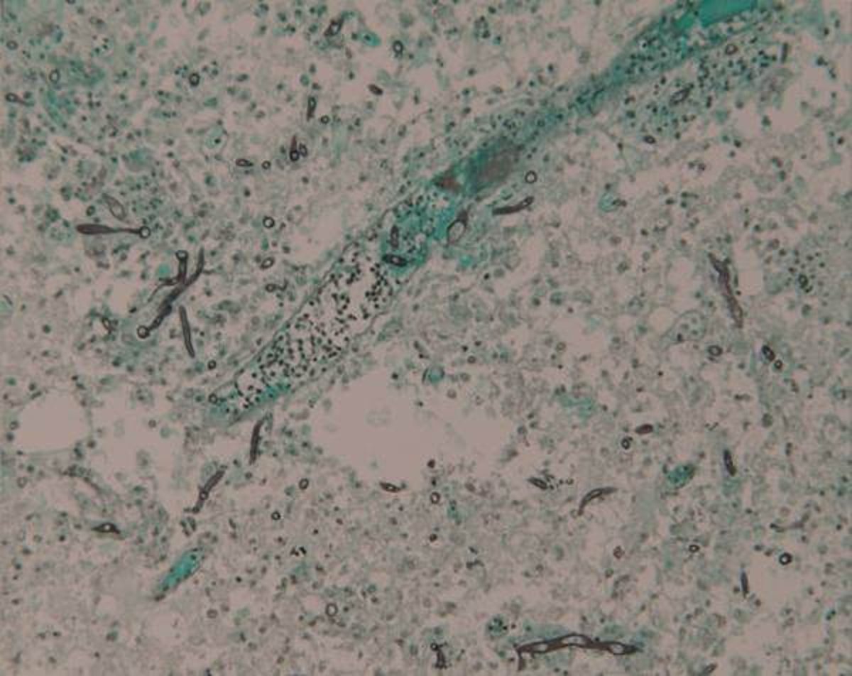
Grocott methenamine silver (GMS)-stained histomicrograph demonstrating branching, septate fungal hyphae consistent with Aspergillus species in the cerebellum from a young chick exhibiting neurologic signs.
Courtesy of Dr. S.M. Williams.
In these topics