

Sex determination of the gonads is important for development of the sex phenotype (internal and external genitalia, secondary characteristics) and sexual behavior. A sex chromosome genotype of XY leads to the development of testes due to the sex-determining region of the Y chromosome (SRY) gene. The SRY gene induces downstream factors such as SRY-box containing gene 9 (SOX9), anti-Müllerian hormone, and glial cell line–derived neurotrophic factor in Sertoli cells.

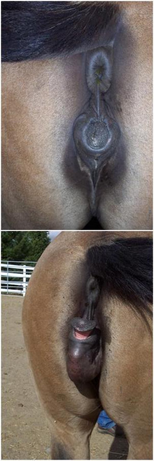
SRY negative XX sex reversal in a horse. External genitalia before (top) and after (bottom) sexual stimulation.
Courtesy of Dr. Ahmed Tibary.
Intersex conditions have been described in several domestic animal species. True hermaphrodites are rare and have both ovarian and testicular tissue and exhibit anomalies of the external genitalia. The karyotype is variable and may be a chimera, mosaic, or unknown. Pseudohermaphroditism, often referred to as sex reversal syndrome, is more common. Animals have one or the other type of gonad and external genitalia of the opposite sex. Animals may be XY SRY negative or XX SRY negative. In horses, the most common type is 64XY SRY negative. Some cases of sex reversal are believed to be due to a recessive autosomal gene mutation.
The most common intersex condition, the male pseudohermaphrodite, has testicular tissue in the abdominal cavity or beneath the skin in the scrotal region, and external genital organs that resemble those of females. Miniature Schnauzers, Basset Hounds, and rarely, Persian cats may present with pseudohermaphroditism when affected by persistent paramesonephric (Müllerian) duct syndrome.
Undescended testes are attached to the uterine horns, and the vasa deferentia are located in the wall of the uterus. There are bilateral oviducts, a complete uterus with a cervix, and a cranial portion of the vagina. Bilateral scrotal testes or unilateral or bilateral cryptorchidism may be present. Affected animals can present clinically with pyometra, urinary tract infection, prostate infection, or Sertoli cell tumor. The diagnosis is confirmed by presence of a 78, XY chromosome constitution, bilateral testes, and the presence of all paramesonephric (Müllerian) duct derivatives. Androgen-dependent masculinization is that of a normal male. Treatment is limited to castration and hysterectomy. The defect is inherited as an autosomal recessive trait in Miniature Schnauzers, and both females and males can be carriers. Homozygous affected dogs with a descended testis are generally fertile and capable of transmitting the trait to all offspring.
Polled intersex syndrome is well described in goats. Polled homozygotic males have decreased fertility due to segmental aplasia of the epididymides. They are often XX SRY negative.
Freemartinism Syndrome in Animals

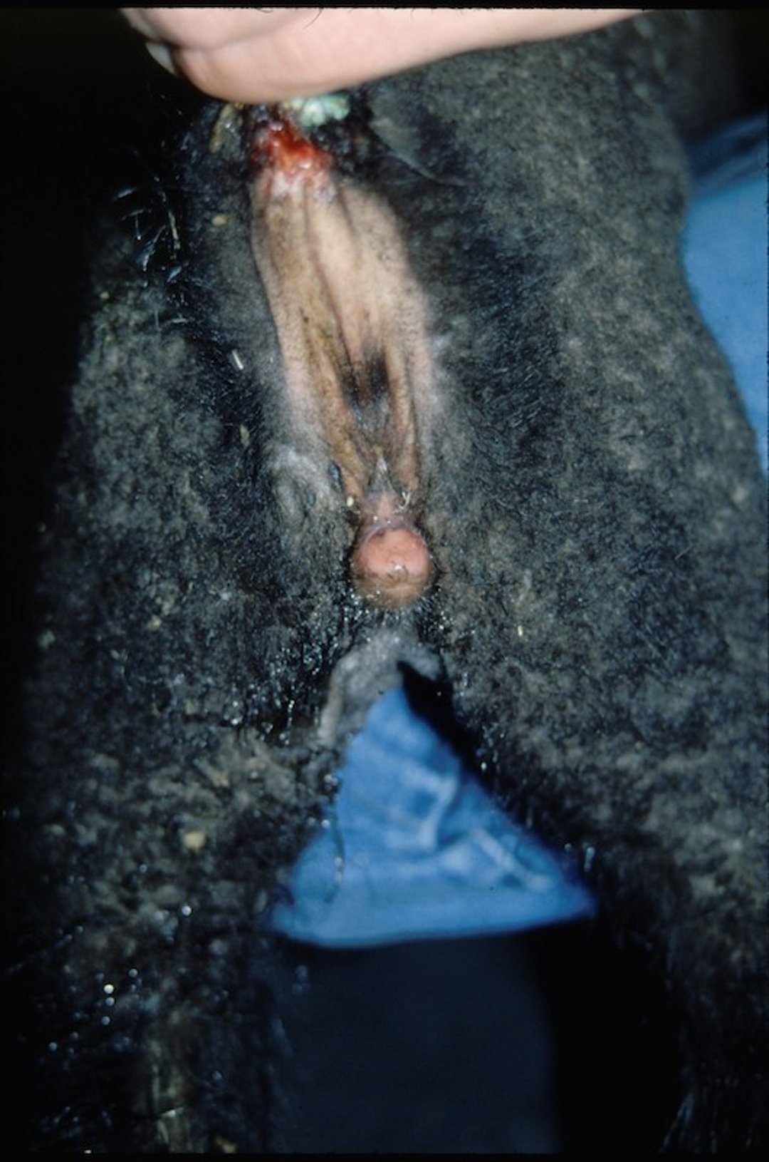
External genitalia of a freemartin ewe lamb.
Courtesy of Dr. Ahmed Tibary.

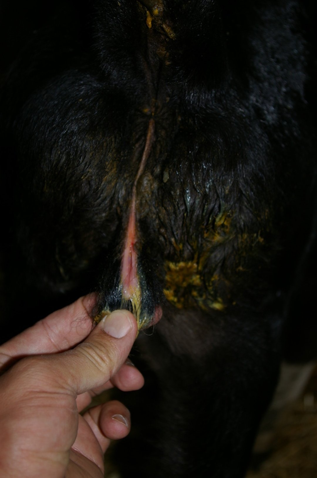
External genitalia of a freemartin heifer.
Courtesy of Dr. Ahmed Tibary.
Freemartinism syndrome is well known in cattle but has also been described in sheep, goats, and camelids. It causes sterility in females born co-twin to males; ~92% of all heifers born co-twin to bull calves are sterile. Single-born freemartin females have been reported and are believed to result from the in utero death of a male co-twin. These animals exhibit varying degrees of female-to-male sex reversal of the internal and external genitalia. The tubular genital organs in affected animals range from cordlike bands to near-normal uterine horns. Freemartins have a short vagina that ends blindly without communication with the uterus. The cervix is absent. The ovaries usually fail to develop and remain small. Vascular anastomosis of the chorionic placentas of the two fetuses results in transfer of anti-Müllerian hormone from the male to the female fetus, which inhibits development of the female tract.
Normal and freemartin cattle can be differentiated based on length of the vagina and on the presence or absence of a cervix. In calves 1–4 weeks old, the normal vaginal length is 13–15 cm, whereas in a freemartin vaginal length is 5–6 cm. Vaginal length is easily measured by gently inserting a well-lubricated probe with a blunt end into the vagina. Cytogenetic examination can demonstrate XX and XY chromosome patterns in freemartins. The interchange of cells that occurs in the placental circulation between the fetuses can also be demonstrated by detecting two different blood types in a single animal.
Other Chromosomal Abnormalities of Animals
Chromosomal abnormalities (XXY) have been described in males with azoospermia due to hypoplasia of the testes, epididymis, and vas deferens. Tortoise-shell or calico male cats possess two X chromosomes (XX/XXY, XY/XXY, or other chimeric or mosaic combination) and are sterile.


