honeypot link
skip to main content
MSD MANUAL
Veterinary Manual
VETERINARY PROFESSIONALS
PET OWNERS
RESOURCES
QUIZZES
ABOUT
VETERINARY PROFESSIONALS
PET OWNERS
<
Veterinary Topics
Respiratory System
Expand all
Collapse all
Respiratory System Introduction
The Respiratory System in Animals
For More Information
Principles of Therapy for Respiratory Disease in Animals
For More Information
Clinical Signs of Respiratory Disease in Animals
For More Information
Causes of Respiratory Disease in Animals
For More Information
Diagnostic Techniques for Respiratory Disease in Animals
For More Information
Control of Respiratory Disease in Animals
For More Information
Aspiration Pneumonia in Large Animals
Aspiration Pneumonia in Large Animals
Etiology
Clinical Findings
Lesions of Aspiration Pneumonia in Large Animals
Diagnosis
Treatment and Prevention
Key Points
For More Information
Bovine Respiratory Disease Complex
Viral Infections Associated with Bovine Respiratory Disease Complex in Cattle
Bovine Herpesvirus 1 (Infectious Bovine Rhinotracheitis Virus, Infectious Pustular Vulvovaginitis)
Etiology and Epidemiology
Clinical Findings
Diagnosis
Treatment and Control
Key Points
For More Information
Bovine Respiratory Syncytial Virus
Etiology
Clinical Findings and Lesions
Diagnosis
Treatment and Prevention
Key Points
For More Information
References
Parainfluenza-3 Virus
Bovine Viral Diarrhea Virus
Other Bovine Respiratory Viruses
Bacterial Pneumonia in Cattle with Bovine Respiratory Disease Complex
Bacterial Pathogens Associated With Bovine Respiratory Disease Complex
Etiology
Clinical Findings
Diagnosis
Treatment
Control
Key Points
For More Information
Overview of Bovine Respiratory Disease Complex
Etiology
Control and Prevention
Key Points
For More Information
Diaphragmatic Hernia
Diaphragmatic Hernia in Animals
Etiology
Clinical Findings
Diagnosis
Treatment
Key Points
For More Information
Fungal Pneumonia in Small Animals
Fungal Pneumonia in Small Animals
Etiology
Clinical Findings
Lesions
Diagnosis
Treatment
Key Points
For More Information
Infectious Respiratory System Diseases in Cattle
Sinusitis in Cattle
Etiology
Clinical Findings
Diagnosis
Treatment
Control
Key Points
Necrotic Laryngitis in Cattle
Etiology
Transmission, Epidemiology, and Pathogenesis
Clinical Findings
Lesions
Diagnosis
Treatment and Control
Key Points
Enzootic Pneumonia of Calves
Etiology
Diagnosis
Thoracic Ultrasonography
Culture and Sensitivity Testing
For More Information
Control and Prevention
Key Points
For More Information
Contagious Bovine Pleuropneumonia
Etiology
Clinical Findings
Lesions on Post-Mortem Examination
Diagnosis
Control
Key Points
For More Information
Bacterial Pneumonia Due to
Mycoplasma bovis
Infection in Cattle
Laryngeal Disorders
Laryngeal Disorders in Animals
Clinical Findings
Diagnosis
Treatment
Key Points
Lungworm Infection
Lungworm Infection in Animals
Epidemiology
Dictyocaulus
spp
Other Species
Pathogenesis
Clinical Findings
Diagnosis
Treatment
Control
Key Points
For More Information
Non-Infectious Respiratory System Diseases in Cattle
Allergic Rhinitis and Enzootic Nasal Granuloma in Cattle
Tracheal Stenosis Syndrome of Feeder Cattle
Pulmonary Emphysema, Edema, and Interstitial Pneumonia in Cattle
Etiology
Clinical Findings
Lesions on Post-Mortem Examination
Diagnosis
Treatment
Control
Key Points
Anaphylaxis in Cattle
Hypersensitivity Pneumonitis in Cattle
Etiology
Clinical Findings
Lesions on Post-Mortem Examination
Treatment and Control
Key Points
Diffuse Fibrosing Alveolitis in Cattle
Acute Respiratory Distress Syndrome of Feedlot Cattle
4-Ipomeanol Toxicosis (Moldy Sweet Potato) and
Perilla
Ketone Toxicosis (Purple Mint Toxicosis) in Cattle
Respiratory Disease in Cattle due to Exposure to Toxic Gases
Vena Caval Thrombosis and Metastatic Pneumonia in Cattle
Etiology
Clinical Findings
Lesions on Post-Mortem Examination
Treatment and Control
Key Points
Pharyngitis
Overview of Pharyngitis in Animals
Clinical Findings
Diagnosis
Treatment
Key Points
For More Information
Pharyngeal Trauma in Animals
Pulmonary Emphysema
Pulmonary Emphysema in Animals
Epidemiology and Pathogenesis
Cattle
Horses
Dogs and Cats
Clinical Findings and Diagnosis
Treatment
Key Points
For More Information
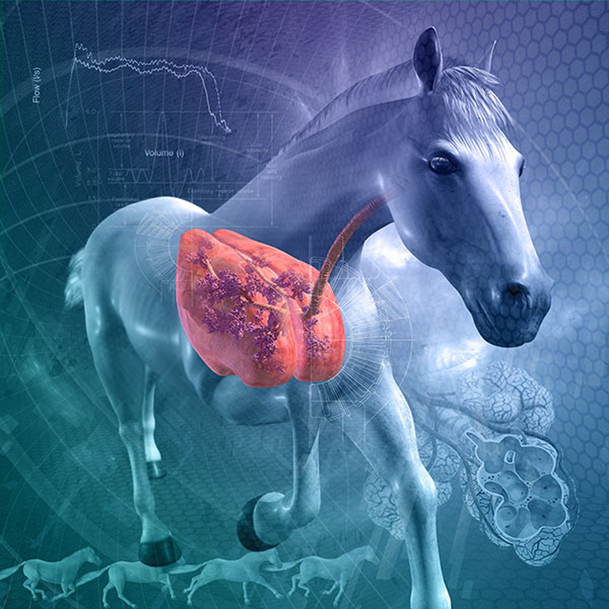
Respiratory Diseases of Horses
Overview of Respiratory Diseases of Horses
Diagnosis and Clinical Findings
Prevention and Treatment
For More Information
Equine Herpesvirus Infection
Etiology and Epidemiology
Clinical Findings
Lesions
Diagnosis
Treatment
Control
Infection by other Herpesviruses
Key Points
References
For More Information
Equine Influenza
Etiology and Epidemiology
Clinical Findings and Lesions
Diagnosis
Treatment and Prevention
Key Points
References
For More Information
Equine Viral Arteritis
For More Information
Hendra Virus Infection in Horses
Etiology and Pathogenesis
Epidemiology and Transmission
Clinical Findings
Lesions in Hendra Virus Infection
Diagnosis
Treatment, Prevention, and Control
Zoonotic Risk
Key Points
For More Information
References
Pleuropneumonia in Horses
Etiology and Pathogenesis
Clinical Findings and Lesions
Diagnosis
Treatment
Key Points
For More Information
Rhodococcus equi
Pneumonia in Foals
Etiology and Pathogenesis
Clinical Findings and Lesions
R equi pulmonary disease
Nonpulmonary R equi
Diagnosis
Treatment and Prognosis
Prevention
Key Points
References
For More Information
Acute Respiratory Distress Syndrome (Acute Bronchointerstitial Pneumonia) in Foals
Etiology, Epidemiology, and Pathogenesis
Clinical Findings and Lesions
Diagnosis
Treatment
Key Points
References
For More Information
Strangles in Horses
Etiology and Pathogenesis
Clinical Findings
Diagnosis
Treatment
Prevention
Control
Key Points
For More Information
Asthma in Horses
Pathophysiology
Etiology
Clinical Findings
Severe Asthma (RAO)
Mild-moderate Asthma (IAD)
Diagnosis
Treatment
Key Points
References
For More Information
Exercise-Induced Pulmonary Hemorrhage in Horses
Etiology and Pathogenesis
Diagnosis
Treatment and Control
Key Points
References
For More Information
Laryngeal Hemiplegia in Horses
Etiology and Pathogenesis
Clinical Findings and Diagnosis
Treatment
Key Points
For More Information
Pharyngeal Lymphoid Hyperplasia in Horses
For More Information
Dorsal Displacement of the Soft Palate in Horses
Etiology and Pathogenesis
Diagnosis
Treatment
Key Points
References
For More Information
Epiglottic Entrapment in Horses
Clinical Signs
Diagnosis
Treatment
References
For More Information
Subepiglottic Cyst in Horses
Diagnosis
Treatment
For More Information
Laryngeal Dysplasia in Horses
For More Information
Diseases of the Nasal Passages in Horses
Epidermal Inclusion Cyst
Redundant Alar Fold
Diseases of the Nasal Septum
Nasal Polyps
Choanal Atresia
Cleft Palate
For More Information
Diseases of the Paranasal Sinuses in Horses
Sinusitis
Primary Sinusitis
Secondary Sinusitis
Ethmoid Hematoma
Paranasal Sinus Cyst
Neoplasia of the Paranasal Sinuses
Key Points
For More Information
Guttural Pouch Disease in Horses
Empyema
Clinical Signs
Diagnosis
Guttural Pouch Mycosis
Guttural Pouch Tympany
Miscellaneous Transient Obstructions of the Larynx During Exercise in Horses
Arytenoid Chondropathy in Horses
Etiology and Pathogenesis
Clinical Findings and Diagnosis
Treatment
Key Points
For More Information
Respiratory Diseases of Pigs
Overview of Respiratory Diseases of Pigs
Atrophic Rhinitis in Pigs
Etiology
Clinical Findings
Lesions
Diagnosis
Control
Key Points
Mycoplasmal Pneumonia in Pigs
Etiology and Epidemiology
Clinical Findings
Lesions
Diagnosis
Control
Key Points
For More Information
Necrotic Rhinitis in Pigs
Etiology
Clinical Findings and Lesions
Diagnosis
Prevention and Treatment
Key Points
Pasteurellosis in Pigs
Pleuropneumonia in Pigs
Etiology
Clinical Findings
Lesions
Diagnosis
Treatment and Control
Key Points
Influenza A Virus in Swine
Etiology
Transmission and Epidemiology
Pathogenesis
Clinical Findings
Lesions
Diagnosis
Treatment and Control
Key Points
For More Information
Respiratory Diseases of Sheep and Goats
Overview of Respiratory Diseases of Sheep and Goats
Diseases of the Upper Respiratory Tract
Diseases of the Lower Respiratory Tract
Sheep Nasal Bot Myiasis
Clinical Findings
Treatment
Mycoplasma
Pneumonias in Goats
Etiology
Clinical Findings
Diagnosis
Control
Key Points
For More Information
Bacterial Bronchopneumonia in Sheep and Goats
Etiology
Pathogenesis
Clinical Findings
Lesions
Diagnosis
Treatment and Control
Key Points
Lentivirus Pneumonia in Sheep and Goats
Etiology and Pathogenesis
Epidemiology
Clinical Findings
Lesions
Diagnosis
Control
Key Points
For More Information
Ovine Pulmonary Adenocarcinoma
Etiology
Clinical Findings
Lesions
Diagnosis
Control
Key Points
For More Information
Respiratory Diseases of Small Animals
Canine Influenza (Flu)
Etiology, Epidemiology, and Transmission
Clinical Findings and Diagnosis
Treatment, Prevention, and Control
Key Points
For More Information
Laryngeal Paralysis in Dogs and Cats
Key Points
Overview of Respiratory Diseases of Dogs and Cats
For More Information
Allergic Pneumonitis in Dogs and Cats
Etiology
Clinical Findings
Diagnosis
Treatment
Key Points
For More Information
Canine Nasal Mites
Etiology and Epidemiology
Clinical Findings
Diagnosis
Treatment
Key Points
For More Information
Feline Respiratory Disease Complex
Etiology
Clinical Findings
Lesions
Diagnosis
Treatment
Prevention
Key Points
For More Information
Lung Flukes in Dogs and Cats
For More Information
Lung Nematodes in Small Animals
Aelurostrongylus abstrusus
Capillaria aerophila (Eucoleus aerophilus)
Filarids
For More Information
Neoplasia of the Respiratory System in Dogs and Cats
Tumors of the Nose and Paranasal Sinuses
Tumors of the Larynx and Trachea
Primary Lung Tumors
Clinical Findings
Diagnosis
Treatment
Metastatic Tumors of the Lungs
For More Information
Pneumonia in Dogs and Cats
Clinical Findings
Diagnosis
Treatment
For More Information
Pulmonary Thromboembolism in Dogs and Cats
Clinical Findings
Diagnosis
Treatment
Key Points
For More Information
Rhinitis and Sinusitis in Dogs and Cats
Etiology
Clinical Findings
Diagnosis
Treatment
For More Information
Tonsillitis in Dogs and Cats
Etiology
Clinical Findings and Diagnosis
Treatment
For More Information
Feline Bronchial Asthma
Kennel Cough
Etiology
Clinical Findings
Diagnosis
Treatment
Prevention
Key Points
For More Information