


Disease in amphibians can best be minimized through prevention or early treatment. Proper care and housing are important, in part because ideal environments for amphibians are often moist and warm—the same conditions that lead to the growth of many bacteria and molds. Amphibians are sensitive to their environments and easily become ill due to poor environmental conditions, such as poor water quality, poor diet, improper tank setup, overcrowding, and improper or too-frequent handling. Owners of amphibians must pay close attention to sanitation and hygiene to prevent illness.
Diseases Caused by Bacteria
Bacterial infection is usually caused by bacteria that are normally found in the environment. Such bacteria can become a problem when the balance in the enclosure is disrupted by changes in various elements such as temperature, diet, crowding, or cleanliness.
Chlamydiosis
Chlamydiosis is a serious infection caused by several species of Chlamydia bacteria that can lead to death in amphibians. Infected frogs may die suddenly or show signs of lethargy, loss of balance, loss of skin color, tiny red spots on the skin, and swelling due to excess fluid in body tissues. An examination of the animal’s tissue by your veterinarian may show enlargement of the liver and inflammation within the liver, spleen, kidney, and other organs. Chlamydiosis may be accompanied by other bacterial infections, which must also be treated appropriately. Antibiotics are often prescribed to treat chlamydial infection.
Mycobacteriosis
Mycobacteriosis is caused by several bacterial species in the Mycobacterium family. It occurs most commonly in injured amphibians with weakened immune systems. Although mycobacteriosis is often an infection of the skin, ingesting organisms in food or water may also lead to gastrointestinal disease and infection throughout the body. Affected amphibians may have small gray lumps in the skin or in body organs such as the liver, kidneys, spleen, and lungs. Infected amphibians may eat well but still lose weight. Your veterinarian can look for the bacteria on the amphibian’s skin or test for it in feces and in the mucus of the throat.
Treatment is not recommended for this disease. Because mycobacteriosis is a serious disease that is transmittable to humans, people with impaired immune system function (including the elderly, infants, or those undergoing chemotherapy) should not keep amphibians diagnosed with this illness. If you have more than one animal, immediately isolate the suspect amphibian from any companions by removing it from the tank. The best defense is prevention. Mycobacteria typically live in the slime layer that builds up in aquatic habitats over time. For this reason, weekly cleaning and removal of this film is recommended.
Mycobacteriosis can pass from animals to humans and can result in skin infection. Always wear protective eyewear and gloves when handling infected animals or cleaning their environment.
Red-leg Syndrome
Red-leg syndrome is a common condition in which there is a reddening of the lower body—usually the legs and sometimes the abdomen—due to dilation of capillaries (tiny blood vessels) under the skin. It accompanies widespread infection in frogs, toads, and salamanders. Red-leg syndrome is most often associated with Aeromonas bacteria, although other bacteria can also cause this syndrome. Viruses and fungi may also cause similar reddening.
Red-leg syndrome

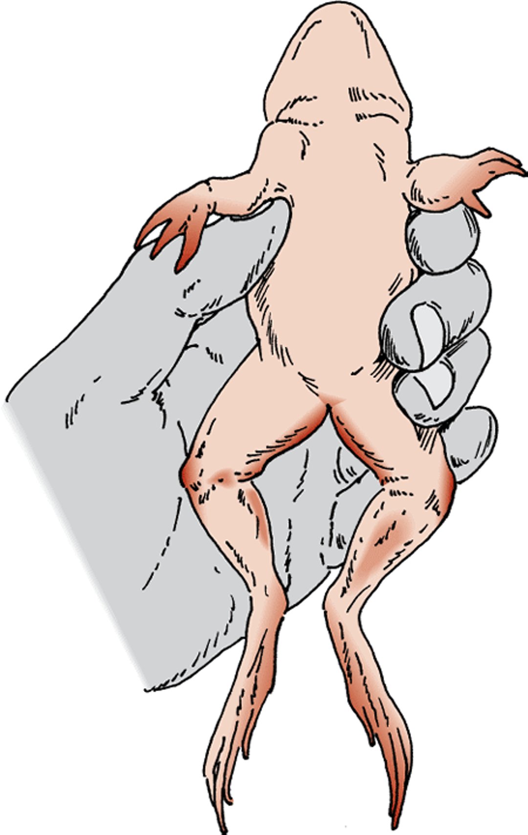
Underfed, newly acquired amphibians that are kept in poor-quality water or other less-than-ideal environmental conditions are particularly susceptible. Signs include lethargy; extreme thinness; open sores that do not heal on the skin, nose, and toes; and the characteristic reddening of the legs and abdomen. Bleeding may also occur in the skeletal muscles, tongue, and “third eyelid,” or protective fold of skin in the eyes of amphibians. When the onset of disease is sudden, however, these signs may be absent. Your veterinarian will look for signs of widespread infection—which could include inflammation or dead cells localized in the liver, spleen, and other abdominal organs—and will likely test the blood or body fluids for bacteria before beginning therapy. Your veterinarian may start treatment with antibiotics for a presumptive bacterial infection while test results are pending. Because a fungal infection may cause similar signs to the bacterial infection that causes red-leg syndrome, the veterinarian also may start anti-fungal medications. Follow your veterinarian’s instructions for the most favorable results.
Maintaining a high-quality environment, including thorough sanitation, will go a long way toward preventing red-leg syndrome. If an animal does become affected, be sure to isolate it from other amphibians in the home and seek immediate veterinary care.
Diseases Caused by Fungi
Fungal infections are common in amphibians. Many of the fungi that infect amphibians are difficult to tell apart without laboratory tests because they produce similar signs, including lethargy and open sores on the skin. Sometimes white or yellow furry growths on the skin may be seen. Your veterinarian can identify some fungi by examining a skin scraping under a microscope. Diagnosis of other fungal infections requires that tissue samples be prepared using special stains and then examined under a microscope. Your veterinarian may take samples to try to grow, or culture, the fungus. This is done so that the fungus can be identified and the best possible treatment selected. Treatment includes proper hygiene and the use of antifungal agents, often given in a dip or bath. Carefully follow your veterinarian’s prescribed dosage and treatment schedule.
Chytridiomycosis
Chytridiomycosis is the most serious fungal infection in amphibians and is thought to play a role in the decline of frog populations in many parts of the world. It is caused by Batrachochytrium dendrobatidis, a fungus related to water molds. The fungus feeds on keratin, a protein found in the outermost layers of the skin. It can survive in most environments even without a host. Death is common in infected animals. Signs include a persistent loss of appetite, lethargy, excessive shedding of skin, constriction of the pupil of the eye, and an inability to coordinate the muscles. Placing an infected animal in a shallow dish of water will often confirm the sloughing of skin associated with this disease.
If you suspect this disease, seek medical attention immediately. Entire collections have been lost in a matter of days. Veterinarians diagnose the disease by examining skin scrapings that are stained and put under a microscope. Treatment includes applying a prescription antifungal medicine (usually as a bath) to the surface of the skin and making sure that animals are kept well within their preferred temperature range.
Chromomycosis
Chromomycosis is caused by several types of pigmented fungi that may be found in organic substances such as topsoil and decaying plant matter. These fungi are capable of growing on tank walls. Amphibians with cuts or sores are at risk because the fungus typically infects the body through broken skin.
Infection is common and often fatal. Signs may include loss of appetite, weight loss, skin wounds or open sores that do not heal and become inflamed, swelling of the abdominal area, and evidence of neurologic impairment such as head tilt or the inability to move correctly.
Confirmation of this disease usually requires examination of the animal’s internal organs and tissues after it has died. Amphibians that are suspected of having chromomycosis may be treated with antifungal drugs, but the chance of survival is poor once the infection has spread to the central nervous system.
The best defense is to improve sanitation. Chromomycosis can cause skin lesions in humans, so be sure to wear powderless gloves and follow careful sanitary procedures when cleaning your pet’s cage, handling any items in the cage, and handling your pet.
Saprolegniasis
Saprolegniasis is a disease caused by several kinds of fungi or “water molds” that infect the gills and/or skin of aquatic and immature amphibians. It commonly affects newts, mudpuppies, aquatic frogs, and tadpoles. When in water, newly affected animals appear to have a whitish cotton-like growth on their skin. As time goes on, the growth may become greenish due to the presence of algae. This growth, sometimes called a fungal mat, can be difficult to see when the amphibian is not in the water. Other signs of saprolegniasis include lethargy, difficulty breathing, lack of appetite, and weight loss. Sores on the skin that do not heal may occur as the infection progresses.
Veterinarians diagnose the disease by identifying the fungus in a scraping taken from the animal’s skin. Treatment with a medicated dip, used as directed by your veterinarian, may be effective. Poor water quality is often the root cause of saprolegniasis. Changing water frequently and providing a correct and constant temperature for your amphibian are necessary for successful treatment. All water containers should be cleaned and sanitized daily.
Other bacterial and fungal infections, such as zygomycosis and mesomycetozoan infection, may be present in amphibians with open sores on the skin. Be sure you understand the correct procedures for applying any medication prescribed by your veterinarian. Ask for a demonstration if you are new to the procedures.
Diseases Caused by Parasites
Many of the single-celled organisms and other microorganisms found in and on amphibians are not associated with disease unless the amphibian becomes stressed or its immune system is weakened. Recently caught or transported amphibians are particularly susceptible to parasites, as are those kept in unhealthy conditions or in environments outside their preferred temperature range.
Whether or not a parasite becomes a threat to the health of an amphibian in an enclosed environment often depends on whether it needs another animal host to properly develop. Amphibians may act as temporary or intermediate hosts for some parasites; in other words, they help the parasite mature before it moves on to its final host. Or amphibians may act as the final destination after the parasite developed on another host. Parasites that require more than 1 host are said to have an indirect life cycle. They tend to die out when wild-caught amphibians are brought into captivity, as long as the other host is not present. The opposite is also true. Parasites that do not need a third party to fully develop—those with a direct life cycle—may thrive in a closed environment. Good hygiene is essential for parasite control and includes the prompt removal of sloughed skin, droppings, uneaten food, and carcasses from animal enclosures.
Parasites of the skin can sometimes be seen by close examination of amphibians using magnification and a bright, cool light. Your veterinarian may need to do a skin scraping or biopsy to identify parasites causing lumps or other skin abnormalities. Fresh fecal samples may be needed to identify internal parasites. (It is a good practice to bring fresh droppings, if available, any time your pet visits the veterinarian.) Some small frogs are translucent enough to allow your veterinarian to see parasites inside the body by shining a bright light through the body of the frog, a procedure called transillumination. In some cases, parasites are found only on examination after death.
It is common for your veterinarian to find various microorganisms in the droppings. Some types of microorganisms do not cause disease and do not require treatment in healthy amphibians. Although many immature roundworms found in the feces do not cause disease, treatment is recommended because disease-causing and non-disease-causing worms cannot be readily distinguished from one another.
Pseudocapillaroides xenopi Infection
The roundworm Pseudocapillaroides xenopi burrows into the skin and is known to affect colonies of the aquatic African clawed frog. Signs of infection include blotchy, rough, and pitted skin and skin sores. As the infection progresses, lethargy, loss of appetite, and sloughing of the skin occur. The disease may also make amphibians more susceptible to bacterial infections that can lead to death.
Diagnosis is made by finding small, white roundworms beneath the mucus on the skin. A veterinarian can confirm the presence of these parasites by performing a skin scraping that is examined immediately under a microscope. Treatment with anthelmintics, drugs that destroy parasitic worms, may be effective. Follow the dosage and treatment schedule prescribed by your pet’s veterinarian. Frequent water changes with removal of shed skin containing the parasite are required to prevent the infection from getting worse and spreading to other amphibians in the enclosure.
Rhabdiasis
Rhabdiasis is caused by the lungworm Rhabdias, which damages the lungs of captive amphibians. Associated infections may occur as a result of this damage. The lungworm has a direct life cycle with free-living phases. Adult worms live in the lungs where they deposit eggs that are coughed up, swallowed, and then excreted into the environment. Larvae then burrow through the skin of a new host where they mature and migrate to the lungs, repeating the cycle. Affected animals may appear thin, are generally weak, and lose appetite.
Your veterinarian may diagnose this disease by examining fresh feces or secretions from the mouth and nose for lungworms or their eggs. When rhabdiasis is suspected, treatment usually involves 1 of 2 commonly prescribed drugs (fenbendazole or ivermectin) that are used to remove worms or other parasites. Following the second day of each 2-day fenbendazole treatment or each dose of ivermectin, the animals should be moved into a newly established environment to prevent reinfection from free-living life stages that have become established in the material (such as moss or mulch) in their enclosure. The relocation to a clean environment is critical to the recovery of your pet and any instructions from your veterinarian should be followed precisely.
Diseases Caused by Viruses
Like other species, amphibians are susceptible to a variety of viral infections. Some viruses that affect amphibians can also cause cancer. No specific treatments are available for viral infections in amphibians, so general supportive care is provided.
Ranaviruses
Ranaviruses have been identified in many wild populations of amphibians across the world. Some of these cause signs very similar to those seen with bacterial skin infections. Ranaviruses are transmitted through exposure to contaminated water or soil, contact with infected animals, or eating tissues from infected animals. The original viral infection may lead to secondary infections; many outbreaks of red-leg syndrome (see above) may have been caused by an underlying and undiagnosed ranavirus infection. Your veterinarian may diagnose a ranavirus infection through blood testing or a series of tests on infected tissue, including culture, special staining and microscopic analysis, or PCR testing. There is no treatment for ranaviral disease other than supportive care and appropriate treatment for the secondary bacterial or fungal infections.
Renal Adenocarcinomas (Lucké Tumors)
Renal adenocarcinomas (Lucké tumors), caused by a herpesvirus, may occur in leopard frogs (Rana pipiens) that are wild-caught in the northeastern and north central United States. Few frogs with tumors are seen in the summer because the virus needs cold temperatures to grow. The virus is thought to be transmitted through breeding ponds and matures while infected frogs are hibernating at 41°F to 50°F (5°C to 10°C). Tumors are most common in the early spring when frogs emerge from hibernation.
The spread of the tumor to the liver, lungs, and other organs is common; both the original tumor and its offshoots can become very large. Signs are lethargy and bloating. Your veterinarian will likely diagnose this tumor by microscopic analysis of a needle aspirate or biopsy of the pet’s kidney. There is no known treatment.
Metabolic Bone Disease
Metabolic bone disease often results from an imbalance of calcium, phosphorus, and vitamin D3 in the diet or from lack of exposure to ultraviolet light, which is critical to the body’s formation of vitamin D3. It is frequently seen in amphibians that are fed a diet of invertebrates without supplementation ( see Diet for Amphibians). With the exception of earthworms, most invertebrates used as food are deficient in calcium. Crickets are often a culprit; amphibians fed a cricket-only diet are at high risk for developing metabolic bone disease.
The disease results in deformity of the lower jaw, fractures, and scoliosis (curvature of the spine). In severe cases, muscle spasms and bloating occur. Your veterinarian will diagnose the condition by examining x-ray images for the thinning of the outer layers of leg bones, deformities of the lower jaw and the bone at the base of the tongue, fractures caused by disease, and in severe cases, gastrointestinal gas. Your veterinarian also may take blood to measure levels of calcium and phosphorus.
Treatment includes correcting the diet and administering a calcium supplement as directed by your veterinarian. Full-spectrum lighting with biologically active ultraviolet-B light should be provided. Vitamin D3 can also be administered in severe cases. Follow your veterinarian’s treatment program carefully for the best results.
Thiamine Deficiency
Thiamine deficiency is typically seen in amphibians that are fed frozen fish. Many frozen fish contain the enzyme thiaminase, which destroys thiamine (vitamin B1). Thiamine is required by many tissues in the body, especially those of the central nervous system. Signs of thiamine deficiency include tremors, seizures, and severe arching of the back and neck. Initial treatment is the injection of thiamine by your veterinarian, followed by thiamine supplements with each meal for a prescribed period of time. Thiamine deficiency can be prevented by routinely supplementing diets with 250 mg of thiamine per kilogram of fish fed.
Obesity
Obesity is also considered a disease. Overfeeding is the primary cause of obesity. Owners often fail to realize that many amphibians will continue to consume prey as long as it is available and without regard for their energy needs. As in mammals, it is assumed that obesity in amphibians can cause stress on certain organs. Your veterinarian can examine the body using gentle finger pressure to feel for fat deposits; however, in females, ultrasonography may be necessary to differentiate fat deposits from egg masses.
Treatment for active species includes increasing the size of the enclosure to allow increased activity. Maintaining the amphibian at the upper end of its preferred temperature range will accelerate metabolic rate and increase caloric use. However, you should never exceed the maximum recommended temperature. Reducing the amount of food given to an animal will help control its weight. Ask your veterinarian for guidance in determining the appropriate amount of food for your pet.
Trauma
Injuries are common in captive amphibians and include cuts, broken bones, internal bleeding, excessive drying (desiccation), and the loss of toes, limbs, or tail. Prompt evaluation by your veterinarian and supportive care are required for a successful outcome. Desiccation is common in amphibians that escape their enclosure or do not receive proper care. For smaller amphibians (those weighing less than 30 grams), most fractures can be managed conservatively with cage rest. For larger amphibians, the use of casts or internal pins may be helpful. Pain management must be considered for amphibians with traumatic injuries. Because amphibians have opioid receptors, using opioid drugs may help reduce pain. Anti-inflammatory drugs such as meloxicam may also be used to provide pain relief.
For More Information
Also see professional content regarding diseases of amphibians.


