


Fasciola hepatica is one of the most important flukes of domestic ruminants worldwide, causing liver fluke disease (liver rot, fascioliasis). Chronic liver fluke disease is more common in cattle and rarely fatal. Acute and subacute disease is more common in sheep and camelids and is often fatal. Traumatic hepatitis occurs as immature flukes migrate through the liver tissue before entering and remaining in the bile ducts. Anemia, submandibular edema, decreased weight gains, and decreased milk production can occur. Fecal sedimentation is an easy and inexpensive diagnostic tool, but it can detect only patent infections. ELISA tests are available in some countries for individual or bulk milk tank detection of antibodies. Control should focus on removing parasites from the animal, reducing the intermediate host snail population, and excluding production animals from snail-infested pastures. Various anthelmintics are available to treat animals with liver flukes; however, anthelmintic resistance to some compounds does occur.
Etiology of Fasciola hepatica in Ruminants

Courtesy of Dr. Raffaele Roncalli.
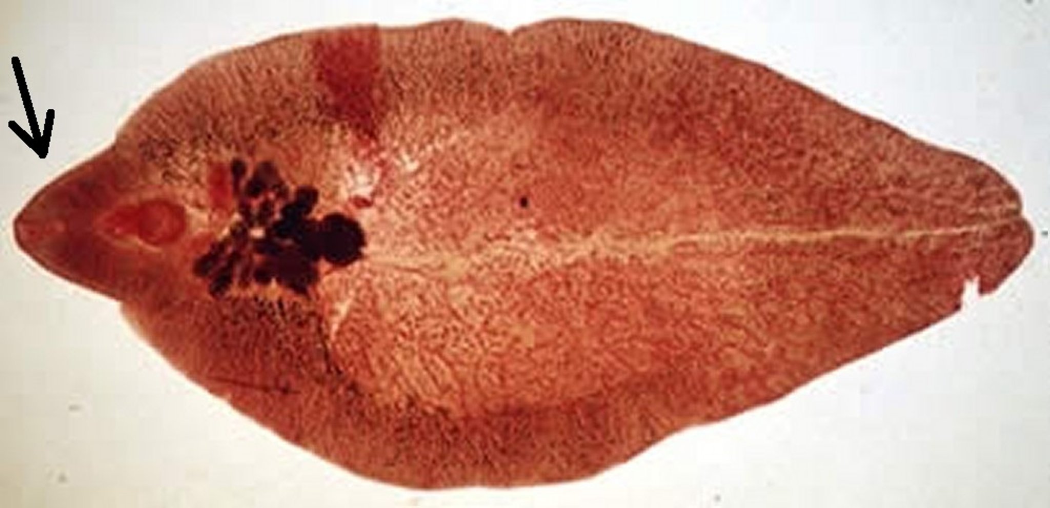
F hepatica (30 × 2–12 mm and leaf-shaped) is distributed worldwide and has a broad host range, including humans. Economically important infections occur in cattle, sheep, alpacas, and llamas in three forms: 1) chronic, which is rarely fatal in cattle but often fatal in sheep, alpacas, and llamas; 2) subacute or acute, which occurs primarily in sheep, alpacas, and llamas, and often fatal; and 3) in conjunction with black disease ( see Infectious Necrotic Hepatitis in Animals), which is most common in sheep and usually fatal.

Courtesy of Dr. Lora Ballweber.
Eggs are passed in the feces, and miracidia develop within as little as 9–10 days (at 22°C–26°C [71.6°F–78.8°F]; little development occurs below 10°C [50°F]), although eggs can survive for at least 2 years. Eggs hatch only in water, and miracidia are short-lived (~3 hours). Miracidia infect lymnaeid snails, in which asexual development and multiplication occur through the stages of sporocysts, rediae, daughter rediae, and cercariae. After 6–7 weeks (or longer if temperatures are low), cercariae emerge from snails, encyst on aquatic vegetation, and become metacercariae. Infected snails may extend the developmental period by hibernating during the winter. Metacercariae may remain viable for many months, unless they become desiccated. They can survive in damp hay for a short time, depending on temperature and humidity during drying and storage; this can be from 2 to 3 weeks, but with high humidity and cold temperatures it can extend to 4 months.
After ingestion by the host, usually with herbage, young flukes excyst in the duodenum, penetrate the intestinal wall, and enter the peritoneal cavity, where they migrate to the liver. The time required for this transit can vary and results in delayed development rates. Such delays affect the efficacy of some treatments because many are effective against flukes only later in their development. The young flukes penetrate the liver capsule and tunnel through the parenchyma for 6–8 weeks, growing and destroying tissue. They then enter small bile ducts and migrate to the larger ducts and, occasionally, the gallbladder, where they mature and begin to produce eggs. The prepatent period is usually 2–3 months, depending on the fluke burden. The minimal period for the completion of one entire life cycle is ~17 weeks. Adult flukes may live in the bile ducts of sheep for years; most are shed from cattle within 5–6 months.
Clinical Findings of Fasciola hepatica in Ruminants
Fascioliasis ranges in severity from a devastating disease in sheep, alpacas, and llamas to a subclinical infection in cattle. The course usually is determined by the number of metacercariae ingested. Acute disease occurs 2–6 weeks after the ingestion of large numbers of metacercariae (usually >2,000) over a short period. In sheep, acute fascioliasis occurs seasonally and is manifest by a distended, painful abdomen; anemia; and sudden death, which occurs 2–6 weeks after infection. The acute syndrome can be complicated by concurrent infections with Clostridium novyi, resulting in black disease (clostridial necrotic hepatitis), although this is now less common because of vaccination against clostridial diseases.
In subacute disease, large numbers (500–1,500) of metacercariae are ingested over longer periods of time; survival is longer (7–10 weeks), even in cases with extensive hepatic damage, but deaths occur because of hemorrhage and anemia.
Chronic fascioliasis can occur in any season, but it manifests primarily in late fall and winter. It occurs as a result of ingesting moderate numbers (200–500) of metacercariae over much longer periods of time. Clinical signs include anemia, unthriftiness, submandibular edema, and reduced milk production, but even heavily infected cattle may show no clinical signs. However, their immunity to other pathogens (eg, Salmonella spp) may be reduced, and reactions to the single intradermal test for tuberculosis modified. Heavy chronic infection is fatal in sheep, alpacas, and llamas.
Sheep do not appear to develop resistance to infection, and chronic liver damage is cumulative over several years. In cattle, a partial acquired resistance develops beginning 5–6 months after infection.
Lesions
The severity of fascioliasis depends on the number of metacercariae ingested, the phase of development in the liver, and the species of host involved. During the first phase, immature, wandering flukes destroy liver tissue and cause hemorrhage. The second phase occurs when the flukes enter the bile ducts, where they ingest blood and damage the mucosa with their cuticular spines. In acute fascioliasis, damage is extensive; the liver is enlarged and friable, with fibrinous deposits on the capsule. Migratory tracts can occur, and the surface has an uneven appearance. In chronic cases, cirrhosis develops. The damaged bile ducts become enlarged, or even cystic, and have thickened, fibrosed walls. In cattle but not sheep, the duct walls become greatly thickened and often calcified. Aberrant migrations occur more commonly in cattle, and encapsulated flukes may be found in the lungs. Mixed infections with Fascioloides magna can occur in cattle.
Tissue destruction by wandering flukes may create a microenvironment favorable for the activation of clostridial spores.
Diagnosis of Fasciola hepatica in Ruminants
Fecal sedimentation test
Commercial ELISA tests
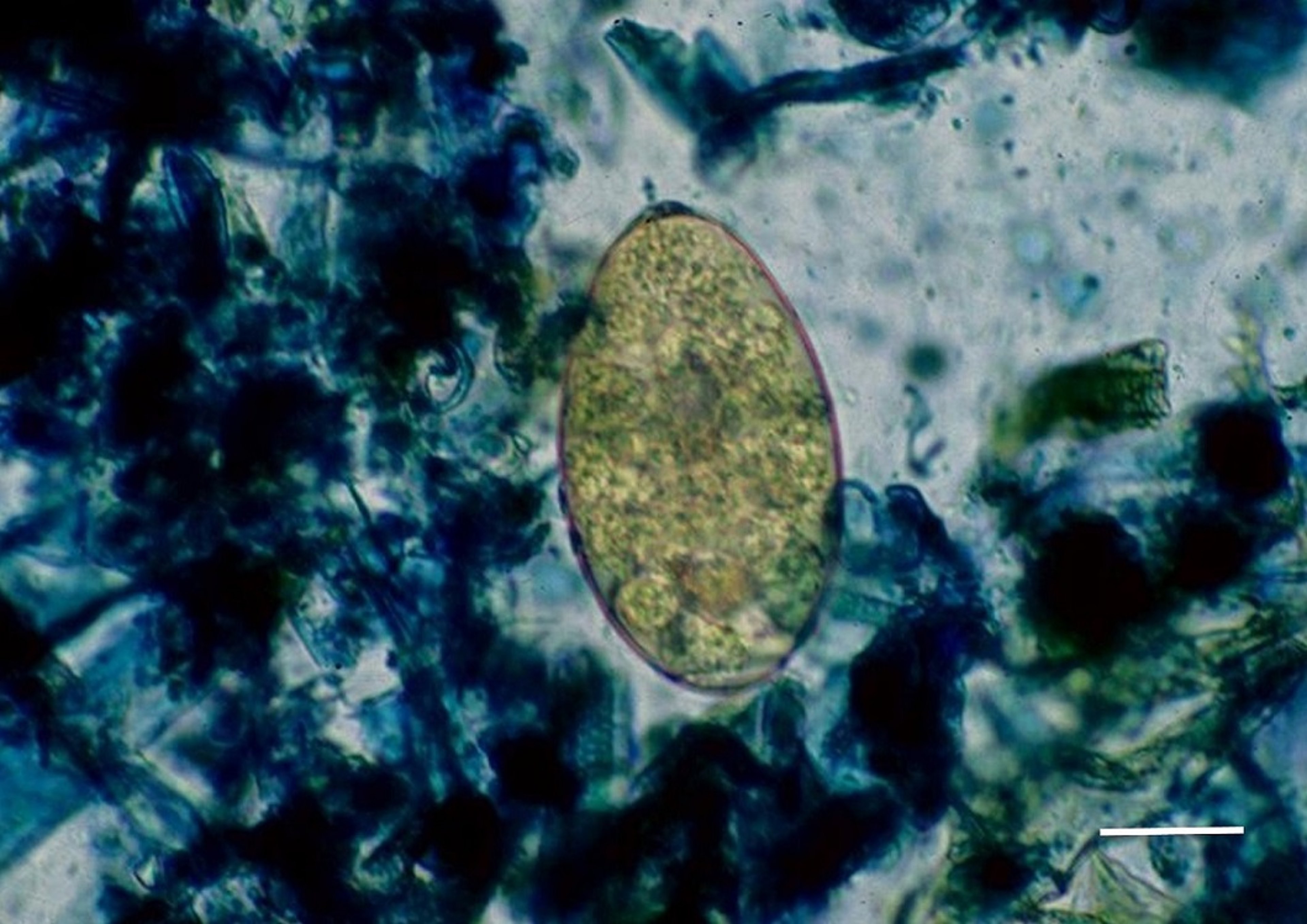
The oval, operculated, golden-brown eggs (130–150 × 65–90 mcm) of F hepatica can be distinguished from those of paramphistomes (rumen flukes), which are larger and clear. In areas where both F hepatica and F gigantica occur, their eggs cannot be reliably distinguished. Eggs of F hepatica cannot be found in feces during acute fascioliasis. In subacute or chronic disease in cattle, the number varies from day to day, and repeated fecal sedimentation may be required. Fecal sedimentation is easy and inexpensive; however, it can detect only patent infections. Accuracy depends on the number of eggs being shed and the amount of feces used. Commercial ELISA antibody tests are available in some countries; however, they are validated only for F hepatica. These tests enable detection 2–4 weeks after infection and well before the patent period; however, antibody levels do not directly correlate to parasite numbers, and antibodies remain for weeks after the infection is terminated, so they are not necessarily indicative of an active infection.
Bulk milk antibody detection is useful as a herd screening procedure; a positive result suggests that at least 25% of the herd is infected.
Coproantibody ELISA, which is available in some countries to test fecal samples, can detect antibodies 6–8 weeks after infection. The antibodies disappear within a couple of weeks of resolution of the infection; however, the reliability of the ELISA test under field conditions has been questioned, especially when mixed-age infections may be present.
Plasma concentrations of gamma-glutamyltransferase, which are increased with bile duct damage, are also helpful during the late maturation period when flukes are in the bile ducts.
At necropsy, the nature of the liver damage is diagnostic. Adult flukes are readily observed in the bile ducts, and immature stages may be squeezed or teased from the cut surface.
Control of Fasciola hepatica in Ruminants
Anthelmintics
Pasture management
Control measures for F hepatica ideally should involve removing flukes in infected animals, reducing the intermediate host snail population, and excluding production animals from snail-infested pastures. In practice, only the first of these is used in most cases. Historically, snail control using molluscicides was used to reduce snail populations on pasture; however, these practices are currently banned in most countries because of the negative environmental impact. Draining low-lying swampy areas, if possible, to reduce snail habitat can be an effective alternative to chemical control. It is recommended that production animals be kept out of snail-infested pastures, but such exclusion is frequently impractical because of the size of the areas involved and the consequent expense of erecting adequate fencing. When possible, however, grazing high-risk pastures should be avoided.
Ruminants infected with F hepatica can be treated with a number of anthelmintics, including triclabendazole, clorsulon (cattle and sheep only), albendazole, netobimin, nitroxinil, closantel, rafoxanide, and oxyclozanide. Not all are approved in all countries—eg, only clorsulon (2 mg/kg; available only combined with ivermectin for cattle) and albendazole (10 mg/kg for cattle and goats; 7.5 mg/kg for sheep) are approved in the US, and none are approved for alpacas and llamas—and most have long withdrawal periods before slaughter if used in meat-producing animals and before milk from treated production animals can be used for human consumption.
Anthelmintic resistance by F hepatica to various compounds, including albendazole, clorsulon, and triclabendazole, has been demonstrated, further complicating control programs based only on anthelmintic usage.
The timing of treatment is of critical importance so that the pharmacokinetics of the drug used will result in the optimal removal of flukes; each flukicide has varying efficacy against different ages of fluke. Timing of treatments is determined by local epidemiologic factors, and additional treatments are administered when conditions are most favorable for parasite multiplication. For example, in the Gulf Coast states of the US, cattle should be treated before the fall rainy season and again in the late spring. In the northwestern US and in northern Europe, cattle should be treated at the end of the pasture season and, if not housed, again in late January or February. In European countries with large susceptible sheep populations, computerized prediction systems based on rainfall, evapotranspiration, number of wet days per month, and/or prevalence are used to predict the timing and severity of disease. In areas where heavy infections are expected, sheep may require treatment in September or October, January or February, and again in April or May to reduce both the chances of acute or chronic infections and the output of fluke eggs for development of future disease.
Key Points
Liver fluke disease is most often associated with F hepatica. The disease can manifest in acute, subacute, or chronic forms; sheep, alpacas, and llamas are prone to the acute/subacute form, and cattle are more prone to the chronic form. The form is greatly influenced by the number of metacercariae ingested, which, in turn, is affected by grazing management.
Treatment options suffer from the ineffectiveness of most compounds against immature flukes during the migratory phase, the phase in which the most severe damage occurs.
Detection of liver flukes still depends on the fecal sedimentation procedure, which can detect only mature, patent infections. Egg-shedding varies daily; thus, repeated examinations may be necessary. An antibody ELISA test is available in some countries; however, the interpretation of results can be problematic. A coproantigen ELISA is also available in some countries; however, its reliability has been questioned.
For More Information
Howell AK, Williams DJL. The epidemiology and control of liver flukes in cattle and sheep. Vet Clin Food Anim. 2020;36:109-123.
Fairweather I, Brennan GP, Hanna REB, Robinson MW, Skuce PJ. Drug resistance in liver flukes. Int J Parasitol Drugs Drug Resist. 2020;12:39-59.
Sustainable Control of Parasites (SCOPS): Liver Fluke
Also see pet health content regarding lung flukes in cats and dogs.


