


The success of CPR depends on many factors, including the underlying cause of the arrest, the timeliness and effectiveness of the intervention, and the preparedness of the team administering CPR. Overall prognosis of recovery from cardiopulmonary arrest (CPA) with CPR efforts is as high as 35%–44%; however, < 10% of animals survive to hospital discharge. Animals with CPA associated with anesthesia have a better prognosis.
The American College of Veterinary Emergency and Critical Care developed the first set of guidelines for veterinary CPR in 2012; this effort was termed the Reassessment Campaign on Veterinary Resuscitation (RECOVER). Recently, online and hands-on courses have been developed to certify veterinary staff as rescuers and instructors of Basic Life Support (BLS) and Advanced Life Support (ALS). Further information is available at https://recoverinitiative.org/.
CPR is divided into 5 domains:
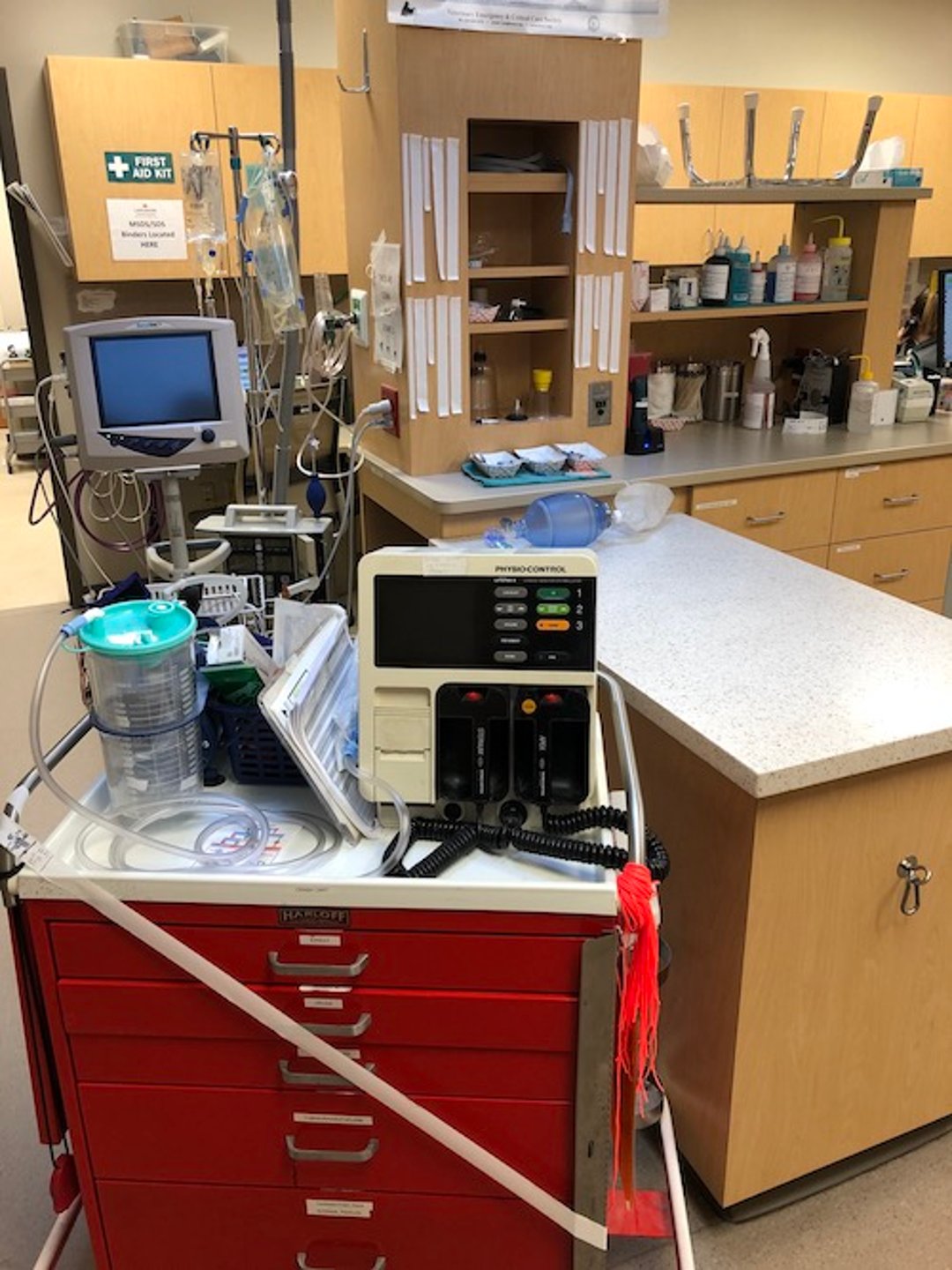
Prevention and Preparedness

Courtesy of Dr. Andrew Linklater.
In an effort to have the entire veterinary team prepared for CPR on any animal, RECOVER guidelines recommend standardization and regular audit of resuscitation equipment (a ready area and a crash cart) as well as immediate availability of cognitive aids and descriptive CPR algorithms (eg, dose charts, checklists), which are available through the RECOVER website (see above).
Cognitive skill training and didactics should be incorporated for all veterinary team members on a regular basis. Assigning a leader and having specific leadership training, including debriefing after any CPR efforts, are recommended as well. Each team member should be familiar with available medical equipment and their role during CPR and should exercise clear, closed-loop communication. CPR training should be performed at least every 6 months with staff members that may be involved in CPR.
Every patient admitted into the ICU should have a CPR code status. Closely monitoring patients under anesthesia is essential to prevent anesthesia-related CPA. Monitoring patients that are sedated or anesthetized is essential to identify trends and prevent CPA. Patients that present to the ER should have an immediate triage to identify those with life-threatening problems and thus avoid CPA.
Basic Life Support
When CPA is recognized, CPR efforts should begin immediately. Early recognition and intervention is essential. Palpation of pulses is not recommended before initiating compressions because this will delay intervention. Mouth-to-nose resuscitation should be performed until endotracheal intubation and positive-pressure ventilation with 100% oxygen can be accomplished. The compression to ventilation ratio in mouth-to-nose should be 30:2. In the hospital, intubation should occur early; however, thoracic compressions should be not discontinued to facilitate placement of an endotracheal tube.
Once the airway is established, it is imperative to confirm placement with thoracic auscultation, visualization, palpation, and ETCO2 monitoring, as well as to secure the tube in place. Ventilations should be provided at a rate of 10 breaths/minute (1 breath every 6 seconds), with a volume of 10 mL/kg and an inspiratory time of 1 second. Ideally, these breaths are provided with a portable bag-valve-mask apparatus.
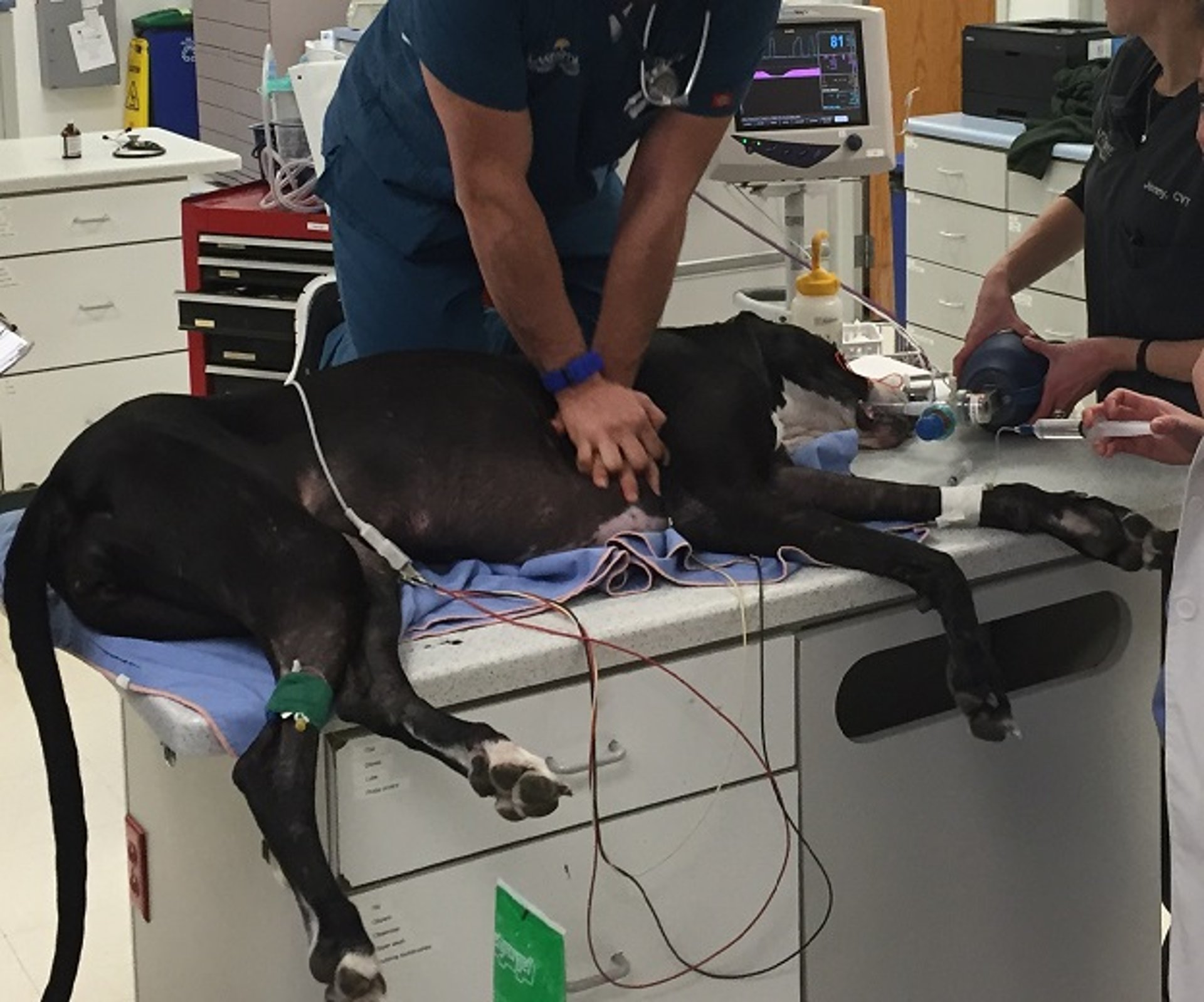
Simultaneous with ventilation, circulation should be promoted in small animals by compressing the chest externally. The following are key points in regard to performing chest compressions:

Courtesy of Dr. Andrew Linklater.
The animal is in lateral recumbency (or dorsal recumbency for barrel-chested animals, such as Bulldogs).
Elbows should be locked, with one hand on top of the other and with shoulders directly above the hands. Core muscles (compressing with movement from the waist) rather than biceps/triceps should be engaged; a step-stool should be used if needed.
Compressions should be performed over the widest part of the thorax using the "thoracic pump" technique in animals with a thoracic conformation that is equally wide and tall.
Compressions may be performed directly over the heart (at the fourth and fifth intercostal space) using the "cardiac pump" technique in animals with a thoracic confirmation that is taller than it is wide.
The compression rate should be 100–120 compressions/minute regardless of the size of the animal.
Each compression should be delivered quickly, compressing 1/3 to 1/2 of the width of the thoracic wall and allowing full recoil between compressions.
Thoracic compressions should be done for a total of 2 minutes without interruption because it takes ~1 minute of continuous thoracic compressions before myocardial perfusion pressure reaches its maximum.
When the cardiac pump technique is used, direct compression of the ventricles of the heart contribute to forward blood flow; in the thoracic pump technique, changes in thoracic pressure are the important mechanism to generate forward blood flow. Simultaneous ventilations and compressions should be done in 2-minute cycles; individuals performing the ventilation and compressions should change roles every 2 minutes to prevent fatigue and less-effective compressions. Interruptions to chest compressions to assess ECG, palpate for pulses, or auscult the heart should be minimal and only done between 2-minute cycles. Interposed abdominal compressions may be added for animals without abdominal disease if adequately trained staff are available. This is performed by placing both hands on the abdomen and compressing quickly, timing the compression to be done between chest compressions.
The goal is to improve venous return to the heart during the diastolic phase of the compression cycle. Monitoring CPR (see below) may necessitate a change in CPR technique.
Advanced Life Support
Several steps must occur to institute ALS:
an ECG is obtained to characterize arrhythmias
end-tidal CO2 is measured to monitor quality of CPR efforts (see below)
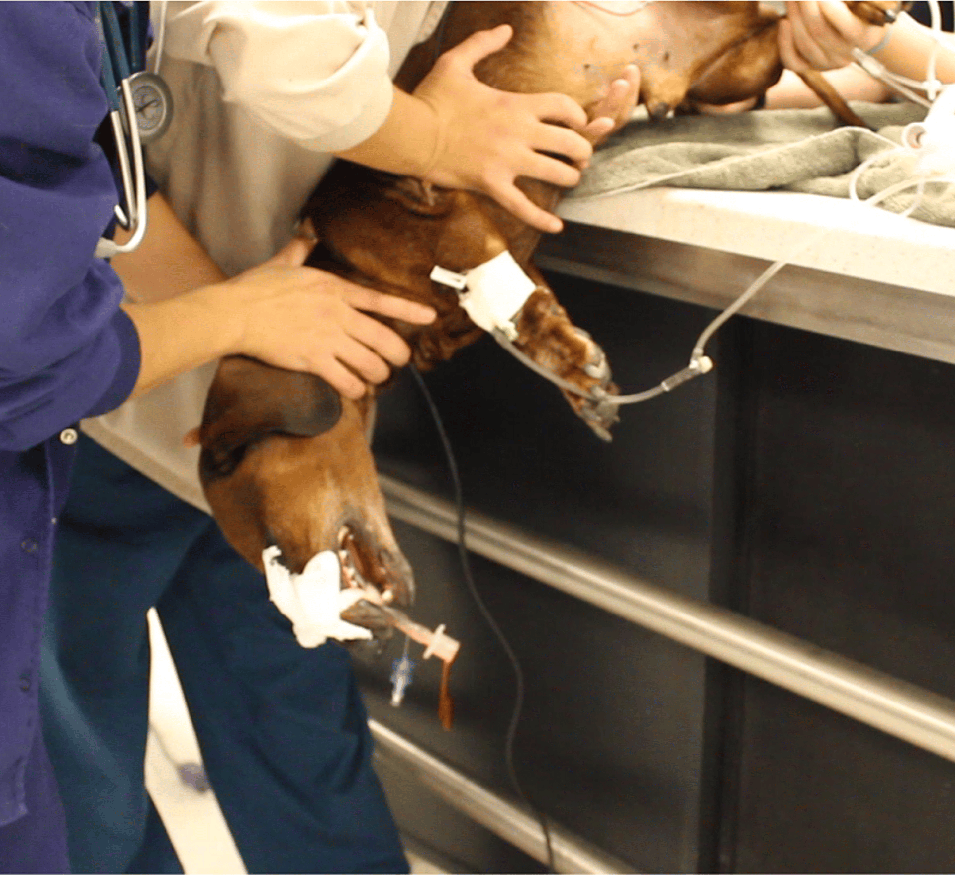
IV access is obtained (intraosseous [IO] or intratracheal [IT] routes may be used as alternatives)
drugs or defibrillation are administered, based on the identified rhythm
The purpose is to reestablish electrical and mechanical activity of the heart. The ECG is evaluated and pulses palpated only at the 2-minute cycle intervals, when changing compressors. The major arresting rhythms in veterinary medicine include sinus bradycardia, asystole, pulseless electrical activity (PEA, previously termed electromechanical dissociation), pulseless ventricular tachycardia, and ventricular fibrillation or flutter.
Drugs or defibrillation are selected based on the arrhythmia or known/suspected underlying disease ( see Table: Drugs and Defibrillation Used in Cardiopulmonary Resuscitation). Drugs are administered through the following route priority: central IV, peripheral IV, IO, then IT. Drugs that can be administered via the IT route include naloxone, atropine, vasopressin, epinephrine, and lidocaine (best remembered by the acronym NAVEL). The dosage for all drugs is usually doubled when administration is IT. Intracardiac administration of drugs is no longer recommended because this may result in arrhythmias, myocardial hemorrhage, or myocardial vessel laceration. Poster algorithms are available through the RECOVER initiative (see above).
If the patient received medications that have a reversal agent, it should be administered: naloxone for opioids, atipamezole for dexmedetomidine, flumazenil for benzodiazepines, and yohimbine for xylazine. Inhalant anesthetics (such as isoflurane) should be discontinued and the anesthetic circuit flushed with oxygen.

Courtesy of Dr. Andrew Linklater.
If the animal is known or suspected to be hypovolemic, isotonic balanced crystalloid solutions should be rapidly infused to restore volume and promote perfusion. Overzealous fluid administration can result in fulminant pulmonary edema due to poor myocardial contractility and arrhythmias. Fluids should not be administered to euvolemic animals; the increase in central venous pressure may reduce myocardial and cerebral blood flow. Metabolic alterations such as hyperkalemia, hypocalcemia, and severe acidosis should be treated when evident.
In cardiac arrests known or suspected to be associated with hyperkalemia, calcium gluconate should be administered. Regular insulin at 0.2 U/kg, followed by glucose at 1–2 g/U of insulin, diluted to 25%, temporarily reduces serum levels of potassium and should be considered.
Arrhythmias of Cardiac Arrest
Asystole in Cardiac Arrest of Animals
Asystole appears as a flat line on the ECG and suggests complete absence of electrical activity. Epinephrine or vasopressin is administered every second cycle of CPR. Atropine may be considered every second cycle as well. Fine ventricular fibrillation may look like asystole, and for this reason, open-chest heart massage and direct observation of myocardial activity are warranted early with this arrhythmia; if fibrillation is visualized, defibrillation is indicated.
Pulseless Electrical Activity (PEA) in Cardiac Arrest of Animals
The ECG tracing can be normal or show an arrhythmia (commonly a bradyarrhythmia of ventricular or supraventricular origin), but the heart has no mechanical activity associated with the electrical activity: no contractions, no cardiac output, and subsequently, no pulses. In this arrhythmia, it is vital that thoracic auscultation be performed in tandem with central pulse (femoral arterial) palpation and ECG evaluation between BLS cycles. Heart sounds and pulses are absent. Severe hypovolemia, pericardial effusion, an obese patient, and significant accumulation of fluid or air in the pleural cavity can prevent detection of normal heart sounds; the ECG associated with these conditions typically demonstrates tachyarrhythmias, in contrast to the usually normal or slow rate of PEA. Epinephrine or vasopressin are the drugs of choice with this arrhythmia and are administered every second cycle; atropine may be considered, alternating with epinephrine.
Sinus Bradycardia in Cardiac Arrest in Animals
Sinus bradycardia has P, QRS, and T waves that appear normal, except they occur at a much slower rate. This arresting rhythm may be caused by many disease processes, such as high vagal tone due to GI, urinary, ocular, or thoracic disease, and hyperkalemia due to urinary obstruction or rupture and prolonged CPA with CPR efforts. Treatment of known or suspected hyperkalemia with calcium gluconate, insulin, and dextrose with or without sodium bicarbonate may be necessary. Atropine is indicated in this arrhythmia.
If the CPA is believed to be associated with drug administration, a reversal agent should be administered in addition to treating arrhythmias in ALS.
Ventricular Flutter in Cardiac Arrest in Animals
This rhythm is more chaotic than ventricular tachycardia and is prefibrillatory. Lidocaine is the drug of choice to block the excited focus. If lidocaine is ineffective after two boluses and perfusion is absent, defibrillation may be required.
Ventricular Fibrillation and Pulseless Ventricular Tachycardia in Cardiac Arrest of Animals
Ventricular fibrillation implies that multiple foci within the ventricles are firing rapidly and independently, resulting in no coordinated mechanical activity. There are no ventricular contractions and no cardiac output. With pulseless ventricular tachycardia, the QRS is wide, the rate is fast (usually >180 bpm), and there is no or limited coordinated cardiac output.
The goal is to abruptly stop the abnormal electrical activity and allow a normal, coordinated electrical rhythm to take over. Electrical defibrillation is more successful when there are few, strong foci (coarse fibrillation) than when there are multiple, weak foci (fine fibrillation). Electrical defibrillation is most successful shortly after fibrillation starts (< 20-second duration) because, for every minute of ventricular fibrillation, the likelihood of a successful electrical defibrillation attempt decreases by 10%. One full cycle of BLS should be done before defibrillation on a patient that has been in ventricular fibrillation for > 4 minutes to allow blood flow and oxygen delivery to myocardial cells. This also allows determination of dose, preparation, and charge of the defibrillator.
After defibrillation, a BLS cycle is immediately started, and the ECG and patient are evaluated after this two-minute cycle. If defibrillation was unsuccessful, another shock may be administered; increasing the dose by up to 50% may be considered. If a defibrillator is not available, a precordial thump may be delivered. If defibrillation is unsuccessful, amiodarone, lidocaine, or epinephrine may be administered.
Open-Chest Cardiopulmonary Resuscitation
If closed-chest BLS is unsuccessful (as determined by lack of spontaneous respiration or inability to generate detectable forward blood flow) after 5–10 minutes, open-chest CPR (see below) is indicated. Instances when open-chest CPR is indicated during initial BLS include:
unwitnessed arrest
recent abdominal or thoracic surgery
suspected pleural or pericardial disease
trauma or pathology of the chest or abdominal wall with blood loss
diaphragmatic hernia
larger dogs in which external compressions are unlikely to generate an adequate forward blood flow
If possible, a quick clip of the hair along the intended incision site is helpful. There is no time for an aseptic preparation of the area. A scalpel blade or Mayo scissors are used to incise the skin, subcutaneous tissues, and muscle layers along the cranial border of the fourth or fifth rib from the spine to sternum. Guarded by the thumb and forefinger to prevent injury to the heart and lungs, closed Mayo scissors or Carmalt forceps are used to bluntly enter the pleural space while ventilations are temporarily discontinued. After the pleura is entered, Mayo scissors are used to incise the intercostal muscles along the entire length of the intercostal space on the cranial aspect of the rib. Care should be taken to avoid incising the internal thoracic vessels running parallel and lateral to the sternum. To improve visualization, Finochietto retractors may be used; suction or temporarily placing the patient in sternal recumbency may be necessary to rapidly remove blood.
After the thoracic cavity is opened, manual ventilations should resume. The pericardiodiaphragmatic ligament should be elevated with a finger or instrument and incised with scissors, extending the incision dorsally, taking care to avoid causing injury to the phrenic nerve. The heart is then lifted out of the pericardial sac and observed for any coordinated spontaneous contractions. If no cardiac contractions are noted, the heart is grasped with one or both hands and compressed progressively from the apex to the base. The compression is then released to allow the cardiac chambers to fill with blood. If fine or coarse fibrillation of the heart muscle is noted, internal defibrillation should be performed. Any active bleeding can be clamped at this time.
The descending aorta is located on the dorsal midline and can be isolated and temporarily cross-clamped to direct blood flow to the brain. Aortic cross-clamping can be performed with atraumatic vascular clamps or by using a modified Rommel tourniquet, passing a rubber tube, latex tube, or umbilical tape around the aorta with the assistance of curved hemostats and then clamping on the tube to occlude aortic flow. Aortic cross-clamping can be performed for 10 minutes without serious complications (from lack of blood flow to the spinal cord) and then should be released for 2 minutes.
The ECG is evaluated and drugs given as indicated during ALS procedures. Return of spontaneous circulation allows lavage of the thorax with large quantities of sterile, warm, isotonic saline; placement of a thoracostomy tube; and surgical closure of the thorax. Cardiovascular support is frequently required to maintain circulation while the underlying cause of the arrest is treated.
Monitoring

Courtesy of Dr. Andrew Linklater.
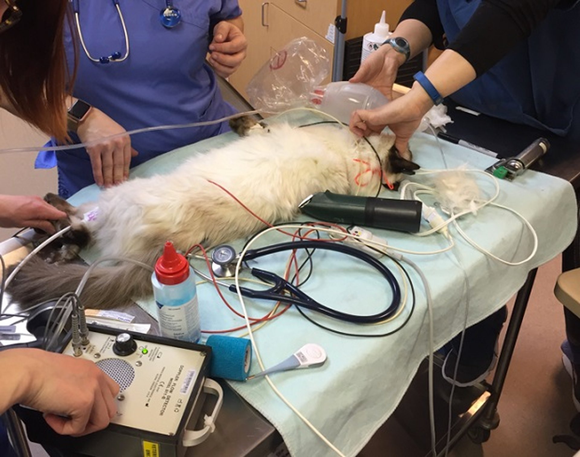
End-tidal CO2 (ETCO2) should be measured in intubated patients, particularly those at risk of having CPA and is a necessary monitoring tool during CPR efforts. Using an ETCO2 reading along with visualization, palpation, and auscultation can help confirm endotracheal intubation. ETCO2 may also be an early indicator of return of spontaneous circulation (ROSC) and effectiveness of CPR efforts (when minute ventilation is consistent). An ETCO2 of < 10 mmHg indicates esophageal intubation, ineffective CPR technique, incorrect placement of endotracheal tube, or hyperventilation (if adequate perfusion is established). When intubation is confirmed and ETCO2 remains low, efforts should be made to improve CPR technique (confirm placement of tube, CPR technique such as hand placement, compression depth, etc). An ETCO2 reading of 12–18 mm Hg indicates adequate CPR efforts. A sharp increase of ETCO2 of 20 mm Hg or a reading of > 45 mm Hg usually indicates ROSC as CO2 delivery to the lungs increases; the patient should be monitored closely for hypoventilation.
Routine monitoring of ECG is essential during CPR to allow identification and specific therapy of arrhythmias. The ECG should only be evaluated at the end of a 2-minute BLS cycle. Palpation of pulses either to detect CPA or to monitor effectiveness of CPR efforts is not recommended because of the insensitive nature of this test, but it may be used to monitor for ROSC between cycles. Use of Doppler monitoring (on eyes or peripheral arteries) to detect CPA or monitor efforts of CPR is not recommended.
Use of blood samples may help guide therapy in some instances during CPR. Centrally collected samples are ideal; however, many patients do not have a central catheter. Peripheral blood samples do not necessarily reflect the central circulation but may help guide therapy in some instances (such as hyperkalemia or severe acidosis). It is not recommended to monitor with arterial gas samples or pulse oximetry; these require pulsatile arterial flow, which is inadequate during CPR.
Postresuscitation Care
Close monitoring of an animal after CPA and ROSC is essential because significant abnormalities of acid-base and electrolytes (especially hyperkalemia and acidosis) are common and may require additional treatment. Rearrest is common, and the underlying etiology that led to CPA must be identified and corrected. Parameters such as ECG, blood pressure, neurologic status, pulse oximetry, ETCO2, and venous blood gases should be monitored closely. Blood pressure support with dopamine, norepinephrine, positive inotropes such as dobutamine, or other pressor agents may be indicated to maintain cardiac output. Body temperature, glucose, PCV/total solids, and lactate provide additional information.
With anaerobic metabolism that occurs during shock and cardiopulmonary arrest, blood lactate levels rise dramatically (normal levels are < 2 mmol/L). With ROSC, lactate levels rise dramatically and then resolve with appropriate treatment.
Routine use of large volumes of fluids is not recommended and should be avoided in animals with congestive heart failure. It is important to use resuscitation endpoints during post-CPA care to normalize venous oxygen content, lactate, blood pressure, central venous pressure, PCV, and oxygen saturation ( see Fluid Therapy in Animals). Medications to help reduce cerebral edema, such as mannitol and furosemide are often recommended to help decrease cerebral edema. Routine mechanical ventilation is not routinely recommended but reasonable in animals that are hypercapneic or hypoxemic. Animals with open-chest CPR will require control of hemorrhage, pleural lavage, placement of a thoracostomy tube, perioperative antibiotics, and closure of the thoracic cavity. Analgesics may be used cautiously as the patient becomes more stable. A large percentage of animals that sustain a CPA will have another episode of CPA.
Investigation into and treatment of the underlying condition that led to the CPA is essential to help prevent recurrence.
Key Points
Initiation of CPR should occur without delay when cardiopulmonary arrest occurs.
Medications that may have contributed to cardiopulmonary arrest should be reversed.
ECG placement is essential for advanced life support.
There are several indications for open-chest CPR efforts.
Assessing and monitoring a patient with return of spontaneous circulation should be done as per any critically ill animal, because re-arrest is common, and the underlying cause must be addressed.
For More Information
Also see pet health content regarding emergency care for dogs and cats and emergency care for horses.


