

Phaeohyphomycosis refers to chronic cutaneous, subcutaneous, mucosal, cerebral, or systemic infection due to pigmented (dematiaceous) septate molds. Fungi in this category are saprophytic, widely distributed organisms found in soil, water, and decaying vegetable matter. Infection is believed to result from fungal implantation into tissue at the site of an injury.
Humans and other animals have been reported to be affected by several fungal genera, including Alternaria, Bipolaris, Cladophialophora , Cladosporium, Curvularia, Exophiala, Fonsecaea, Moniliella, Phialophora, Rhinocladiella, and Ulocladium.
Clinical Findings and Lesions of Phaeohyphomycosis in Animals
Phaeohyphomycosis has been described in cows, cats, horses, and dogs. The most common clinical presentations include ulcerated cutaneous nodules of the digits, pinnae, nasal planum, and nasal/paranasal tissues, especially in cats. Widespread skin lesions may occur.
The nodules may ulcerate and have draining fistulous tracts. These pyogranulomas contain pigmented, septate hyphae with irregular enlargements and thin-walled, budding, yeastlike forms. Granulomatous meningoencephalitis due to pigmented fungi has been reported in dogs and cats.
Dogs treated with multiple immunosuppressive agents, especially cyclosporine, appear to be predisposed to developing multifocal cutaneous lesions. Systemic dissemination is most likely in patients treated with immunosuppressive drugs.
Diagnosis of Phaeohyphomycosis in Animals
Cytologic and/or histopathologic evaluation
Lesions may resemble other diseases

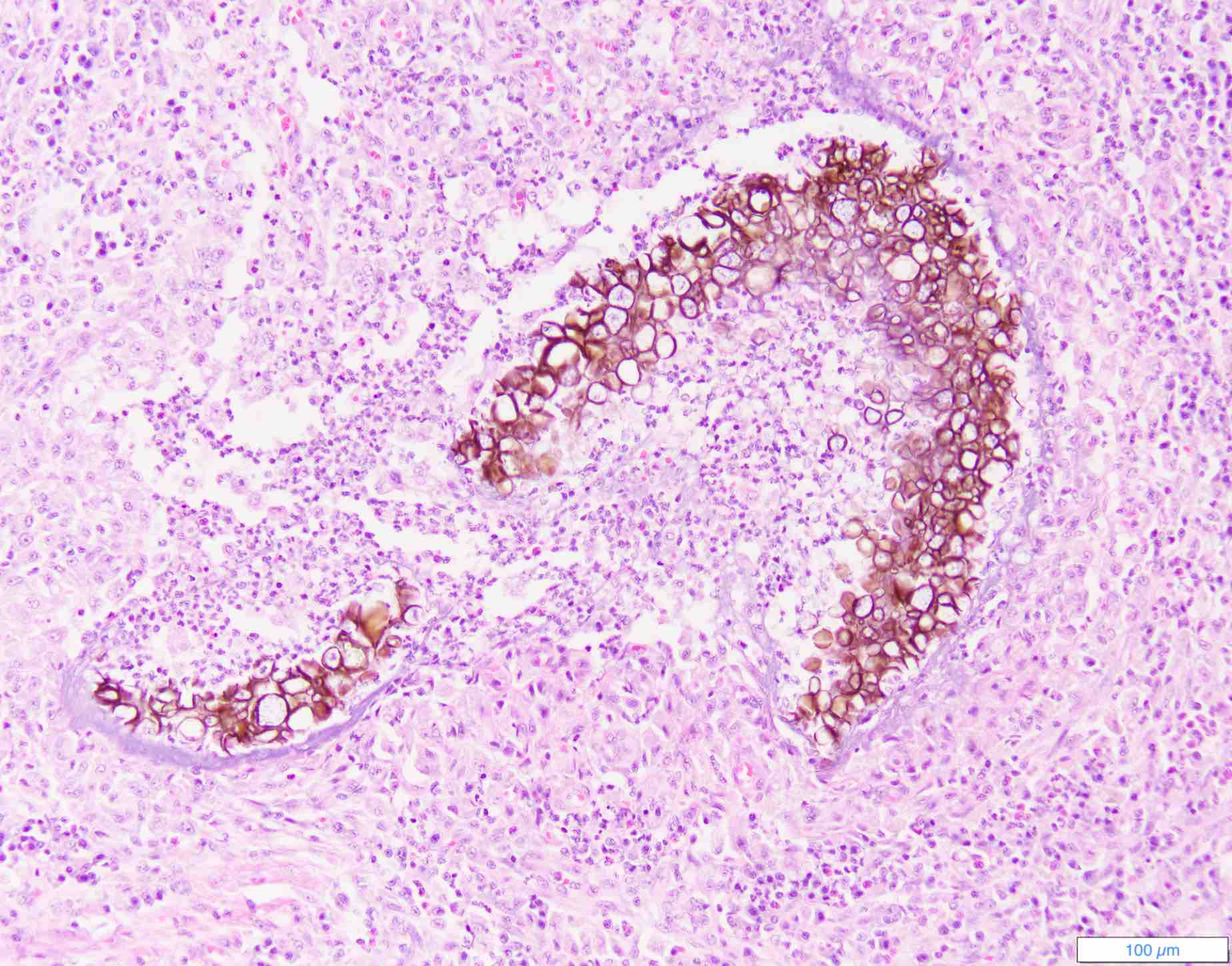
Deep dermal/subcuticular phaeohyphomycosis, dog. Note the pigmented fungal hyphae. H&E stain.
Courtesy of Dr. Rosalie Ierardi.
Phaeohyphomycosis can be diagnosed by microscopic examination of exudate and biopsy specimens, revealing pigmented, dark-walled, irregularly septate filamentous hyphae (2–6 mcm in diameter) or yeastlike cells. Infected tissues may be grossly pigmented, giving an appearance of melanoma. The several causative fungi cannot be identified by their histologic features in tissues; culture isolation and/or PCR assay is required. The differential diagnosis should include neoplasia, other granulomas, and epidermoid cysts.
Treatment of Phaeohyphomycosis in Animal
Surgical excision of lesions may be required
Immunosuppression should be reversed, if possible
Phaeohyphomycosis is generally poorly responsive to treatment. Wide excision of cutaneous or subcutaneous lesions is recommended, followed by 6–12 months of treatment with itraconazole (10 mg/kg every 24 hours). Nonresectable disease should be treated with itraconazole. Voriconazole or posaconazole may be more effective; however, voriconazole is not recommended in cats.
In dogs being treated with immunosuppressive treatment, the prognosis may be better if the immunosuppressive drugs (especially cyclosporine) can be discontinued. The author has observed one case of widespread cutaneous Bipolaris skin nodules in a severely neutropenic horse that resolved without specific treatment as the horse's immune status improved.
Key Points
Phaeohyphomycosis is typically a subcutaneous granulomatous lesion due to a pigmented mold.
Histopathologic evaluation and culture and PCR assay are used to diagnose phaeohyphomycosis.
Phaeohyphomycoses are poorly responsive to antifungal treatment; surgical resection may be required.


