


Bursitis is an inflammatory reaction within a bursa. The causes can range from overuse, mild trauma, or severe trauma to sepsis. Bursitis in its various forms is more common and a more important cause of dysfunction in horses than in other species. It can be classified as true bursitis or acquired bursitis. True bursitis is inflammation in a normal bursa that is naturally present. Some examples are trochanteric bursitis, supraspinous bursitis (fistulous withers), and bicipital bursitis. Acquired bursitis is development of a subcutaneous bursa where one was not previously present and inflammation of that bursa. Examples of acquired bursitis include capped elbow over the olecranon, and capped hock over the tuber calcaneus (when a natural bursa was not present). Accumulation of fluid in acquired bursitis is termed hygroma.
Bursitis may manifest as acute or chronic inflammation. Examples of acute bursitis include bicipital bursitis and trochanteric bursitis in the early stages. Bursitis is generally characterized by swelling, local heat, and pain on palpation. Chronic bursitis usually develops in association with repeated trauma, fibrosis, and other chronic changes (eg, capped elbow, capped hock, and carpal hygroma). Excess bursal fluid accumulates, and the wall of the bursa is thickened by fibrous tissue. Fibrous bands or a septum may form within the bursal cavity, and generalized subcutaneous thickening usually develops. These bursal enlargements develop as cold, painless swellings and, unless greatly enlarged, do not severely interfere with function. Septic bursitis is more serious and is associated with pain and lameness. Infection of a bursa may be hematogenous or follow direct penetration.
The pain in acute bursitis may be relieved by rest, bandaging when possible, application of cold packs, aspiration of the bursa contents, injection of intrabursal medication, and, most important, removal of the underlying cause, such as repeated trauma. Treatment of chronic bursitis may require surgery, which is often done with an arthroscope inserted into the affected bursa. In infected bursitis, local and systemic antimicrobials are indicated. Drainage of infected fluid and debridement of infected bursal tissue are required. Bursoscopy is helpful in bursae that can be examined with an arthroscope.
Capped Elbow and Capped Hock
Capped elbow and capped hock are inflammatory swellings of subcutaneous bursae in horses. The conditions are often termed hygromas. Usually, capped hock and capped elbow occur in acquired bursae, but some horses have naturally occurring subcutaneous calcaneal bursae. The bursae are located over the olecranon process and tuber calcaneus for the elbow and hock, respectively. Frequent causes of capped elbows include trauma from lying on poorly bedded hard floors, kicks, iron shoes projecting beyond the heels and then damaging the elbows while the horse is lying down, prolonged recumbency, and horses hitting their own elbows while trotting. Capped hocks are usually the result of horses kicking the stall with their hocks, but they may result from riding the tailgate of trailers, or from external trauma.
Clinical Findings and Diagnosis of Capped Elbow and Capped Hock in Horses

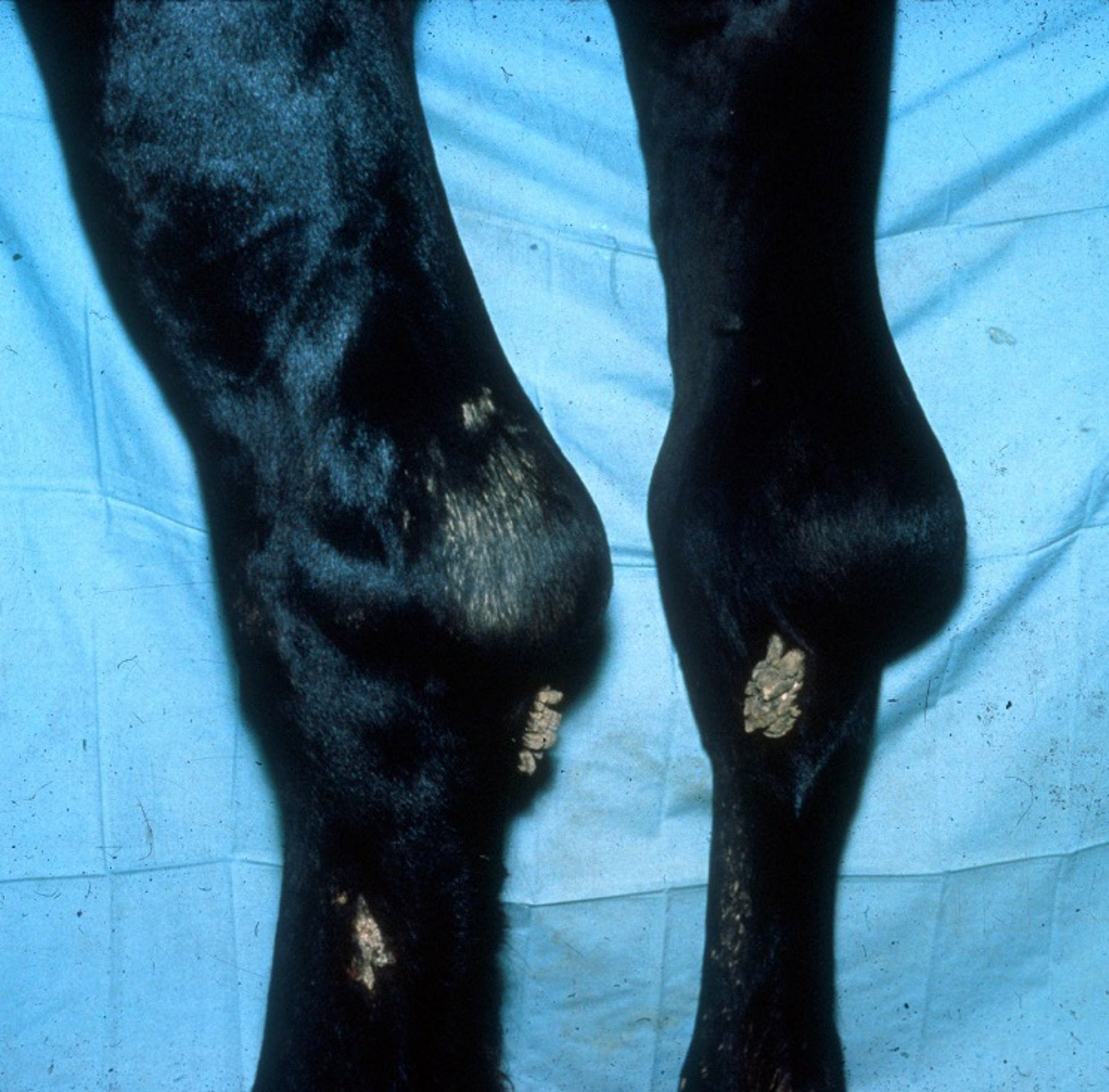
Courtesy of Dr. Stephen Adams.
Circumscribed edematous swelling develops over and around the affected bursa in capped elbow and capped hock. Lameness is rare. The affected bursa may be fluctuating and soft at first; within a short time, however, a firm fibrous capsule forms, especially if there is a recurrence of an old injury. Initial bursal swellings may be hardly noticeable or quite sizable. Chronic cases may progress to abscess formation.
Treatment of Capped Elbow and Capped Hock in Horses
Many horses with capped hock or capped elbow do not need to be treated. The horses are usually not lame. However, the initiating cause should be removed. With capped hock, behavioral modification so that the horse does not kick the stall offers the best hope of permanently resolving the problem. A shoe-boil roll placed around the pastern should be used to prevent recurrence of a capped elbow if the condition has been caused by the hoof or the shoe of the horse striking the elbow. Acute early cases may respond well to applications of cold water and administration of NSAIDs. When cosmetic results are important, bursae may be aseptically aspirated followed in a few days by aseptic aspiration and injection of a corticosteroid. This procedure carries a risk of introducing infection. Older encapsulated bursae are more refractory. Surgical treatment (usually curettage and drainage) is recommended for advanced chronic cases that are causing lameness or for cases that become infected.
Fistulous Withers and Poll Evil
Fistulous withers and poll evil are rare, inflammatory conditions of horses that differ essentially only in location: the supraspinous bursa in fistulous withers or the atlantal (cranial nuchal) bursa in poll evil. This discussion focuses on fistulous withers; except for anatomical details, however, it also applies to poll evil. In the early stage of the disease, a fistula is not present. The disease usually takes on a true fistulous character if the bursal sac becomes infected by pyogenic bacteria after the sac ruptures or is opened for surgical drainage.
Etiology of Fistulous Withers and Poll Evil in Horses
Fistulous withers and poll evil may be traumatic or infectious in origin. Of concern, Brucella abortus and Brucella suis have been associated with fistulous withers and poll evil. Brucella abortus can sometimes be isolated from the fluid aspirated from the unopened bursa. If horses are from areas endemic for Brucella, precautions should be taken to avoid zoonotic infections.
Clinical Findings of Fistulous Withers and Poll Evil in Horses

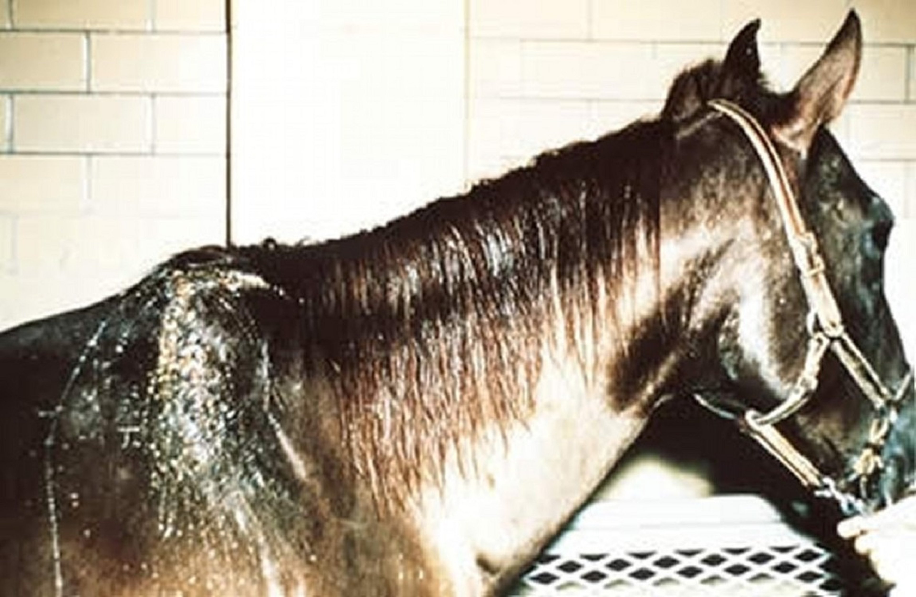
Courtesy of Dr. Thomas Lane.
The inflammation leads to considerable thickening of the bursa wall. The bursal sacs are distended and may rupture when the sac has little covering support. In more chronic, advanced cases, the nuchal ligament and the dorsal vertebral spines are affected and may become necrotic.
In the early stage of the disease, the supraspinous bursa distends with a clear, straw-colored, viscid exudate. The swelling may be dorsal, unilateral, or bilateral to the withers, depending on the arrangement of the bursal sacs between the tissue layers. It is an exudative process from the beginning; however, no true suppuration or secondary infection occurs until the bursa ruptures or is opened.
Treatment and Prevention of Fistulous Withers and Poll Evil in Horses
The earlier treatment is instituted, the better the prognosis. The most successful treatment is complete dissection and removal of the infected bursae, nuchal ligament, and associated necrotic tissues. Ventral drainage should be established. Surgery for fistulous withers can be done using local anesthesia in the standing horse. Culture of the drained fluid and selection of antimicrobial drugs based on susceptibility patterns of isolated microorganisms can be useful. The expense of the protracted treatment required in chronic cases often exceeds the value of the animal. Vaccines have not proven helpful against Brucella. Treatment of infected atlantal bursae is similar, but surgery is generally done with the horse anesthetized. Atlantal bursitis that is not infected and has no drainage has been treated successfully using bursoscopy for debridement of proliferative synovial tissues and lavage of the bursa.


