

Babesia spp blood parasites are transmitted by ixodid ticks. Babesiosis is typically characterized by fever and intravascular hemolysis leading to progressive anemia, hemoglobinuria, and jaundice, and may result in death. Diagnosis is principally by light microscopic evaluation of blood smears. The most common treatments are imidocarb dipropionate and diminazene aceturate. Control is via chemotherapy, vector control, and vaccination in some countries.
Babesiosis is the clinical disease associated with infection with protozoa of the genus Babesia, which are blood parasites transmitted by ixodid ticks. Among other wild and domestic mammals, cattle, horses, sheep and goats, swine, and cats and dogs are susceptible. In addition, babesiosis is a zoonotic disease affecting humans. The disease, which may be fatal, is typically characterized by fever and intravascular hemolysis leading to progressive anemia, hemoglobinuria, and jaundice. Diagnosis is principally by light microscopic evaluation of blood smears. The most common treatments are imidocarb dipropionate and diminazene aceturate. Control is via chemotherapy, vector control, and vaccination in some countries.
Etiology and Pathogenesis of Babesiosis in Animals
Babesia are intraerythrocytic protozoan parasites from the phylum Apicomplexa, order Piroplasmida. More than 100 species of Babesia exist, affecting domestic animals (cattle, horses, sheep, goats, pigs, dogs, and cats), wildlife, and, occasionally, humans.
Traditionally, Babesia spp were classified based on morphology and vector and host specificity; however, recent molecular characterizations suggest greater complexity.
Babesia bovis is a much more virulent organism than B bigemina. With most strains of B bigemina, the pathogenic effects relate more directly to erythrocyte destruction. With virulent strains of B bovis, a hypotensive shock syndrome, combined with generalized nonspecific inflammation, coagulation disturbances, and erythrocytic stasis in capillaries, contribute to the pathogenesis.
The following are indicative of Babesia spp affecting domestic animals; however, the list is far from complete.
Cattle
Babesia divergens and B major are two temperate-zone species with features comparable to those of B bovis and B bigemina, respectively. Babesia divergens is a small, pathogenic Babesia of considerable importance in the British Isles and northwest Europe, whereas B major is a large Babesia of lower pathogenicity. Babesia divergens is transmitted by Ixodes ricinus, and B major by Haemaphysalis punctata.
Horses
Equine piroplasmosis is caused by Theileria (formerly Babesia) equi or B caballi. Theileria equi is a small parasite and is more pathogenic than B caballi. Theileria equi was reclassified as a Theileria in 1998. Equine babesiosis is found in Africa, Europe, Asia, South America, Central America, and the southern US. It is transmitted by ticks of the genera Rhipicephalus, Dermacentor, and Hyalomma. Intrauterine infection, particularly with T equi, is also relatively common.
Sheep and Goats
Although small ruminants can be infected by several species of Babesia, the two most important species are B ovis and B motasi, transmitted by Rhipicephalus bursa and Haemaphysalis spp, respectively. Infection is of importance in the Middle East, southern Europe, and some African and Asian countries.
Pigs
Babesia trautmanni has been noted to cause severe disease in pigs. This parasite has been reported in Europe and Africa. Another species, B perroncitoi, is of similar pathogenicity but apparently has a limited distribution in the areas mentioned above. The vectors of these Babesia have not been clarified, although Rhipicephalus spp have been shown to transmit B trautmanni.
Dogs and Cats
Babesia spp have been reported in dogs from most geographic areas. These include B canis, B vogeli, and B rossi. Babesia canis is transmitted by Dermacentor reticularis in Europe, B vogeli by Rhipicephalus sanguineus in tropical and subtropical countries, and B rossi by Haemaphysalis elliptica in South Africa. Consequences of Babesia infection vary from a mild, transient illness to acute disease that rapidly results in death.
Babesia gibsoni is the other important Babesia of dogs and is a much smaller parasite. It has a more limited distribution and characteristically causes a chronic disease with progressive, severe anemia that is not readily treated with normal babesiacides.
Illness of varying severity due to B felis in domestic cats has mostly been reported in southern Africa, as well as B leo, and B lengau and less-well defined species. Sporadic cases associated with other Babesia species have been reported elsewhere. An unusual feature of B felis is its lack of response to the normal babesiacides.
Transmission and Epidemiology of Babesiosis in Animals
The major economic impact of babesiosis is to the cattle industry in tropical and subtropical geographic areas, and is attributed to Babesia bovis and Babesia bigemina; however, the diseases caused by different Babesia have many common features. Babesiosis is a tickborne disease and its geographic distribution is therefore determined by the distribution of tick vectors. The main vectors of B bigemina and B bovis are 1-host Rhipicephalus (Boophilus) spp ticks which are widespread in tropical and subtropical areas. Transmission occurs transovarially. Although these Babesia spp can be readily transmitted experimentally by blood inoculation, mechanical transmission by insects or during surgical procedures has no practical importance. Intrauterine infection has also been reported but is rare.
In Rhipicephalus spp ticks, the blood stages of the parasite are ingested during engorgement and undergo sexual and asexual multiplication in the replete female, infecting eggs and subsequent parasitic stages. Transmission to the host occurs when larvae (in the case of B bovis) or nymphs and adults (in the case of B bigemina) feed. The percentage of larvae infected can vary from 0%–50% or higher, depending mainly on the level of parasitemia of the host at the time the female ticks engorge. Under field conditions, the rate of tick transmission is generally higher for B bigemina than for B bovis.
In endemic areas, three features are important in determining the risk of clinical disease in cattle:
Calves have a level of immunity (related both to colostral-derived antibodies and to age-specific factors) that persists for ~6–8 months, whereas older animals without immunity are susceptible to clinical disease.
Animals that recover from Babesia infections are generally immune for the typical productive lifespan of animals raised in commercial settings.
The susceptibility of cattle breeds to ticks and Babesia infections varies (eg, Bos indicus cattle tend to be more resistant to both ticks and the effects of B bovis and B bigemina infection than Bos taurus–derived breeds).
At high levels of tick transmission, virtually all calves become infected with Babesia by 6–8 months of age, show few if any clinical signs, become immune, and retain this immunity into their adult life. This situation can be altered by a reduction in tick numbers, either natural (eg, climatic) or artificial (eg, acaricide treatment or changing breed composition of herd). Low tick numbers may mean that there is insufficient transmission of Babesia to calves to ensure all are infected. Other circumstances that can lead to clinical outbreaks include the introduction of susceptible cattle to endemic areas and the incursion of Babesia-infected ticks into previously tick-free areas.
Major epizootics occurred in cattle in the US, Australia, and South Africa in the 19th century, associated with the movement of infected cattle and the tick vector. Some countries retain tick-free quarantine zones as a means of controlling the spread of the Rhipicephalus (Boophilus) tick vector.
Bovine babesiosis is an OIE-listed disease and included on the Voluntary 2021 U.S. National Animal Health Reporting System (NAHRS) Reportable Diseases, Infections, and Infestations List.
Zoonotic Risk
Cases of babesiosis in humans have been reported. The rodent parasite B microti and the cattle parasite B divergens are the most commonly implicated species in North America and Europe, respectively. However, B duncani, B venatorum, and some less well-defined species have also been implicated. The reservoir hosts and vectors of some of these species are not necessarily known. Babesiosis infections in humans are acquired via bites from infected ticks or via contaminated blood from an infected transfusion donor. Cases reported in splenectomized or otherwise immunocompromised individuals are often fatal.
Clinical Findings of Babesiosis in Animals
Acute babesiosis generally runs a course of ~1 week or less. The first clinical signs are lethargy, weakness, depression, and fever (frequently ≥106°F [41°C]), which persist throughout, and these are accompanied later by inappetence, anemia, jaundice, and weight loss; hemoglobinemia and hemoglobinuria occur in the final stages. Involvement of the CNS due to adhesion of parasitized erythrocytes in brain capillaries can occur with B bovis infections. Late-term pregnant cows may abort, and temporary infertility due to transient fever may occur in bulls. Many animals recover; however, some may die if not treated.
Animals that recover from the acute disease remain infected for a number of years with B bovis or for a few months with B bigemina. No clinical signs are apparent during this carrier state.
Postmortem Lesions
Postmortem lesions (particularly with B bovis) include the following:
enlarged and friable spleen
swollen liver with an enlarged gallbladder containing thick granular bile
congested, dark-colored kidneys
generalized anemia and jaundice
Most clinical cases of B bigemina have hemoglobinuria; however, this is not invariably the case with B bovis. Other organs, including the brain and heart, may show congestion or petechiae.
Diagnosis of Babesiosis in Animals

Courtesy of State of Queensland, Department of Agriculture and Fisheries, Tick Fever Centre, Wacol, Queensland, Australia.

Courtesy of State of Queensland, Dept of Agriculture and Fisheries, Tick Fever Centre, Wacol, Queensland, Australia.
Physical examination and history
Light microscopic evaluation of blood smears
PCR assay
Serologic testing
Clinical findings and history may provide a presumptive diagnosis of babesiosis; however, other conditions also cause similar clinical signs.
Courtesy of State of Queensland, Department of Agriculture and Fisheries, Tick Fever Centre, Wacol, Queensland, Australia.

Courtesy of Dr. John W. Harvey.

Courtesy of State of Queensland, Department of Agriculture and Fisheries, Tick Fever Centre, Wacol, Queensland, Australia.

Courtesy of State of Queensland, Department of Agriculture and Fisheries, Tick Fever Centre, Wacol, Queensland, Australia.
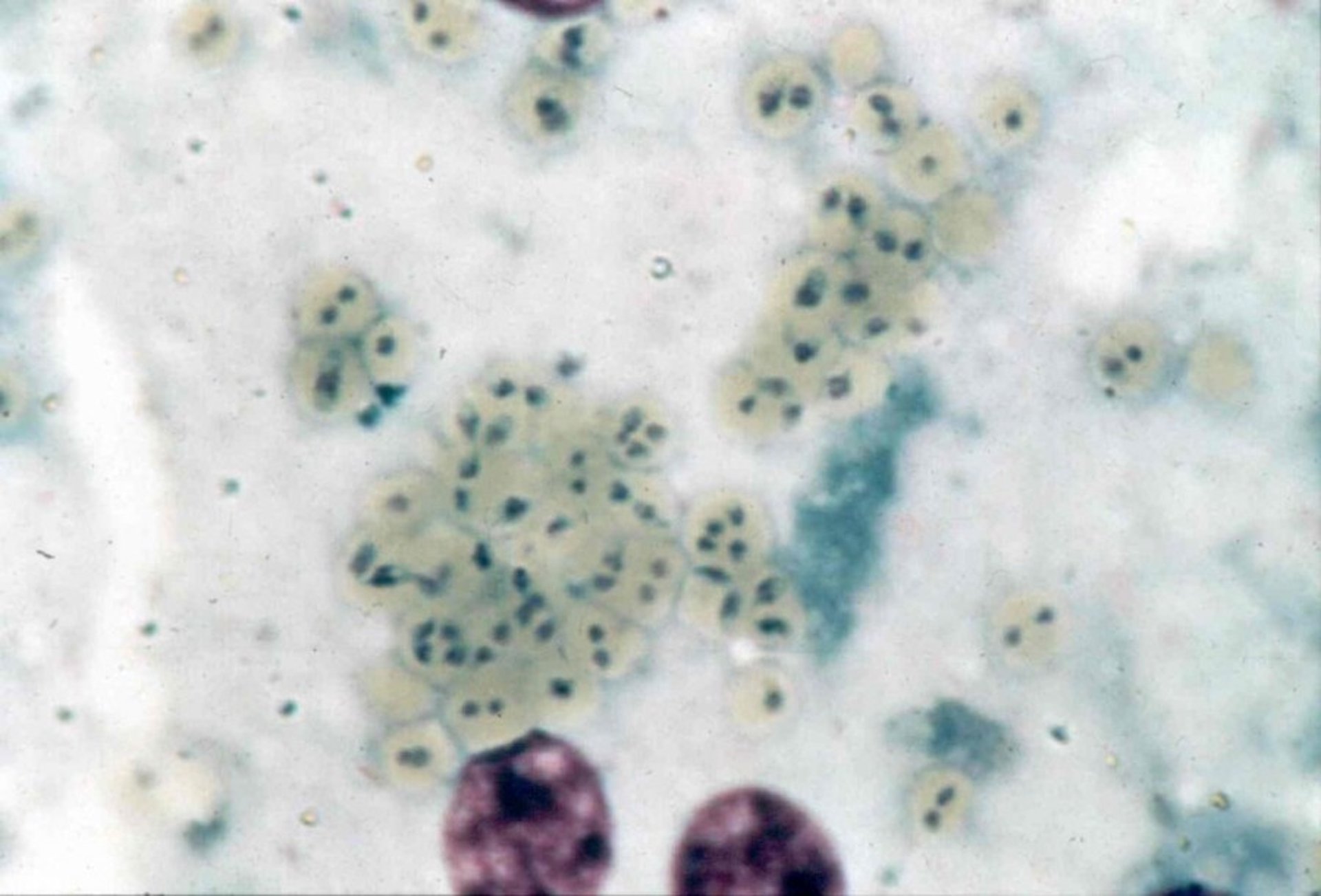
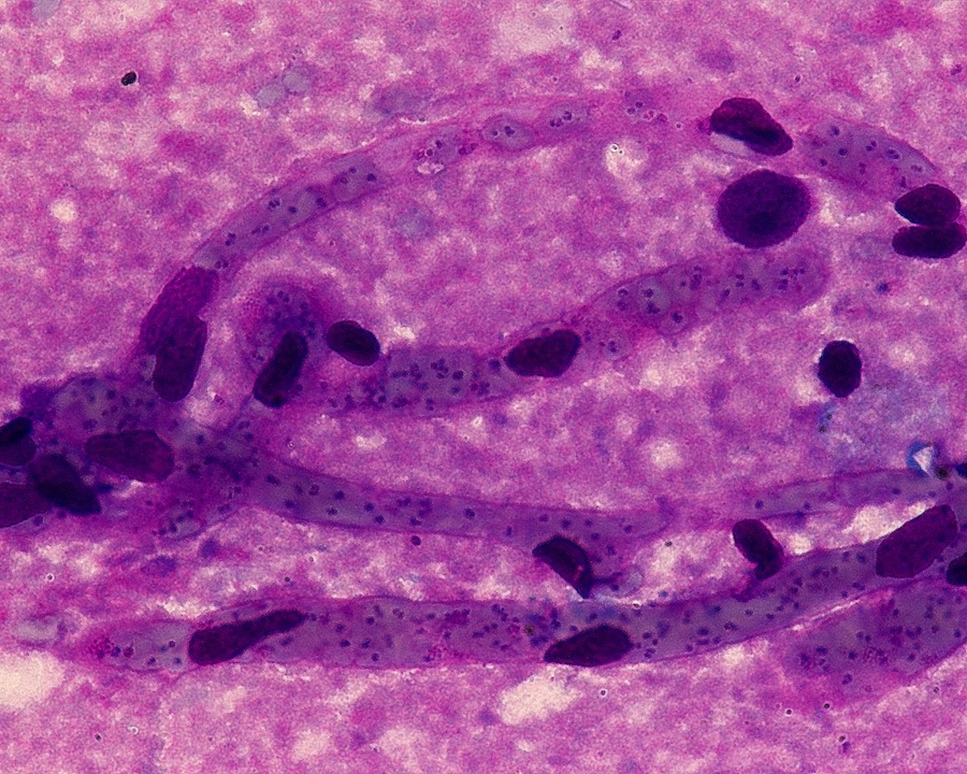
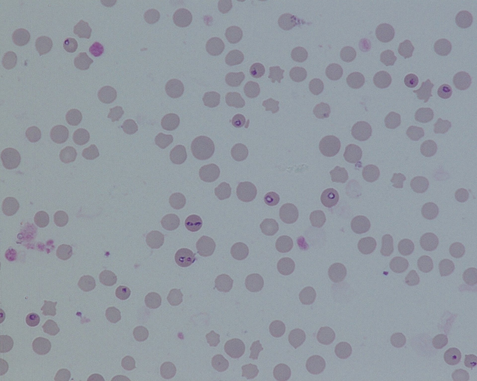
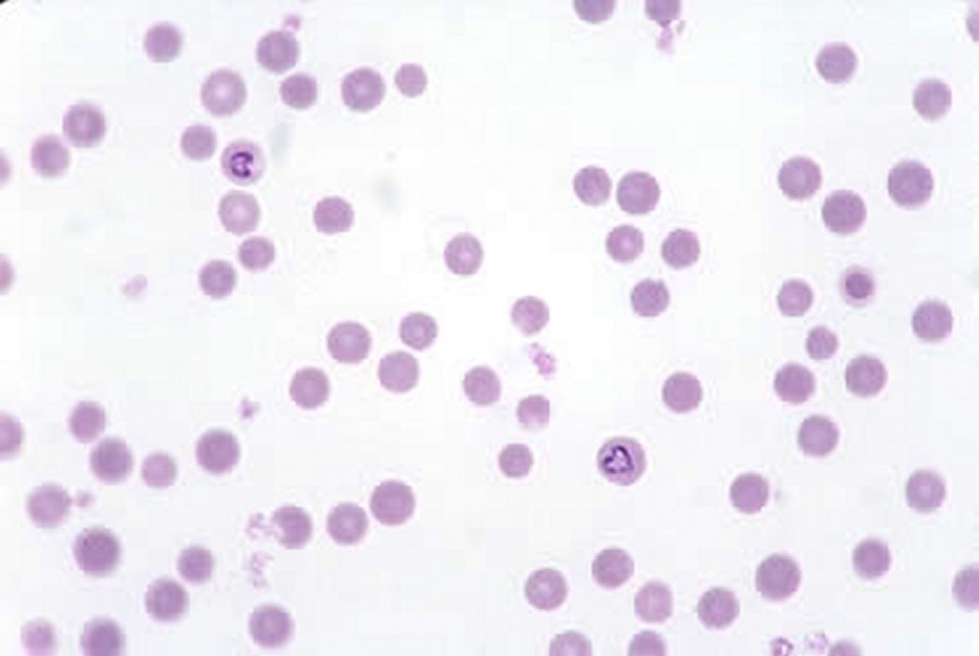
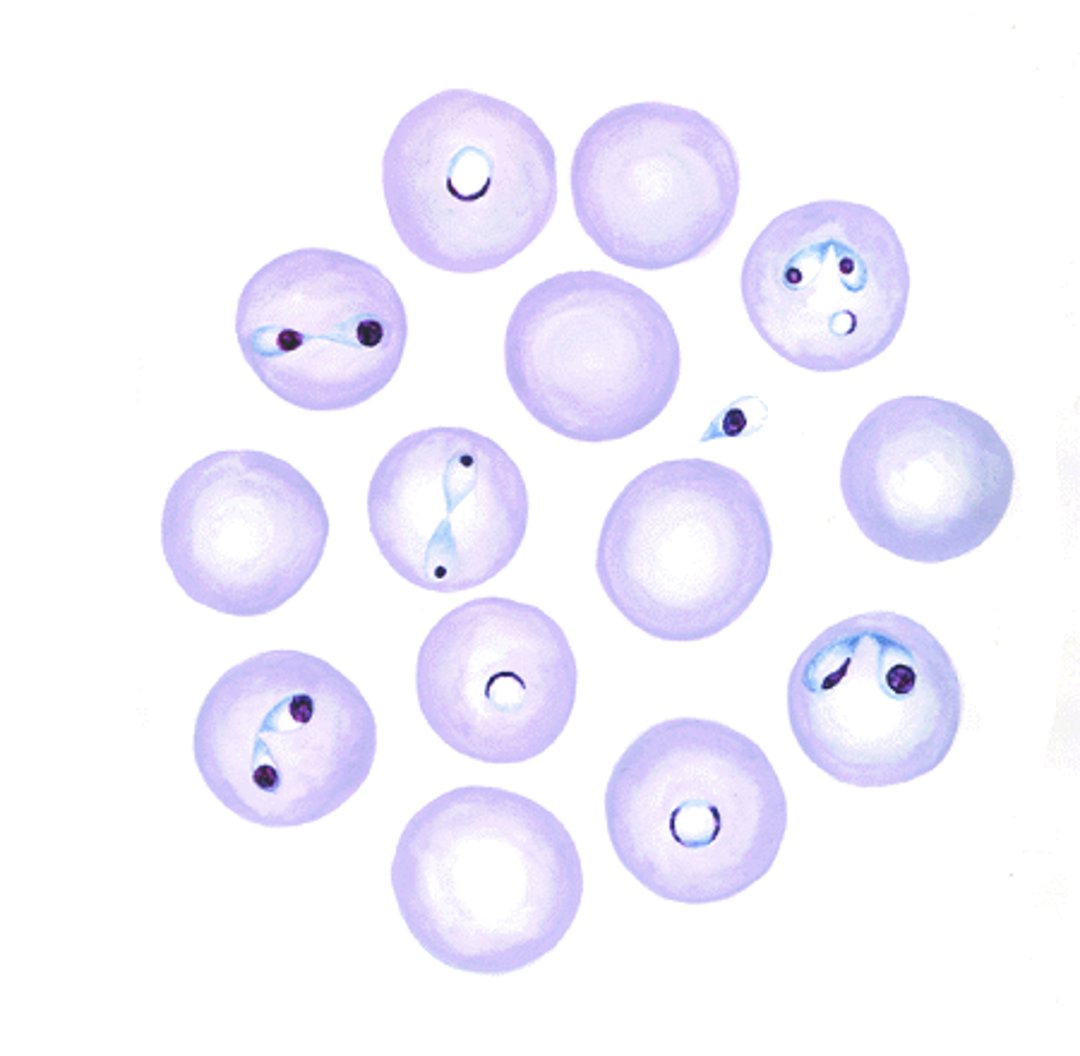
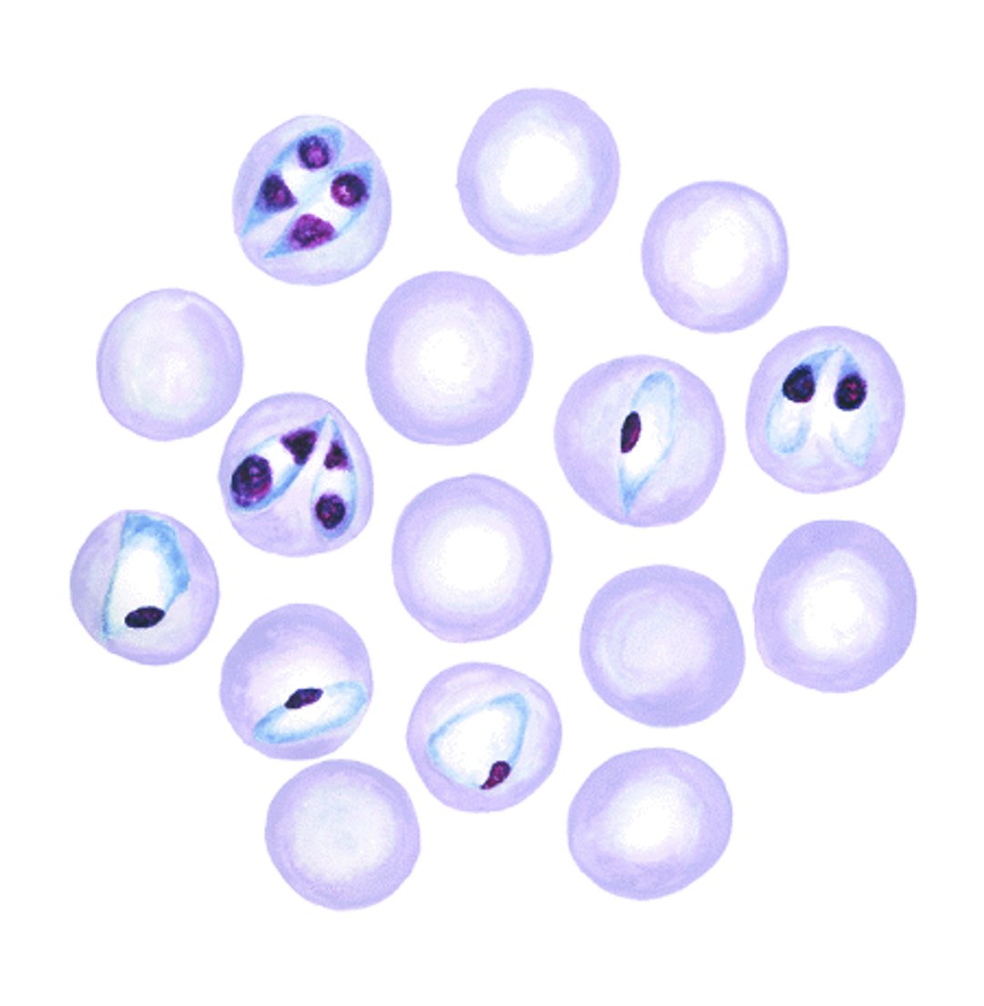
Examination of Giemsa-stained blood or organ smears by light microscopy is essential to confirm the diagnosis. Light microscopy is rapid and inexpensive but does require some expertise. From the live animal, blood smears should be prepared from capillaries, for example from the ear or tail tip, to improve the sensitivity of detection of B bovis, because B bovis-infected erythrocytes adhere to capillary endothelium. Smears of brain, muscle, kidney, spleen, and from a blood vessel in an extremity should be taken at postmortem examination. Note that detection of Babesia spp using light microscopy in the carrier state has poor sensitivity.
Microscopically, the species of Babesia involved can be determined morphologically; however, expertise is required, especially in B bovis infections in which few organisms may be present. Babesia bovis is generally small, with the parasites in paired form at an obtuse angle to each other and measuring ~1–1.5 × 0.5–1 mcm. B bigemina is larger (3–3.5 × 1–1.5 mcm), with paired parasites lying almost parallel or at an acute angle to each other. Single forms of both parasites are also commonly seen.
Other Diagnostic Tests
Molecular methods such as PCR assays are more sensitive than light microscopy and may be useful to detect Babesia spp in the carrier state or during chronic infection. Molecular methods are also used to differentiate and characterize isolates. Because of the persistence of the organisms in the blood, detection of the organisms does not necessarily indicate that they are the cause of the current disease or are having any health impact.
Serologic tests have been described for the detection of antibodies to Babesia spp in carrier animals. The most commonly used are the indirect fluorescent antibody test and ELISA. These are not useful for diagnosis in the acute stage of illness.
Subinoculation of blood (~500 mL) into a fully susceptible animal, preferably a splenectomized calf, and subsequent monitoring of the recipient for infection may occasionally be justified to confirm infection in suspected carrier animals.
Treatment, Control, and Prevention of Babesiosis in Animals
Babesiacides
Supportive treatment
Tick control
Use of resistant breeds
Vaccination
Babesiacides
A variety of babesiacidal drugs have been used to treat bovine babesiosis; however, only diminazene aceturate and imidocarb dipropionate are still in common use. These drugs are not available in all endemic countries, or their use may be restricted. Manufacturers' recommendations for use should be followed. For treating cattle, diminazene is administered at 3.5 mg/kg, IM, once. For treatment, imidocarb is administered at 1.2 mg/kg, SC, once. At a dosage of 3 mg/kg, imidocarb provides protection from babesiosis for approximately 4 weeks and may also eliminate B bovis and B bigemina from carrier animals.
Supportive Treatment
Supportive treatment is advisable, particularly in valuable animals, and may include the use of anti-inflammatory drugs, corticosteroids, and fluid therapy. Blood transfusions may be lifesaving in very anemic animals.
Tick Control
Tick control, via acaricides or management practices, can be useful in reducing tick burdens, which can lower transmission rates. However, this can lead to naive cattle populations, with consequent risk of outbreaks of disease should tick populations increase. Chemical tick control cannot be relied upon to prevent transmission of Babesia, and outbreaks often occur after introduction of susceptible cattle to endemic areas despite acaricide use. Acaricide resistance is also an increasing problem. However, acaricidal tick control before moving animals from tick-infested areas is useful to prevent the introduction of ticks and babesiosis to tick-free areas.
Eradication of the tick vector is rarely feasible on individual premises but may work on a regional level in well-coordinated programs.
Using Resistant Breeds
Bos indicus–based breeds are commonly used to minimize production losses associated with ticks and babesiosis.
Vaccination
Vaccination using live attenuated strains of the Babesia parasites has been used successfully in countries such as Argentina, Australia, Brazil, Israel, South Africa, and Uruguay. The vaccine is available in either a chilled or frozen form. One vaccination produces adequate immunity for the typical productive lifespan of animals raised in commercial settings. Commercial vaccines based on recombinant antigens are not yet available.
Key Points
Babesia spp are widespread in the domestic and wildlife populations of the world, and they occasionally infect humans.
Ixodid ticks are the vectors for transmission of Babesia spp.
Babesiosis is a serious and economically important cause of illness and death in cattle in tropical and subtropical geographic areas.
Control is based on specific drug treatments, tick control strategies, and vaccination in some countries.
For More Information
Bock R, Jackson L, de Vos A, et al. Babesiosis of cattle. Parasitology 2004;129 Suppl:S247-69.doi: 10.1017/s0031182004005190.
Solano-Gallego L, Sainz Á, Roura X, et al. A review of canine babesiosis: the European perspective. Parasit Vectors 2016;9(1):336. doi: 10.1186/s13071-016-1596-0.
Young KM, Corrin T, Wilhelm B, et al. Zoonotic Babesia: A scoping review of the global evidence. PLoS One 2019;14(12):e0226781. Published 2019 Dec 30. doi:10.1371/journal.pone.0226781.
Lempereur L, Beck R, Fonseca I, et al. Guidelines for the detection of Babesia and Theileria parasites. Vector Borne Zoonotic Dis 2017;17(1):51-65. doi:10.1089/vbz.2016.1955.
Gray JS, Estrada-Peña A, Zintl A. Vectors of babesiosis. Annu Rev Entomol 2019;64:149-165. doi:10.1146/annurev-ento-011118-111932.
Also see pet health content regarding babesiosis in dogs, cats, and horses.


