

Esophageal stricture is a pathologic narrowing of the lumen that may develop after anesthesia, trauma (eg, foreign body), ingestion of caustic substances, exposure to certain drugs (such as doxycycline or clindamycin), esophagitis, gastroesophageal reflux, or tumor invasion. Most strictures develop in the thoracic portion of the esophagus. Esophageal tumors are rare; however, esophageal sarcomas may be associated with Spirocerca lupi infection, requiring consideration in areas where this parasite is prevalent. Esophageal compression by vascular ring anomalies or extramural tumors may mimic the clinical signs of strictures.
Clinical signs are similar to those associated with foreign bodies and include regurgitation, ptyalism, dysphagia, and pain.
For diagnosis, an esophagram under fluoroscopy is the preferred tool, because it allows visualization of the number, length, location, and severity of strictures. Esophagoscopy can also be diagnostic but does not allow visualization beyond the stricture unless esophageal balloon dilation is also performed.

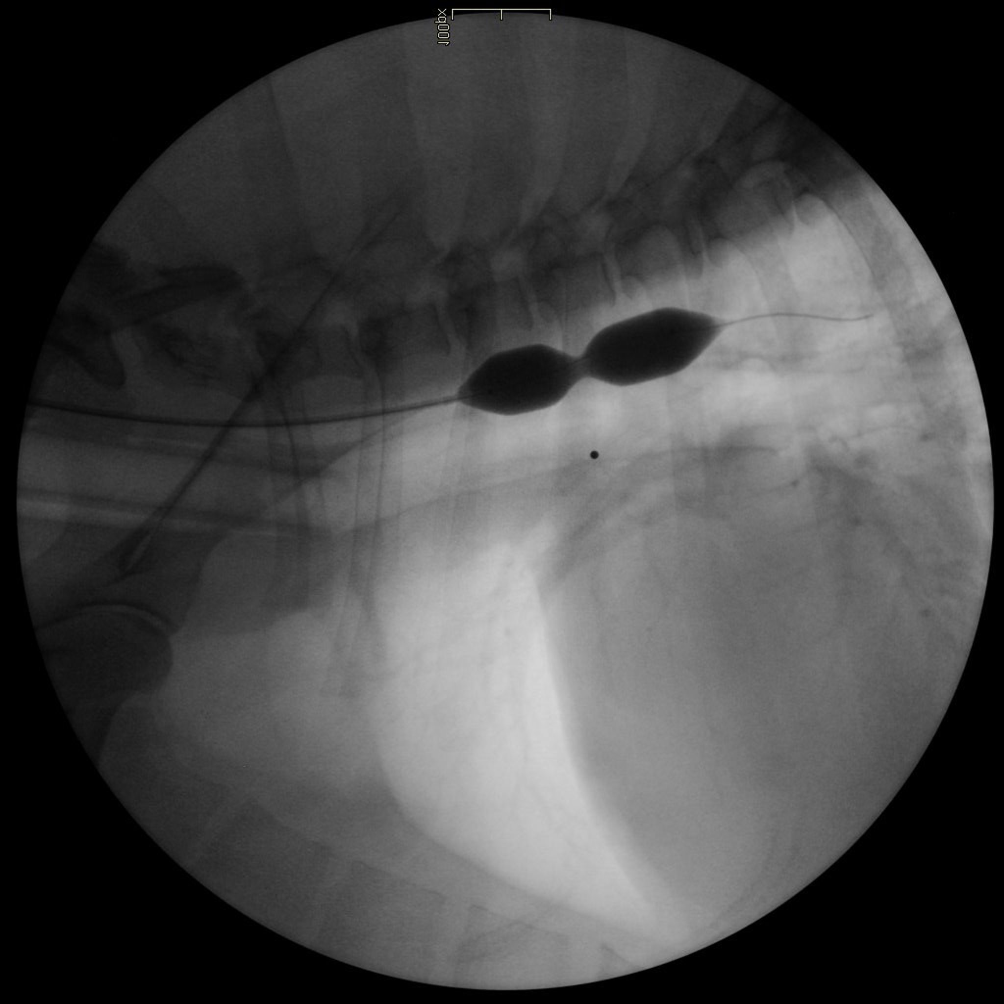
Fluoroscopic (lateral) view of the thorax of a dog showing an esophageal stricture being dilated.
Courtesy of Alex Zur Linden.
Treatment with balloon catheter dilation has been the most successful. Bougienage is another, less available, technique. It theoretically causes more shear stress on the esophagus but has not been shown to have an appreciably different complication rate than balloon dilation. Some cases can require multiple dilation procedures.
To avoid multiple repeated general anesthesias, indwelling esophageal balloon dilation feeding tubes ("B-tubes") have been created for dogs. After initial dilation of the stricture under general anesthesia, the owner is asked to inflate the balloon attached to the esophagostomy tube twice a day. The objective is to keep the esophagus opened and avoid stricture recurrence. This new technique has been successful in 9 dogs and 3 cats. Swallowing and gagging during inflation that resolved immediately after deflation of the balloon were reported by all owners. Minor complications reported included regurgitation, skin infection, and vomiting. Two dogs experienced recurrent regurgitations.1
Esophageal stents have been used in strictures that have been refractory to dilation procedures. However, this method has been limited by a high rate of complications. Surgical resection of a single stricture is another option; however, it is less successful than ballooning or stenting the esophagus.
All mechanical treatments to address strictures are likely to induce some degree of esophagitis, which must be treated to decrease the chance of stricture reformation. The use of corticosteroids or mitomycin to help prevent stricture reformation is controversial. No data exist regarding the success of this adjunct treatment for esophageal strictures in dogs and cats; however, intralesional use has been helpful in decreasing recurrence in humans.
References
Tan DK, Weisse C, Berent A, Lamb KE. Prospective evaluation of an indwelling esophageal balloon dilatation feeding tube for treatment of benign esophageal strictures in dogs and cats. J Vet Intern Med , 2018;32(2):693–700.


