


Benign Oral Tumors
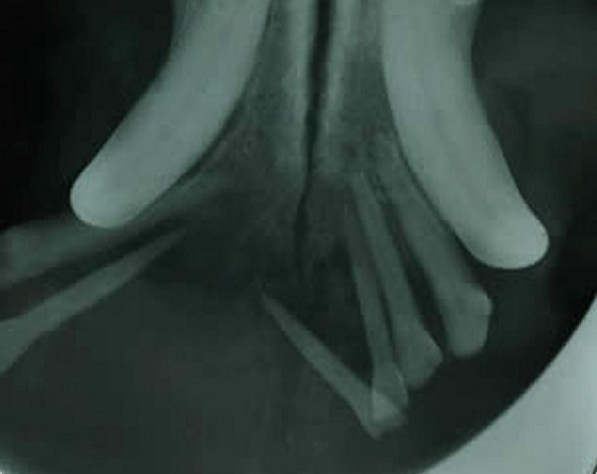
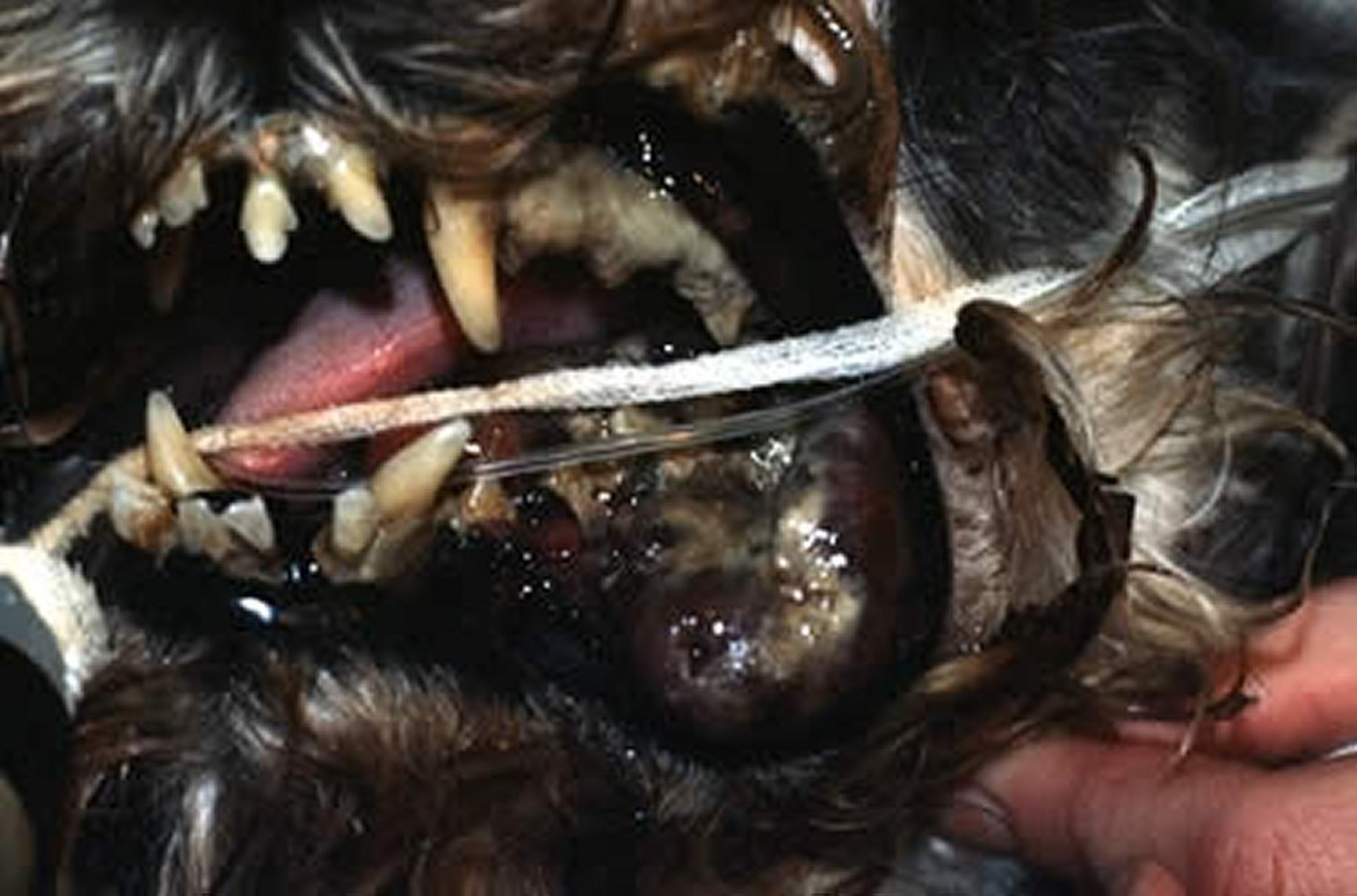
Peripheral odontogenic fibromas (previously called fibromatous epulis or ossifying epulis) are the most common benign oral tumors. These firm masses involve the gingival tissue adjacent to a tooth. They affect dogs of any age but are most common in dogs >6 yr old. Some develop centers of ossification, visible as distinct alveolar bone proliferation extending into the soft-tissue mass. They are generally solitary, although multiple lesions may be present. The tumors do not metastasize but may become quite extensive. They arise from the periodontal ligament of the affected tooth, and complete surgical removal must include tissues up to and including the periodontal ligament. This usually necessitates conservative resection of the neoplastic lesion, extraction of the affected tooth or teeth, and curettage of the extraction sites (ie, removal of remaining periodontal ligament). Complete excision is curative.

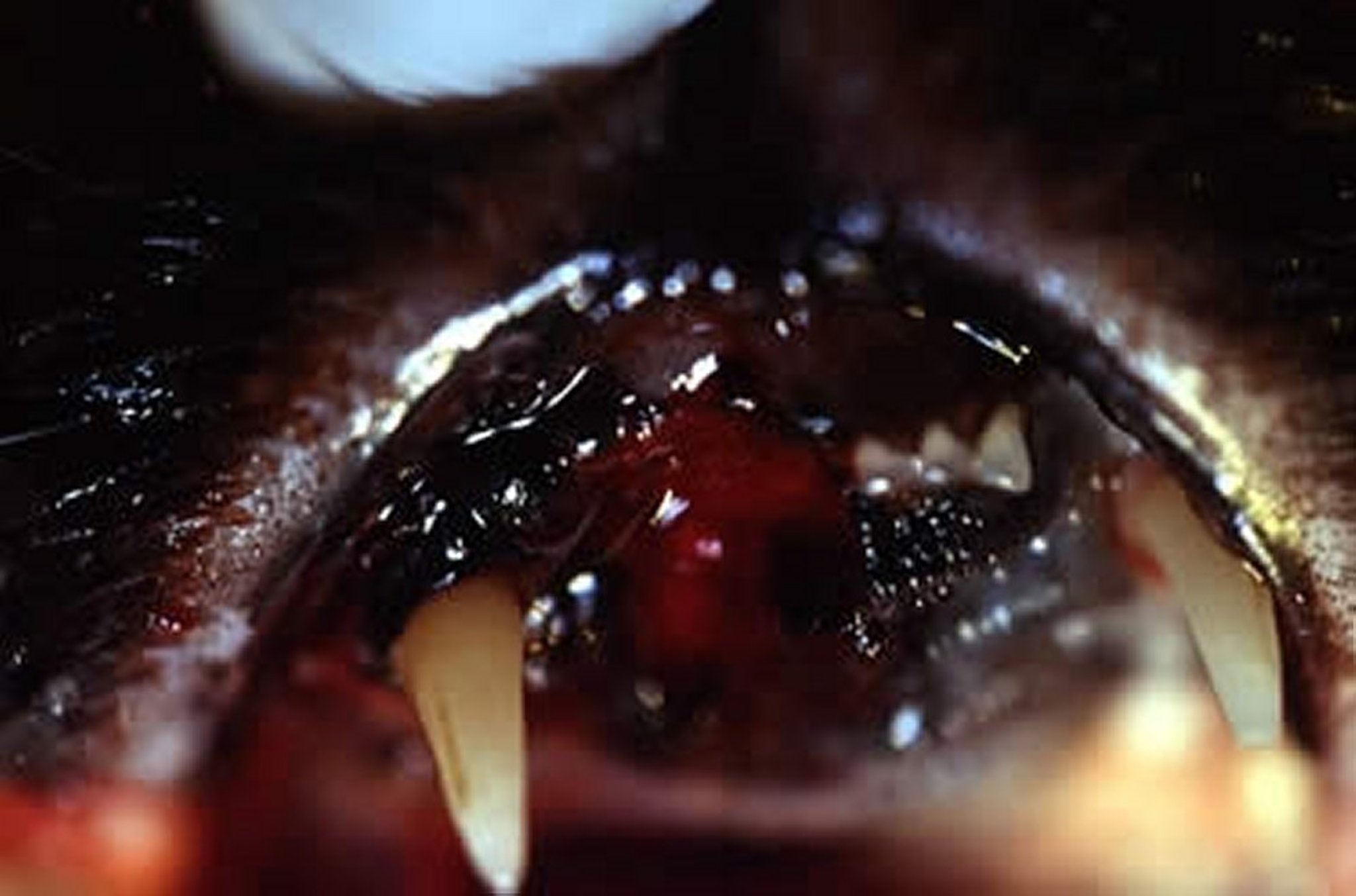
Courtesy of Dr. Ben Colmery III.

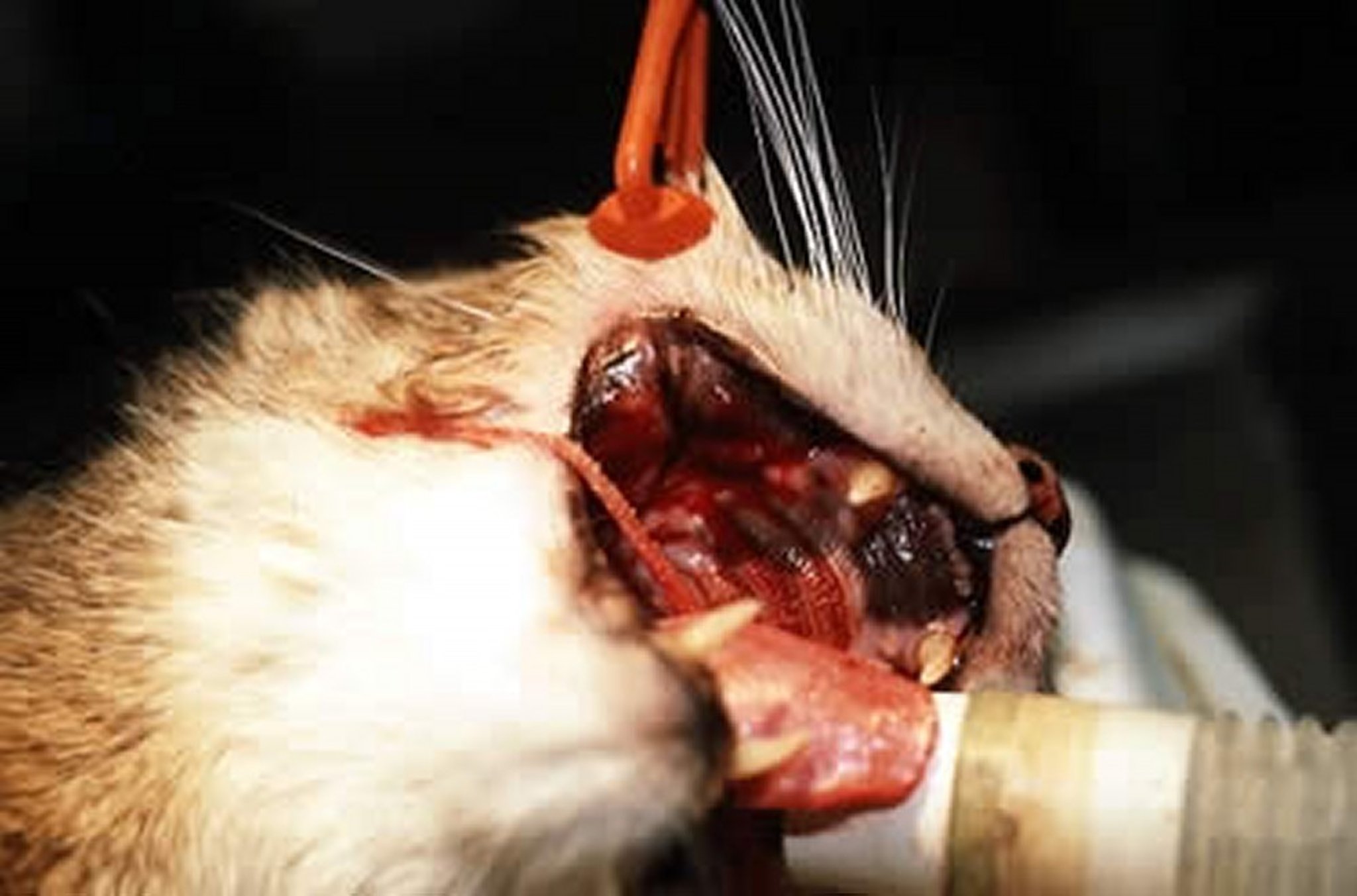
Courtesy of Dr. Ben Colmery III.
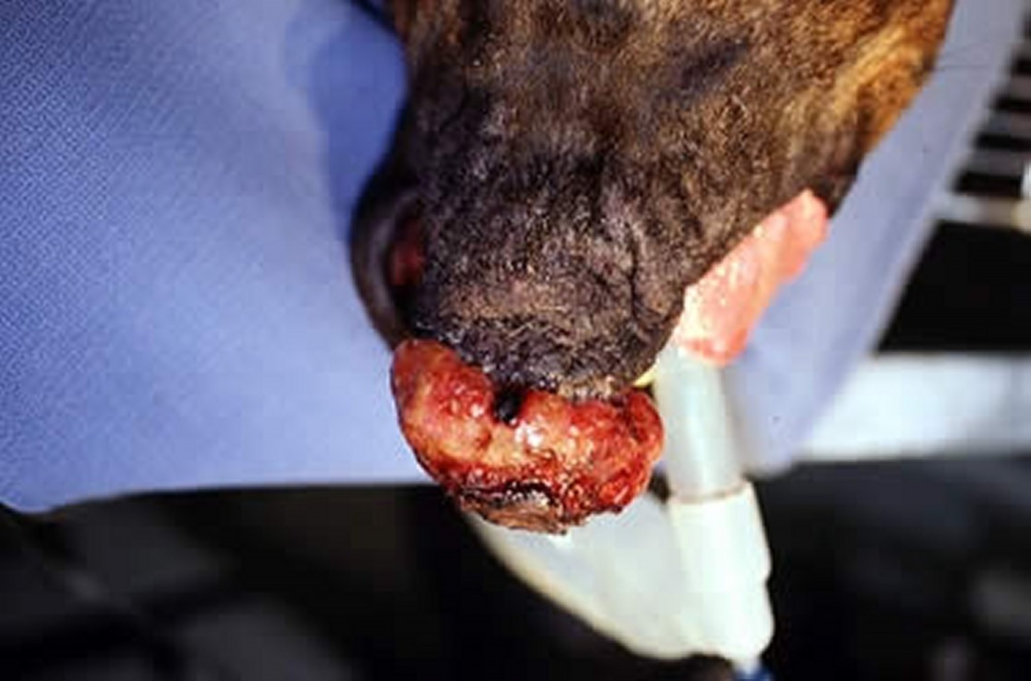
The canine acanthomatous ameloblastoma (previously called acanthomatous epulis) is much more locally aggressive, quickly invading the local tissues including bone. This tumor does not metastasize, but because of its locally aggressive nature, surgical excision should include a 1-cm margin of clinically normal tissue (including bony margins) to prevent recurrence. Radiation therapy may minimize disfigurement when treating large tumors. Adequate surgical removal is curative.
Malignant Oral Tumors
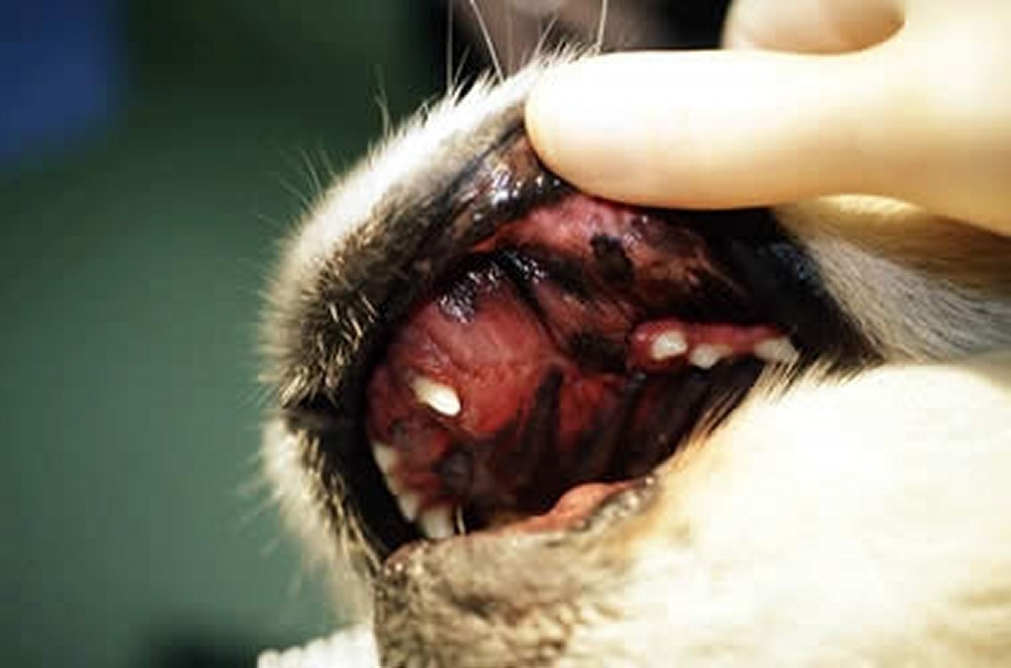
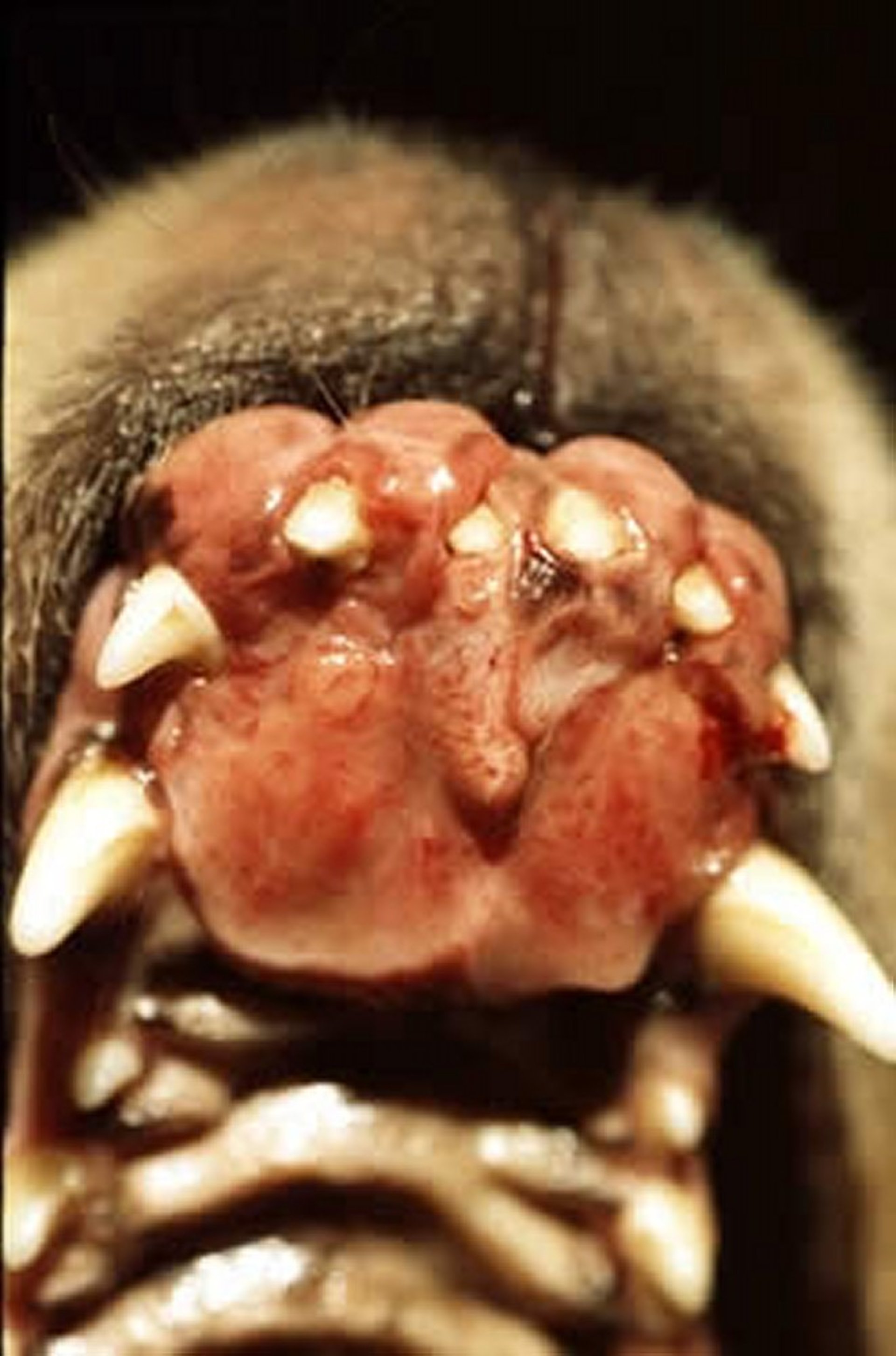
In dogs, the three most common malignant oral tumors are malignant melanoma, squamous cell carcinoma, and fibrosarcoma. The incidence of malignant oral tumors is higher in dogs >8 yr old.
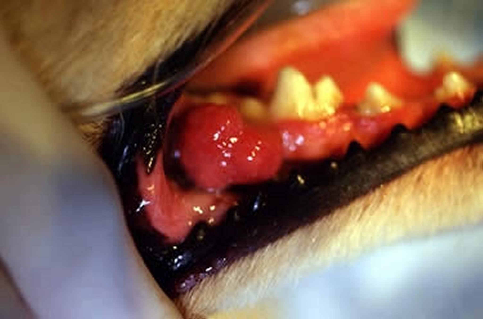
Squamous cell carcinomas are by far the most common malignant oral neoplasms in cats; they commonly involve the gingiva and tongue and are locally highly invasive. Fibrosarcomas are the next most common. In cats, these tumors are locally invasive and, if extensive, carry a poor prognosis.
Clinical Findings:
Courtesy of Dr. Ben Colmery III.
Courtesy of Dr. Ben Colmery III.
Courtesy of Dr. Ben Colmery III.
Courtesy of Dr. Ben Colmery III.

Courtesy of Dr. Ben Colmery III.

Courtesy of Dr. Ben Colmery III.
Courtesy of Dr. Ben Colmery III.
Signs of malignant oral tumors vary depending on the location and extent of the neoplasm. Halitosis, reluctance to eat, and hypersalivation are common. If the oropharynx is involved, dysphagia may be present. The tumors frequently ulcerate and bleed. The face may become swollen as the tumor enlarges and invades surrounding tissue. Regional lymph nodes often become swollen before oral and pharyngeal tumors are seen.
Diagnosis:
Because of the varied behavior of oral and maxillofacial tumors, presurgical characterization is valuable to plan the extent of the required surgery. Biopsy is the most reliable method to obtain a definitive diagnosis; however, a cytologic diagnosis from impression smears of a fine-needle aspirate is possible in many cases. Therefore, a histologic diagnosis is typically necessary to plan for accurate treatment. Malignant melanomas are variable in appearance, pigmented or nonpigmented, and should be considered in the diagnosis of any oral tumor. Squamous cell carcinomas commonly involve the gingiva or palatine tonsils (unilateral), and lymphosarcoma should be a differential diagnosis for bilaterally enlarged tonsils. Lymph nodes and the lungs should be evaluated for regional and distant metastasis.
Treatment:
Malignant melanomas are highly invasive and metastasize readily; consequently, the prognosis is guarded to poor. Surgical resection can extend survival and may be curative, particularly with masses in the rostral areas of the mouth. However, local recurrence is common. Immunotherapy is available as an adjunct treatment to radical surgery and chemotherapy. Nontonsillar squamous cell carcinomas are locally invasive with a low rate of metastasis, and the prognosis in dogs is good with aggressive and complete surgical resection, radiation therapy, or both. Tonsillar squamous cell carcinomas are aggressive and have a poor prognosis. Fibrosarcomas have a guarded prognosis because of their locally aggressive nature. Recurrence of tumor growth after resection is common.
In cats, squamous cell carcinoma has a poor prognosis unless the entire tumor can be removed, and longterm survival is usually seen only if diagnosed and treated early.


