

Neonatal diarrhea in ruminants remains the most important cause of death in calves under one month of age. Various bacterial, viral, and protozoal agents are recognized as causative agents, and failure of transfer of passive immunity is considered an important predisposing factor. Clinical presentation can range from loose stools in an otherwise healthy animal to severe dehydration, recumbency, coma, and ultimately death. Treatment includes eliminating the causative agent and correcting and maintaining the water, acid-base, and electrolyte balance of affected animals through oral and parenteral fluid therapy.
Diarrhea is common in newborn calves, lambs, and kids. The clinical presentation can range from mild diarrhea without systemic disease to profuse, acute diarrhea associated with rapid dehydration, severe disturbance of acid-base and electrolyte balance, and death, sometimes in as few as 12 hours. This discussion emphasizes the disease in calves, but the principles of pathophysiology and treatment also apply to lambs and kids.
Etiology of Diarrhea in Neonatal Ruminants
Several enteropathogens are associated with diarrhea in neonates. Their relative prevalence varies geographically, but the most prevalent infections in most areas are enterotoxigenic Escherichia coli, rotavirus, coronavirus, and Cryptosporidium parvum. Cases of neonatal diarrhea are commonly associated with more than one of these agents, and the cause of most outbreaks is multifactorial. Determining the particular agents associated with an outbreak of diarrhea can be important, because specific therapy and prophylaxis are available for some. Also, some agents have zoonotic risk. Diarrhea is also present in septicemic colibacillosis.
Bacteria

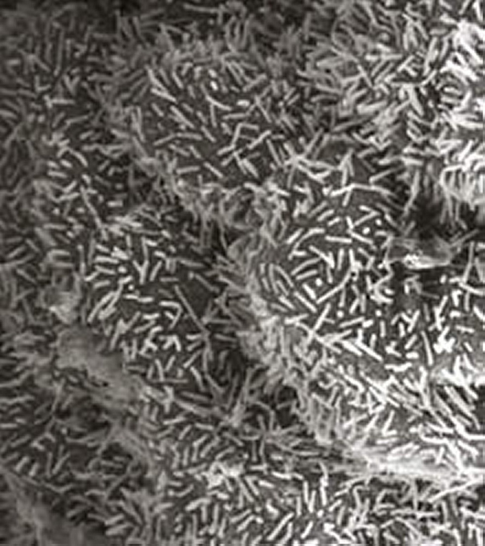
Courtesy of Dr. J. J. Hadad and Dr. Carlton Gyles.
Enterotoxigenic E coli is an important bacterial pathogen associated with neonatal diarrhea in calves during the first week of life. Enterotoxigenic E colihas two virulence factors associated with production of diarrhea that are fimbrial antigens enabling them to attach to and colonize the villi of the small intestine of neonatal calves in the first days of life. Strains in calves most commonly possess F5 (K99) or F41 fimbrial antigens, or both. These antigens are the focus of immunologic protection. Enterotoxigenic E coli produce a thermostable, nonantigenic enterotoxin (Sta) that influences intestinal ion and fluid secretion to produce a noninflammatory secretory diarrhea.
Diarrhea in calves and lambs also has been associated with enteropathogenic E coli that adhere to the intestine to produce so-called attaching and effacing lesions, with dissolution of the brush border and loss of microvillous structure at the site of attachment, a decrease in enzyme activity, and changes in ion transport in the intestine. These enteropathogens are also called “attaching and effacing E coli.” Some produce verotoxin, which may be associated with a more severe hemorrhagic diarrhea. The infection most frequently is in the cecum and colon, but the distal small intestine can also be affected. The damage in severe infections can result in edema and mucosal erosions and ulceration, leading to hemorrhage into the intestinal lumen.
Salmonella spp, especially S Typhimurium and S Dublin, but occasionally other serovars, cause diarrhea in calves 2–12 weeks old. Salmonellae produce enterotoxins but are also invasive and produce inflammatory change within the intestine. In calves, infection commonly progresses to a bacteremia. (Also see Salmonellosis.)
Clostridium perfringens types A, B, C, and E produce a variety of necrotizing toxins and cause a rapidly fatal hemorrhagic enteritis in calves. Infection with type B or C is a common cause of enteritis and dysentery in lambs. In calves, neonatal diarrhea has been associated with C perfringens type A. The disease characterized by peracute hemorrhagic abomasitis and enteritis is seen sporadically, although outbreaks can occur. C perfringens type A forms part of the normal microbiome of the digestive tract of calves and can be isolated from feces of healthy calves. When encountering favorable environmental conditions, some strains of the pathogen can cause clinical disease.
Campylobacter jejuni and Yersinia enterocolitica may be present in the feces of calves and lambs with diarrhea but also may be found in the feces of healthy animals.
Viruses
Rotavirus is the most common viral cause of diarrhea in calves and lambs. Groups A and B rotavirus are involved, but group A is most prevalent and clinically important and contains several serotypes of differing virulence. Rotavirus replicates in the mature absorptive and enzyme-producing enterocytes on the villi of the small intestine, leading to rupture and sloughing of the enterocytes with release of virus to infect adjacent cells. Rotavirus does not infect the immature cells of the crypts. With virulent strains of rotavirus, the loss of enterocytes exceeds the ability of the intestinal crypts to replace them; hence, villous height is reduced, with a consequent decrease in intestinal absorptive surface area and intestinal digestive enzyme activity.
Coronavirus is also commonly associated with diarrhea in calves. It replicates in the epithelium of the upper respiratory tract and in the enterocytes of the intestine, where it produces similar lesions to rotavirus but also infects the epithelial cells of the large intestine to produce atrophy of the colonic ridges.
Other viruses, including Breda virus (torovirus), a calici-like virus, astrovirus, and parvovirus, have been demonstrated in the feces of calves with diarrhea and can produce diarrhea in calves experimentally. However, these agents have also been found in the feces of healthy calves. The importance of these agents in the syndrome of diarrhea in neonates has yet to be determined. The viruses of bovine virus diarrhea and infectious bovine rhinotracheitis are reported to cause calf diarrhea, but this is not a common manifestation of these infections.
Protozoa
Cryptosporidium parvum is a common cause of diarrhea in calves and lambs. The parasite does not invade but adheres to the apical surface of enterocytes in the distal small intestine and the colon. This results in loss of microvilli, decreased mucosal enzyme activity with villous blunting and fusion (leading to a reduced villous surface absorptive area), and inflammatory changes in the submucosa. Mammalian cryptosporidia lack host specificity.
Giardia duodenalis is a common, presumably asymptomatic infection in the intestine of young calves and lambs. It has been found in the feces of poorly growing calves that have a chronic mucoid diarrhea, but there is little evidence for a causative association of this organism with diarrhea in calves or lambs.
Other Causes
Calves fed large amounts of milk or inappropriately formulated milk replacers produce a large volume of feces with a greater than normal fluid content but do not have a fluid diarrhea with weight loss. Similarly, calves sucking high-producing beef cows grazing lush pasture may have loose feces. Milk replacers with poor quality, heat-denatured proteins or with excessive amounts of soybean or fish protein or carbohydrates of nonmilk origin have a higher risk of producing diarrhea. Similarly, incorrectly prepared oral electrolyte solutions or mixtures of milk with electrolyte solutions with excessively high osmolarity of the final solution can result in osmotic diarrhea.
There is some evidence that oral administration of antimicrobials such as neomycin or tetracycline to young calves for 3–5 days can result in villous change with resultant malabsorption and mild diarrhea. Prolonged and high-dose antibiotic treatment of calves can lead to diarrhea associated with intestinal dysbiosis. Colisepticemia and ruminal drinking can also be accompanied by diarrhea.
Epidemiology and Transmission of Diarrhea in Neonatal Ruminants
Enteropathogens associated with diarrhea are commonly found in the feces of healthy calves; whether intestinal infection leads to diarrhea depends on a number of determinants, including differences in virulence of different strains of a pathogen and the presence of more than one pathogen. The resistance of the calf is of major importance and is largely determined by successful transfer of colostral immunoglobulins. Colostrum-deprived calves are highly susceptible to all types of infections, including with enteropathogens, and develop severe and often fatal disease.
The progression of infection, the severity of lesions produced, and the severity of the diarrhea can be modulated by immunoglobulins and other protectives compounds received via colostrum. Immunoglobulins act directly on pathogens in the intestinal lumen during the period of colostrum ingestion as well as after, because significant amounts of circulating immunoglobulins are re-secreted into the intestine, especially when the concentration of circulating immunoglobulin is high. The lack of specific antibodies in dams that have not been exposed to specific pathogens, and the use of specific vaccines in these dams, further modulate this influence. Stress caused by a poor environment, inadequate protection from the weather, or an insufficient or inappropriate diet also increases the risk of disease.
With all of the enteropathogens, healthy adult cattle may be carriers and periodically excrete the organism in feces. Excretion may increase around parturition and be more frequent in primiparous cows. This can lead to contaminated calving areas and infection of the udder and perineum of the dam. Other sources of infection include the feces of healthy calves and the feces of diarrheic calves, which contain large numbers of organisms early in the course of infection. A few scouring calves can result in severe contamination of the calf-rearing area. Transmission is by fecal-oral contact, fecal aerosol, and, in the case of coronavirus, by respiratory aerosol.
Pathogenesis of Diarrhea in Neonatal Ruminants
Diarrhea in neonatal ruminants is usually associated with disease of the small intestine and can be caused by hypersecretion or malabsorption. Hypersecretory diarrhea develops when an abnormal amount of fluid is secreted into the gut, exceeding the resorptive capacity of the mucosa. In malabsorptive diarrhea, the capacity of the mucosa to absorb fluid and nutrients is impaired to the extent that it cannot keep up with the normal influx of ingested and secreted fluids. This is usually the result of villous atrophy, in which the loss of mature enterocytes at the tips of the villi results both in a decrease in villous height (with a consequent decrease in the surface area for absorption) and in loss of the brush border digestive enzymes. The extent and distribution of villous atrophy varies with different pathogens and can explain variation in the severity of clinical disease.
Malabsorptive diarrhea may be aggravated by the colonic fermentation of nutrients that normally would have been absorbed in the small intestine. Fermentation products, especially lactic acid, appear to draw water into the colon osmotically, which contributes to the severity of diarrhea.
Inflammation contributes to the pathophysiology of diarrhea in most intestinal infections, and mediators of inflammation can affect ion flux within the intestine. Inflammation also leads to vascular and lymphatic damage and to structural damage of the crypt-villus unit. Inflammation, leading to necrosis of the enterocyte, submucosal inflammatory infiltration, and villous atrophy, is also a major component of the pathophysiology of diarrhea produced by salmonellae, as well as of diarrhea produced by enteropathogenic E coli and by toxigenic C perfringens.
Enterotoxigenic E coli produce the enterotoxin Sta, which stimulates marked hypersecretion by activating guanylate cyclase and by inducing a net secretion of sodium and chlorine. The membrane-bound sodium-glucose cotransport system remains functional but cannot compensate for the increased secretory activity. Salmonellae also elaborate enterotoxins. Infections with verotoxin-producing enteropathogenic E coli result in accumulation of fluid within the large intestine and extensive damage to the large intestinal mucosa, with edema, hemorrhage, and erosion and ulceration of the mucosa, which results in blood and mucus in the lumen.
Viruses usually produce a malabsorptive diarrhea by destroying the absorptive cells of the mucosa, thus shortening the intestinal villi. The mechanism by which cryptosporidia produce diarrhea is not completely understood, but it appears to have both malabsorptive and inflammatory components.
Most infectious forms of diarrhea have hypersecretory, inflammatory, and malabsorptive components, although one usually predominates. These lead to a net loss of water and electrolytes; if severe, the calf develops hypovolemia, acidemia, hypoglycemia, and prerenal azotemia.
Inappropriately formulated milk replacers may produce diarrhea by two mechanisms, both associated with malabsorption. Vegetable (especially soybean) products are commonly used as protein sources in the manufacture of milk replacers. Depending on the degree of refinement, these products may contain carbohydrates that are indigestible in young calves. Such carbohydrates are not absorbed in the small intestine and may contribute to diarrhea via colonic fermentation. In addition, most calves < 3 weeks old appear to have an allergic reaction to soy proteins that results in villous atrophy, leading to diarrhea that is probably malabsorptive.
Clinical Findings of Diarrhea in Neonatal Ruminants
The clinical presentation of neonatal diarrhea can vary greatly depending on the level of severity and can range from loose stools in an otherwise healthy animal to recumbency and coma in severely dehydrated and acidotic animals. The major signs include:
diarrhea with loose to watery feces
varying degrees of dehydration
dullness and varying degree of weakness
The age at first presentation, the severity of clinical symptoms, and the course of clinical disease can vary considerably depending on the causative agents involved.
Diarrhea due to enterotoxigenic E coli is seen in calves < 3–5 days old, rarely later. However, the age of susceptibility may be extended in the presence of other pathogens. Onset is sudden. Profuse amounts of liquid feces are passed, and the calves rapidly become depressed and recumbent. Calves may lose >12% of body weight in fluid within hours, and hypovolemic shock and death may occur in 12–24 hours. Body temperature may be increased but is commonly normal or subnormal. If fluid and electrolyte therapy is administered early, response is usually good. Disease produced by attaching and effacing E coli is seen predominantly in calves from 4 days to 2 months old and may manifest with diarrhea or primarily as dysentery with blood and mucus in the feces. The clinical course is short.
Diarrhea due to Salmonella spp usually is not seen in calves < 14 days old. It is characterized by feces that are foul smelling and contain blood, fibrin, and copious amounts of mucus. Septicemia, with high fever and depression progressing to prostration and coma, is the salient manifestation of salmonellosis in calves and, although diarrhea is present, calves die from septicemic shock before showing signs of severe dehydration or hypovolemic shock. Calves with salmonellosis usually deteriorate rapidly and often die despite vigorous therapy.
Hemorrhagic enterotoxemia due to C perfringens type A, B, or C is characterized by acute onset of depression, weakness, bloody diarrhea, abdominal pain, and death within a few hours. It usually develops in vigorous calves just a few days old that have large appetites and a ready source of milk. Calves affected with C perfringens usually die before treatment can be instituted.
Diarrhea due to rotavirus, coronavirus, and other viruses usually is seen in calves 5–15 days old but can affect calves up to several months of age. Affected calves are only moderately depressed and often continue to suck or drink milk. The feces are voluminous, soft to liquid, and often contain large amounts of mucus. Diarrhea commonly persists for 3 to several days, with some cases of coronaviral diarrhea becoming chronic. Cases of viral diarrhea that are uncomplicated by other pathogens commonly respond within a few days to fluid and electrolyte therapy and adequate nutritional support.
Cryptosporidiosis is seen in calves 5–35 days old but most commonly in the second week of life. It is characterized by persistent diarrhea that does not respond to therapy. Diarrhea due solely to Cryptosporidium spp is often mild and self-limiting, although the severity may be related to the general strength of the calf and to the intensity of challenge with the organism. Combination infections with cryptosporidia, rotavirus, and coronavirus are common and result in persistent diarrhea often characterized by emaciation and death. Death from hypoglycemia also occurs as a sequela of cryptosporidiosis in calves 3–4 weeks of age that have recovered from diarrhea but are still emaciated. Death often occurs during a bout of cold weather and is more likely to occur on farms where there is a policy of reducing the amount of milk fed to calves during periods of diarrhea.
Dietary diarrheas are seen in calves < 3 weeks old and are characterized by voluminous feces of pasty to gelatinous consistency. Initially, the calves are bright and alert and have good appetites. Eventually, however, they become weak and emaciated if the diet is not corrected. Infectious forms of diarrhea are often complicated by poor-quality diets or insufficient nutritional intake.
Diagnosis of Diarrhea in Neonatal Ruminants
Isolation of specific pathogens in fresh feces or on postmortem examination
A definite etiologic diagnosis for diarrhea cannot be made based solely on clinical findings. However, the history, age of the animal(s) affected, and clinical signs may permit a presumptive diagnosis. Fecal samples can be tested with calf-side test kits using immune chromatography to identify antigens of common pathogens of neonatal diarrhea or submitted to a diagnostic laboratory for isolation and characterization of enteropathogens. Samples should be taken from several untreated calves in the early stages of diarrhea. The interpretation of fecal microbiology can be difficult because of mixed infections and because enteropathogens are commonly present in the feces of healthy calves.
Postmortem examination allows examination of intestinal mucosa for evidence of diagnostic lesions and for the presence of enteropathogens such as cryptosporidia at the site of intestinal lesions. It may be the only way that disease such as that associated with attaching and effacing strains of E coli can be diagnosed. The diagnostic value of a necropsy diminishes quickly with time after death; important lesions can disappear within minutes due to autolysis.
Blood biochemical, blood gas, and hematologic analysis has limited value to make an etiologic diagnosis but can contribute to determining the level of metabolic disturbances such as dehydration, acid-base and electrolyte imbalances, and glycemia. Complete laboratory examination can, however, be expensive, and assessment of the level of dehydration and acidemia with reasonable precision can be based on the findings of a physical examination.
Treatment of Diarrhea in Neonatal Ruminants
Symptomatic therapy that restores hydration and acid-base- and electrolyte balance
Many of the factors involved in disease resistance are nonspecific; thus, important preventive measures can be taken and therapy can be started before an etiologic diagnosis has been established. Treatment includes fluid therapy for water and electrolyte replacement and correction of acid-base disturbances, alteration of the diet, and anti-inflammatory therapy. In severely affected animals, the need for antimicrobial therapy often must be assessed before a definitive etiologic diagnosis is available.
Fluid and electrolyte therapy is most important and should be started as soon as possible regardless of whether clinical evidence of dehydration has developed (clinical signs of dehydration are not apparent until the calf has lost at least 6% of its body weight in fluid). Calves still able to stand and willing and able to suck can often be treated with oral electrolyte solutions alone. Fluids for oral rehydration should promote the cotransport of sodium with glucose and amino acids and should contain sodium, glucose, glycine or alanine, potassium, and either bicarbonate or citrate or acetate as alkalinizing agents. Oral electrolyte solutions should be offered, alternating with whole milk or milk replacer. Numerous commercial preparations are available for this purpose. Solutions containing carbohydrates, as most oral electrolyte solutions do, should not be administered by stomach tube. Repeatedly force-feeding calves with these solutions risks resulting in rumen acidosis and the rumen drinker syndrome.
Feeding milk may increase fecal volume, but it provides energy to the calf and may promote gut healing. Calves have large energy requirements and little reserve. Electrolyte solutions do not meet calf energy requirements, and milk should not be withheld.
Calves that are recumbent, show evidence of water loss of ≥8% of their body weight, or that are unwilling to voluntarily ingest fluids orally require IV fluid therapy. These calves are usually acidotic, and the fluid and base deficits can be corrected initially by rapidly administering a hypertonic solution of sodium bicarbonate (either 500 mL of a 4.2% solution, or 250 mL of an 8.4% solution), followed by a physiologically balanced electrolyte solution administered at up to 40 mL/kg/hour until the volume deficit is corrected. Because diarrheic calves are frequently hypoglycemic, adding 25–50 g of dextrose to the IV fluids is often beneficial in the initial treatment phase. Oral electrolyte solutions should be used concurrently with and after IV fluid therapy to compensate for ongoing fluid and electrolyte losses.
Diarrhea per se is not an indication for antimicrobial therapy, but parenteral antimicrobial therapy should be considered whenever calves are systemically ill. Field studies revealed that at least 30% of diarrheic calves with systemic disease are bacteremic—a clear indication for parenteral antimicrobial therapy. Because the large majority of cases of bacteremia and septicemia in neonatal calves are associated with E coli, the chosen antibiotic should be effective against gram-negative bacteria.
In several studies, severely affected diarrheic calves treated with NSAIDs in conjunction with fluid therapy showed fewer signs of pain, made a faster recovery, and had better weight gains in the reconvalescent period. These effects reported for several NSAIDs have been attributed to their analgesic, anti-inflammatory, antipyretic, and antisecretory properties.
The use of drugs to reduce intestinal motility such as hyoscine-N-butylbromide or atropine is sometimes advocated, because they decrease fecal output. Although reducing fecal production may be interpreted as a positive treatment outcome, it can also be seen as sequestration of gut fluid containing bacteria, toxins, and undigested nutrients in the intestinal tract. The literature does not provide any strong supportive evidence for or against the use of antimotility drugs.
Intestinal gels and adsorbents, such as kaolin and pectin, are in general use, but their only established effect is to increase fecal consistency; they do not reduce the loss of water and ions.
Prevention and Control of Diarrhea in Neonatal Ruminants
Because of the complex nature of diarrhea in neonates, it is unrealistic to expect total prevention—economical control is the major objective. The incidence of clinical disease and the case fatality rate depend on the balance between the levels of exposure to infectious agents and the resistance in the calf. Differences in herd size; availability of facilities, land, and labor; and general management objectives make it impossible to recommend specific management procedures applicable to all situations. However, several broad principles apply in all herds:
Practice good general hygiene, in particular in maternity and calf-rearing areas
Reduce exposure of neonates to pathogens by separating maternity pens, calf-rearing areas, and hospital pens on the premises
Practice good colostrum management
Avoid mingling older calves with neonates
Proper colostrum management deserves particular attention because the prevalence of failure of transfer of passive immunity in calves is still disturbingly high. A significant portion of both naturally sucking dairy calves and calves handfed colostrum do not acquire adequate amounts of immunoglobulin because of delayed sucking or feeding, ingestion of an inadequate volume of colostrum, or ingestion of colostrum of inferior quality. When time constraints on labor preclude an ensured intake of colostrum by nipple-bottle feeding, administration of 4 L of colostrum by esophageal feeder within the first 2 hours of life can be the best colostrum feeding policy. (Also see Management of Reproduction: Cattle.)
Vaccination of dams late in pregnancy to boost the immunoglobulin content against rotavirus, coronavirus, or enterotoxigenic E coli in colostrum can be useful in herds in which these pathogens have been found to contribute to the neonatal diarrhea problem.
The pregnant dam is vaccinated 6 and 2 weeks before parturition to stimulate antibody production to specific pathogens of the neonatal diarrhea complex; these antibodies are then passed on to the newborn through the colostrum (provided the calf ingests it). A single booster is given in subsequent years. Monoclonal F5 (K99) E coli antibody is commercially available for oral administration to calves immediately after birth. This treatment should be considered in herds where enterotoxigenic E coli was previously isolated. It is meant to be administered in combination with good-quality colostrum because this passive immunization only improves resistance to one specific pathogen.
Vaccination of pregnant cows with rotavirus and coronavirus vaccines increase the amount of specific antibody in colostrum and milk, but the concentration of antibodies in milk may be insufficient to provide local antibody in the intestinal lumen during the period of peak prevalence of infection, which, in calves, is 5–15 days of age. Controlled trials of commercial vaccines have shown variable results. The addition of small amounts of immune colostrum to milk fed during the period of susceptibility can provide some protection against disease.
Zoonotic Risk of Diarrhea in Neonatal Ruminants
Several of the agents that produce diarrhea in calves can also produce diarrheal disease in people. Cryptosporidium parvum and S Typhimurium can produce serious disease, particularly in immunocompromised individuals. These organisms are commonly present as subclinical infections in the gut of calves and lambs, which again emphasizes the importance of personal hygiene when handling calves; immunocompromised people should avoid contact with young ruminants and possibly all farm animals.
Cattle, including calves, are one of the reservoirs for the verotoxic E coli serotype O157:H7 associated with human hemorrhagic colitis and the hemolytic uremic syndrome. Infection in people is usually acquired by consumption of contaminated food, but the infective dose is low and the possibility of infection by direct contact exists. Other verotoxic E coli associated with human disease can also be isolated from the feces of healthy cattle. Human disease from infection with enteric livestock pathogens has occurred after seemingly trivial contact associated with visits to livestock fairs, petting zoos, and farm educational tours. Hand cleansing and disinfection should be a component of these visits.
Key Points
Neonatal diarrhea is the most important cause of disease and death in ruminants up to one month of age.
Several viral, bacterial, and protozoal pathogens have been identified as potential causative agents; in most cases, multiple agents are involved.
Failure of transfer of passive immunity is a common and important predisposing factor.
Treatment consists of correcting water, acid-base, and electrolyte imbalances through either oral or parenteral fluid therapy.


