

Many viruses have been identified from sick and healthy reptiles; however, few have been clearly proved as etiological agents of disease. Several viruses have been linked strongly enough for them to be considered the causative agent until proved otherwise. A number of viral PCR assays and serological tests are available.
Inclusion Body Disease of Boid Snakes
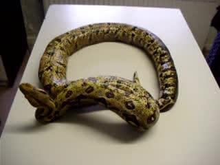
Boas and pythons are most commonly affected by inclusion body disease (IBD), which was originally thought to be caused by a retrovirus but has now been confirmed as a reptarenavirus (see boa constrictor with IBD and ).


Boa constrictor with dysecdysis and a complete loss of normal righting reflex due to inclusion body disease caused by reptarenavirus.
Courtesy of Dr. Stephen Divers.

Boas are considered a more typical host because so many are infected, and they can harbor the virus for months to years with few to no clinical signs. Early signs, possibly precipitated by any factor causing immunosuppression, include a history of unthriftiness, anorexia, weight loss, secondary bacterial infections, poor wound healing, dysecdysis, and regurgitation. In essence, IBD should be considered in every sick boa.
Typical findings in the acute phase of the disease include leukocytosis and a normal chemistry panel. As the disease progresses, WBC counts tend to decline to subnormal levels. Blood chemistry results vary, depending on how debilitated and dehydrated the boa becomes, but organ damage may occur. As the disease becomes chronic, some boas exhibit neurological signs, ranging from mild facial tics and abnormal tongue flicking to failure of the snake to right itself when placed in dorsal recumbency and severe seizures.
Pythons are thought to be a more acutely affected host for the reptarenavirus because the disease course is more acute and neurological signs more often profound, with most pythons developing severe neurological disease. Although the active disease can linger for months or longer in boas, most pythons die within days or weeks after the onset of clinical signs.
Exposure to reptarenaviruses appears to be due to a transfer of body fluids. Breeding, fight wounds, fecal/oral contamination, and snake mites have been implicated as common ways of transfer. A tentative diagnosis is based on patient history and clinical signs. Blood work varies, depending on the stage of the disease; however, few diseases in snakes will cause such increased WBC counts in the early stages. On blood smears, inclusion bodies are frequently found in the cytoplasm of leukocytes. A definitive diagnosis is obtained by PCR assay and biopsy of internal tissues (eg, the liver, kidney, esophageal tonsils, and stomach) in which the characteristic eosinophilic inclusion bodies are found.
Pearls & Pitfalls
|
IBD is not curable, and many owners choose euthanasia. However, individuals may elect to isolate their snakes and treat with supportive and palliative measures. It is essential to educate owners not to sell infected specimens or their offspring, because this has caused the disease to spread worldwide.
Retroviruses of Reptiles
Retroviruses have also been found in Russell vipers, corn snakes, and California kingsnakes in association with malignant tumors. A retrovirus isolated from a sarcoma in a Russell viper was designated as viper virus. A related virus was isolated in a corn snake from a rhabdomyosarcoma and designated corn snake retrovirus.
Adenoviruses of Reptiles
Adenoviruses have been implicated in fatal hepatic or GI diseases in numerous reptiles:
snakes (gaboon vipers, ball pythons, boa constrictors, rosy boas, and rat snakes)
lizards (Jackson chameleons, savannah monitors, and bearded dragons)
crocodilians
In bearded dragons, the route of transmission of adenoviruses appears to be fecal/oral contamination. Clinical signs are more commonly noted in juvenile dragons but can affect adults, usually to a lesser extent. Clinical signs are vague and include lethargy, weakness, weight loss, diarrhea, and sudden death. Morbidity is high in young bearded dragons; however, survival is increased with supportive care. Fluid administration, force feeding, and antimicrobials for secondary infections may be useful.
Clinical signs of disease in bearded dragons are vague and similar to those caused by coccidia and nutritional disorders, and therefore diagnostic confirmation is important. Characteristic intranuclear inclusion bodies are found in several internal organs, primarily the liver. When working with a large breeding group of lizards, it is practical to sacrifice a failing specimen to make a diagnosis. Antemortem diagnosis can be accomplished by liver biopsy. Identification of adenovirus by PCR assay from fresh feces may indicate shedding and infection but not necessarily disease.
Recovered lizards should be quarantined for at least 3 months. Duration of viral shedding after recovery is unknown, so owners should be discouraged from selling or trading previously infected animals.
Herpesviruses of Reptiles
Herpesviruses have been isolated from freshwater turtles, tortoises, and green sea turtles. In freshwater turtles, herpesvirus may be associated with hepatic necrosis. In tortoises, the virus may cause necrosis of oral mucosa, accompanied by anorexia, regurgitation, and oral and ocular discharge.
Treatment in tortoises includes isolation, supportive care, application of 5% acyclovir to oral lesions, and systemic acyclovir. Herpesviral disease is diagnosed by PCR assay, cytological/histological intranuclear inclusion bodies, and histological demonstration of a host pathological reaction.
Herpesvirus infections of farmed green sea turtles have been associated with gray patch disease and lung-eye-trachea disease, whereas fibropapillomas plague certain free-ranging populations. Biopsies of the skin reveal basophilic intranuclear inclusion bodies in epidermal cells; viral particles are noted in the cytoplasm by electron microscopy. PCR assay is frequently the most common approach to identifying the presence of a herpesvirus. No specific treatment exists, but decreased crowding and stress appear to decrease incidence.
Fibropapillomatosis, caused by chelonid alphaherpesvirus 5, has been reported from free-ranging sea turtles, especially around Hawaii (see ). The route of transmission is not known, but leeches have been implicated as mechanical vectors.

Fibropapillomatosis in a green sea turtle. This proliferative condition can affect the integument as well as visceral organs and is caused by an alphaherpesvirus, with likely mechanical vector transmission by leeches.
Courtesy of Dr. Stephen Divers.
The light gray to black masses range in size up to 20 cm (8 inches) in diameter. The location of the masses seems to dictate the severity of clinical signs. Masses occurring on periocular tissue can obscure vision. Growths on the flippers can interfere with swimming and the ability to forage for food. Internal masses also occur, primarily in the lungs, liver, kidneys, and GI tract. Diagnosis is made by characteristic lesions and histological evaluation.
Treatment consists of surgical removal, with wide margins to help decrease recurrence. Some turtles recover spontaneously, whereas those with internal or ocular lesions usually perish.
Ferlavirus and Nidovirus in Reptiles
Ferlavirus (Paramyxoviridae) infections have been reported in various viperid snakes but have also been reported in nonvenomous snakes and lizards. This highly contagious virus causes predominantly respiratory signs; transmission appears to be from respiratory secretions. Secondary bacterial infections are common because of the severe inflammation initiated by the virus, and it is not unusual to note nasal discharge, open-mouth breathing, caseated pus in the oral cavity, and labored breathing. Neurological involvement, including tremors and opisthotonos, is occasionally noted.
Endoscopic biopsies and postmortem samples of lung can be submitted to detect viral particles by histology and electron microscopy. A hemagglutination inhibition test is available to measure antibodies against ophidian paramyxovirus in zoos and private collections; a positive titer should be used as a screening tool to help eliminate infected animals and prevent carriers from entering noninfected collections. PCR assay for detection is also available.
No specific treatment exists, but supportive care and antimicrobials may prove useful. Affected specimens should be isolated and strict hygiene practices followed. Although a vaccine is under development, it is currently not effective.
A nidovirus (Coronoaviridae) has been reported as a cause of pneumonia and stomatitis in pythons and boas. Detection of the virus is possible by PCR assay of oral swabs and tracheal washes.
West Nile Virus in Reptiles
West Nile virus has resulted in epizootic outbreaks in farmed alligators, with the virus causing multiorgan necrosis, heterophilic granulomas, heterophilic perivasculitis, and lymphoplasmacytic meningoencephalitis. Very high viremia titers have been reported in various reptiles, suggesting that reptiles may play a role as an amplification host.
Papillomas in Reptiles
Viral particles appear to be transmitted from one European green lizard to another via bite wounds. The resulting papillomas are 2–20 mm (0.08–0.8 inches) in diameter and may be single or multiple. Although there are no clinical signs in the initial phase, affected lizards may become lethargic and anorectic and die. Diagnosis involves detection of viral particles by electron microscopy.
Treatment consists of surgical removal of single masses, although regrowth is common. Isolation of affected lizards is perhaps the only way to prevent spread.
A papilloma-type virus also appears to affect Bolivian side-neck turtles and appears as oval white skin lesions distributed over the head. Ulcerative shell lesions also occur, primarily on the plastron. Diagnosis is made by identifying viral particles on electron microscopy. Treatment is supportive and palliative, and affected animals should be isolated.
Iridoviruses in Reptiles
Iridoviruses have been reported in various chelonians, snakes, and lizards. An iridovirus was found in a Hermann’s tortoise, which died without prior clinical signs of disease. Progressive anemia in Australian geckos has been linked to an iridovirus. Clinical signs range from none to stomatitis, rhinitis, conjunctivitis, tracheitis, edema, and cutaneous abscessation.
In some cases, the recovered iridoviruses have been closely related to amphibian ranaviruses. Although the clinical importance of several of these viruses remains unclear, iridoviruses deserve close attention, especially in chelonians.
Other Viruses in Reptiles
Many other viruses continue to be isolated from reptiles, but little information exists regarding disease, diagnosis, control, and treatment. Unexplained deaths should be investigated by necropsy, with a complete set of tissues frozen and retained should viral investigations be indicated.
Pearls & Pitfalls
|
For More Information
Divers SJ, Stahl SJ. Mader's Reptile and Amphibian Medicine and Surgery. 3rd ed. Elsevier; 2019.
Available Tests & Panels. Zoological Medicine Diagnostic Laboratory. College of Veterinary Medicine, University of Florida.


