

Larvae of Hypoderma spp cause myiasis characterized by the presence of subcutaneous warbles on the dorsal and lumbar regions of domestic and wild ruminants. Two species—Hypoderma bovis and Hypoderma lineatum—are economically important and primary parasites affecting cattle and water buffalo. A third species, Hypoderma sinense, has beenidentified in cattle and yaks in China. Moreover, Hypoderma tarandi parasitizes reindeer in arctic and subarctic regions, and Hypoderma actaeon and Hypoderma diana are frequently found in deer. Occasional infestations by Hypoderma spp also have been reported in horses, sheep, goats, and humans.Hypoderma spp are found between latitude 25° and latitude 60° in the Northern Hemisphere in >50 countries of North America, Europe, Africa, and Asia. The widespread use of macrocyclic lactones has decreased the prevalence of Hypoderma spp infection to an almost clinically nonexistent extent in North America and some European countries; however, it remains endemic in resource-limited countries in North Africa and Asia.
Life Cycle of Hypoderma spp

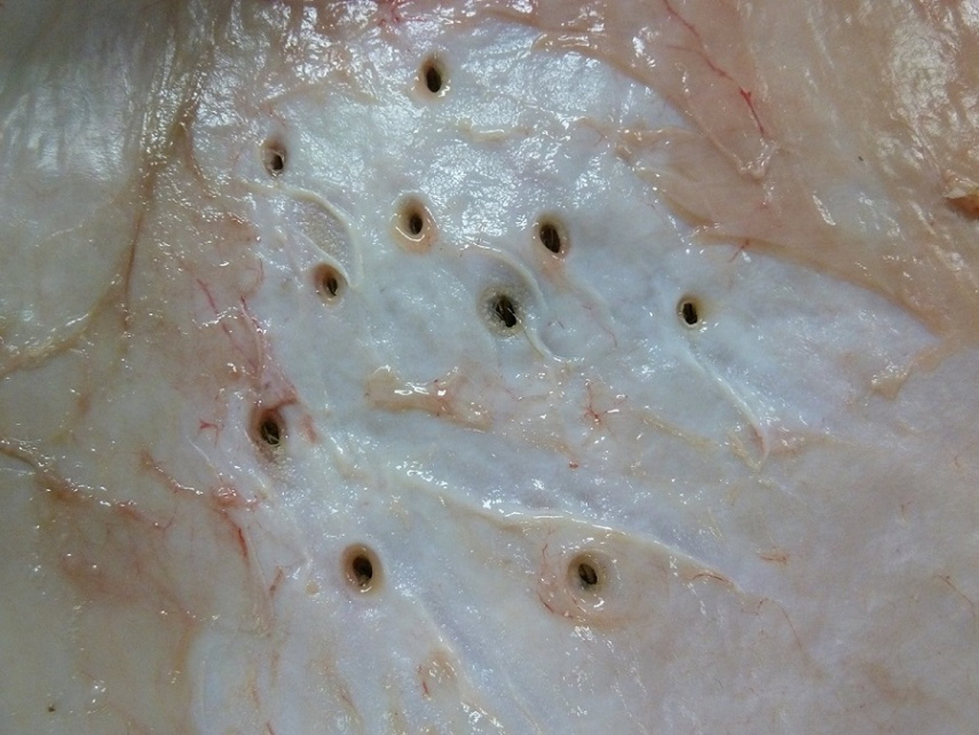
This photograph of the interior part of a cow hide after slaughter shows multiple breathing holes of Hypoderma spp larvae.
Courtesy of Dr. Rosario Panadero-Fontán.

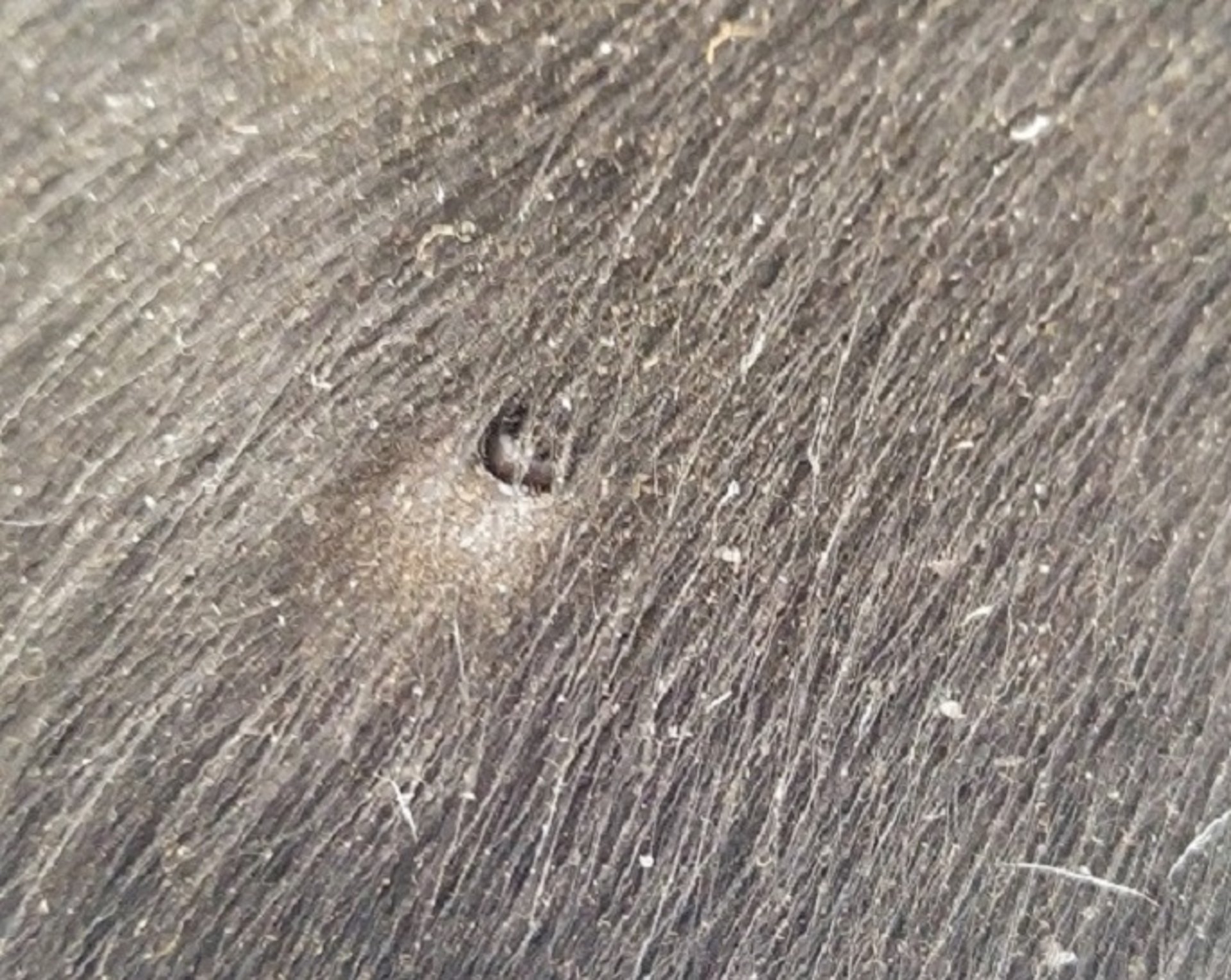
In this photograph of the exterior of the skin of an infected animal, a Hypoderma spp warble breathing hole including the posterior spiracular plate of the larva is visible.
Courtesy of Dr. Rosario Panadero-Fontán.
The critical stages of Hypoderma infestation in a host are:
penetration of the skin (first instar)
migration to resting sites (esophagus for H lineatum, spinal canal for H bovis)
migration to subcutaneous tissue in the back to form warbles (second- and third-stage larvae)
Adult Hypoderma flies are ~15 mm long, hairy, and bee-like in appearance. In spring or early summer, they attach their eggs on the hair of cattle, particularly on the legs and lower body regions. H lineatum deposits eggs in rows of 3–10; H bovis deposits eggs singly on each hair. The eggs hatch in 3–7 days, and first-stage larvae (1 mm long) crawl to the base of the hair shaft and penetrate the skin. Normally, the first-stage larvae (L1) travel through the fascial planes between muscles, along connective tissue, or along nerve pathways. They secrete proteolytic enzymes that facilitate their movement and stimulate an immune response. During the fall, larvae migrate toward two different regions, depending on the species. H lineatum larvae migrate to the submucosal connective tissue of the esophageal wall, where they accumulate for 2–4 months. H bovis larvae migrate to the region of the spinal canal, where they are found in the epidural fat between the dura mater and the periosteum for a similar period. During the rest period, the larvae grow substantially, to triple in size.
Beginning in early winter, the late L1 larvae (~15 mm long) leave their respective resting sites and arrive in the subdermal tissue of the host's back, where they make breathing holes (central puncta) through the skin. A granulomatous reaction, referred to as a warble, forms around the larvae, which undergo two molts, resulting in second and third instars. The warble stage lasts 4–8 weeks. Finally, third-stage larvae emerge through the breathing holes, drop to the ground, and pupate. Flies emerge from the pupae in 1–3 months, depending on weather conditions. Adult flies, which do not feed, live < 1 week. The life cycle is complete in 1 year.
For the two species, seasonal events are similar except that those for H lineatum occur ~6–8 weeks earlier than those of H bovis. These events vary from year to year, but they correlate with local and regional climatic conditions.
Clinical Findings and Pathogenesis of Hypodermiasis

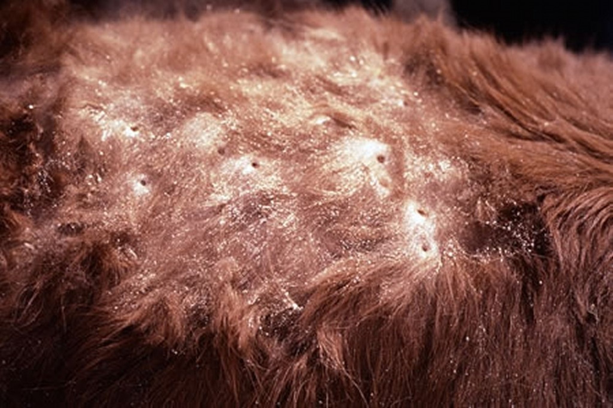
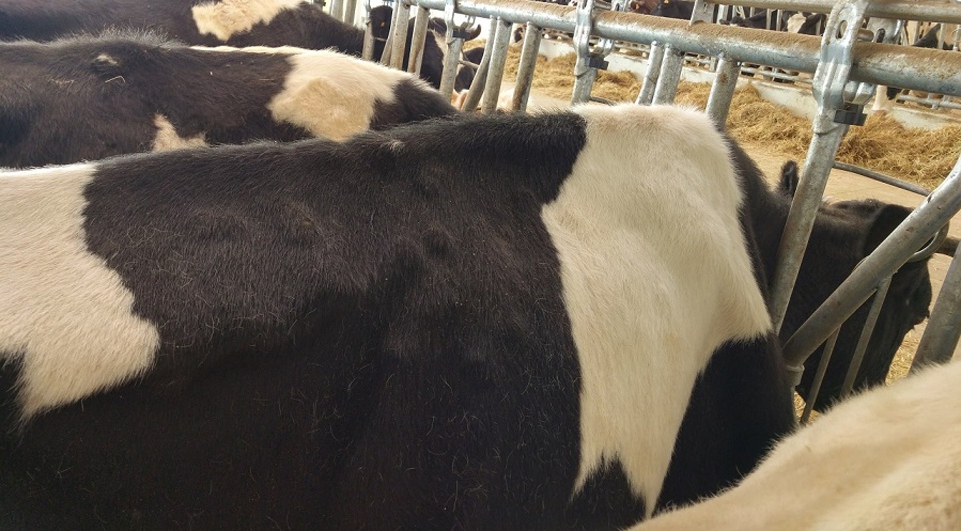
This photograph shows the grubby hide of a cow infested with Hypoderma bovis. Breathing holes made by the larvae are visible.
Courtesy of Dr. Jack Lloyd.

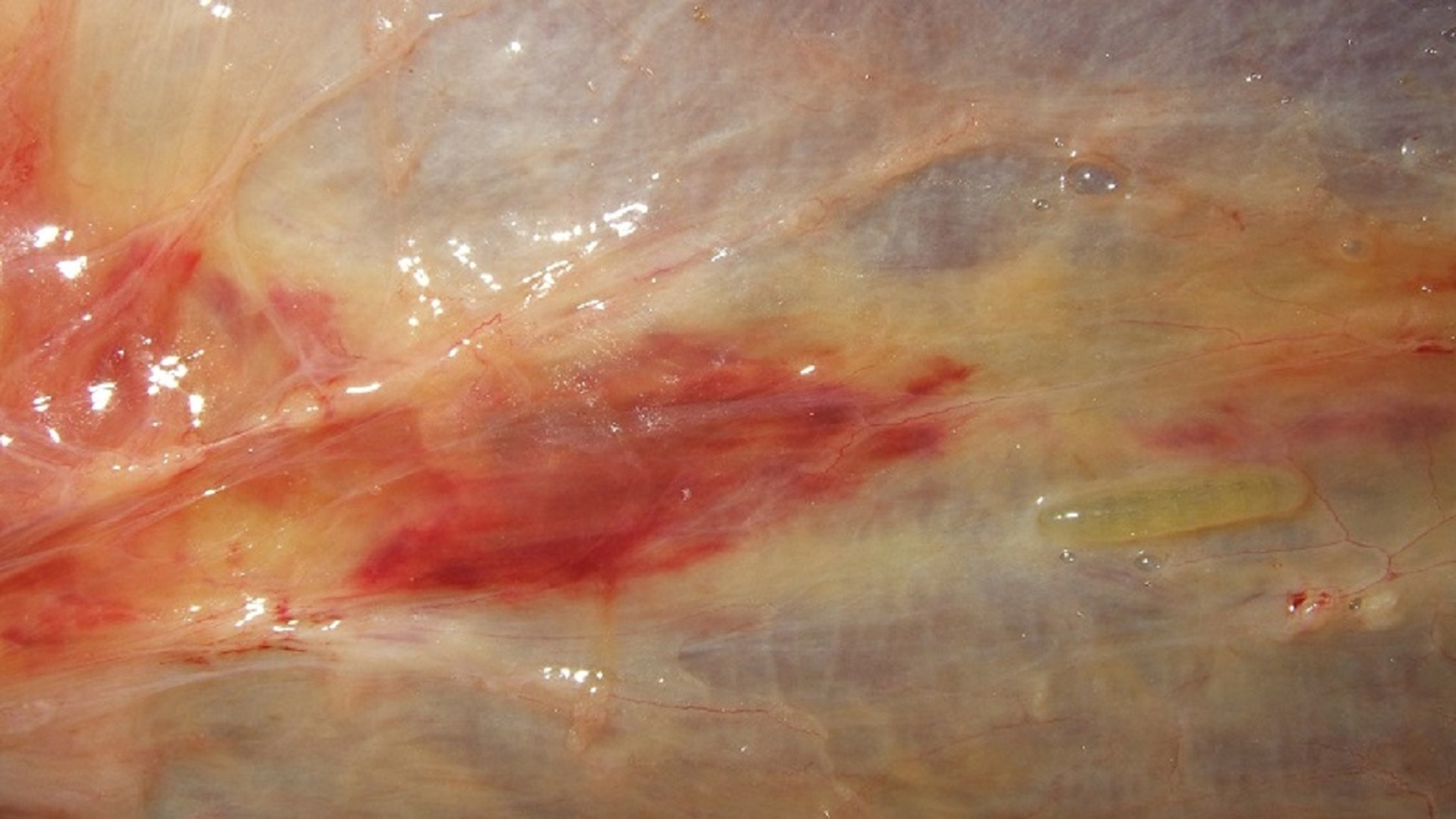
This photograph shows gelatinous and hemorrhagic lesions in the esophageal submucosa that are associated with migrating larvae of Hypoderma lineatum in reinfested cattle.
Courtesy of Dr. Rosario Panadero-Fontán.

The Frisian cow in this photograph is infested with the warble fly Hypoderma lineatum. Several encysted subcutaneous larvae are evident.
Courtesy of Dr. Rosario Panadero-Fontán.
Although bovine hypodermiasis does not induce marked mortality and morbidity, it does affect the productivity and welfare of animals, thus resulting in considerable losses due to:
decreased meat and milk production
poor growth rate and weight gain in primo-infected calves
carcass trimming to remove the damaged tissues
severe depreciation of hides due to the presence of breathing holes
increased susceptibility to other diseases
During periods of sunshine on warm days, cattle may run with their tails high in the air when chased by female heel flies, particularly H bovis. This behavior in cattle, referred to as gadding, is a strategy to avoid female flies and their attempt to deposit eggs. Not all stampeding or gadding of this kind is a response to heel fly attacks; this activity has been observed also in the absence of heel flies. Gadding in cattle may result in loss of production, altered reproduction, self-injury, or death.
Penetration of the skin by newly hatched larvae may produce a hypodermal rash, most often in older, previously infested cattle. The points of penetration are painful and inflamed, and they usually exude a yellowish serum. Larval secretions are implicated in modulatory processes that affect the inflammatory and specific immune responses. This immunomodulation promotes the survival of larvae in primary infestations.
In otherwise healthy cattle, H bovis larvae and their secretions in the epidural fat of the spinal canal are associated with dissolved connective tissue, fat necrosis, and inflammation. Sometimes the inflammation extends to the periosteum and bone, producing a localized area of periostitis and osteomyelitis. Occasionally, the epineurium and perineurium may become involved. In rare severe cases, paralysis or other nervous disorders may occur. Similarly, H lineatum in the submucosa of the esophagus may cause sufficient inflammation and edema in the surrounding tissues to hinder swallowing or eructation. It is unusual, however, for clinical signs of parasitism to be evident during the migratory phase.
Warbles may be found in the infected animal's back from tailhead to shoulders, and from topline to about one-third the distance down the sides. Usually, the warbles are firm and raised considerably above the normal contour of the skin. In each warble is a breathing hole, ranging in size from a small slit to a round hole (3–4 mm in diameter) for more mature larvae. Generally, secondary infection is depressed; however, warbles may occasionally develop in large, suppurating abscesses. The emergence of the third-stage larvae, their forced expulsion, or their death within the cysts usually results in healing of the lesions without complications. Carcasses and hides of cattle infested with cattle grubs show marked evidence of the infestation and have decreased value.
An infested animal may have 1 to > 300 warbles; most, however, have < 100. Infested herds often have individual animals with no larvae. Young animals are the most heavily infested because older animals develop a certain amount of resistance.
If migrating Hypoderma spp larvae die in esophageal tissue (H lineatum) or near the spinal canal (H bovis), they can cause severe reactions and sometimes death in affected animals. These reactions appear to be related to the number of larvae; however, they are rare no matter how many larvae are present.
Death of H lineatum L1 larvae in the submucosal connective tissue of the esophagus causes inflammation of the esophageal wall, dysphagia, drooling, and bloat. Again, recovery is usually rapid and complete (48–72 hours after treatment); in severe cases, however, the bloat may be fatal. Attempts to pass a stomach tube in an affected animal may lead to rupture of the esophagus.
Death of H bovis L1 larvae in the spinal canal of cattle after systemic insecticide treatment has resulted in stiffness, ataxia, muscular weakness, and paralysis of hind limbs. Recovery is usually rapid and complete; occasionally, however, paralysis may be permanent.
Concurrent with the decrease in the prevalences of H lineatum and H bovis in cattle, the widespread use of macrocyclic lactones has led to a decline in the clinical relevance of these parasites in North America. However, serologic surveys have demonstrated that cattle are still exposed to Hypoderma spp. If treatment programs stop or move away from products that have efficacy against Hypoderma spp, clinical signs due to infestation will likely reappear.
Diagnosis of Hypodermiasis
Clinical examination to observe and palpate warbles
Serologic testing to detect specific antibodies against Hypoderma spp

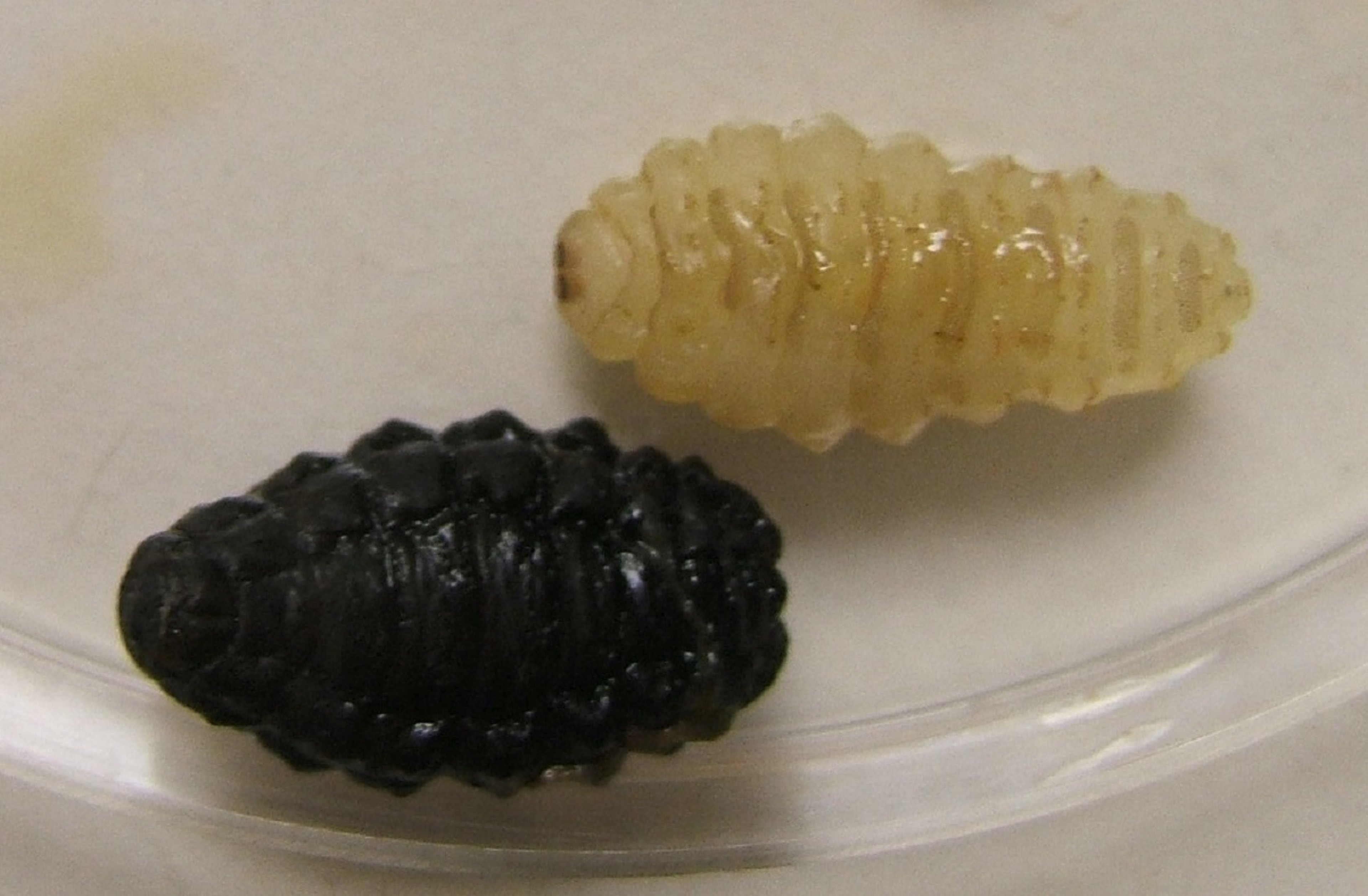
This photograph shows immature larvae (white) and mature, ready-to-pupate third-stage larvae (dark) of Hypoderma lineatum extracted from their warbles. Note that the body is divided into 11 segments and has a characteristic pattern of spiny bands.
Courtesy of Dr. Rosario Panadero-Fontán.
Third-stage larvae of Hypoderma spp are found in warbles, the furuncle-like nodules or cysts along the backs of cattle. After recovery from a warble, third-stage larvae can be easily differentiated. H bovis is 27–28 mm long, has no spines on the tenth segment, and has a funnel-shaped spiracular plate. H lineatum is slightly shorter (25 mm), has spines on the tenth segment, and typically has a flat spiracular plate. In cases of bloat or paralysis, the presence of disintegrating grubs and the associated hemorrhage and tissue damage distinguishes animals that are parasitized from those that are not.
ELISA-based tests have been developed to detect anti-Hypoderma antibodies in sera and milk. These ELISAs are often used to measure risk of exposure and do not correlate well with the observation of clinical hypodermiasis in cattle or the extent of infestation. Early detection of specific antibodies in first-season grazing animals enables the administration of a systemic treatment to avoid the damage due to larvae.
Treatment and Control of Hypodermiasis
Prophylactic/early treatment against L1 larvae before their arrival at resting sites, to avoid economic losses
Therapeutic/late treatment against L2 and L3 larvae in their warbles, to interrupt the life cycle of Hypoderma
Systemic insecticides containing macrocyclic lactones (doramectin, eprinomectin, ivermectin, or moxidectin) in pour-on and injectable formulations are approved for control of Hypoderma and other myiasis-causing flies in many countries. Eprinomectin and moxidectin pour-on formulations are authorized for treatment of both beef and dairy cattle. Otherwise, use of drugs for cattle grub control is prohibited in dairy animals of breeding age. Because residues may be present in cattle for varying periods after treatment, withdrawal times for all treatments must be observed.
Macrocyclic lactones are effective against all larval stages. The salicylanilide closantel is approved in beef cattle for the treatment of dermal stages (L2–L3) of Hypoderma.
Pour-on products are poured evenly along the midline of the back. Some products must not be applied when the skin or hair coat are wet or when rain is expected to wet cattle within 6 hours. The application site should be free of skin lesions, mud, or feces. Cattle stressed by castration, overheating, vaccination, or shipping should not be treated.
In the US and Europe, registrations of most, if not all, systemic organophosphate insecticides for cattle grub control have been canceled, and the practices of dipping or spraying cattle for cattle grub control have been replaced by pour-on and/or injectable treatments.
In areas where Hypoderma spp are prevalent, cattle, especially calves, should be treated as soon as possible after the end of the fly season. They should not be treated later than 8–12 weeks before the anticipated first appearance of grubs on the back, because adverse reactions may occur when migrating larvae are killed.
So-called long-acting or extended-release injectable formulations of ivermectin and eprinomectin, respectively, have been introduced in many countries, providing protection against larval infestation even when the treatment is administered during the fly activity period.
The administration of systemically active insecticides in organized treatment campaigns, combined with immunosurveillance strategies, has led to the eradication of bovine hypodermiasis in some European countries. Untreated animals remain a reservoir of the disease, causing rapid reinfestation of herds.
Where systemic insecticides cannot be used, Hypoderma spp larvae can be controlled by the application of tetrachlorvinphos dust to the warbles on the animal's back. The dust should be applied to the back and worked into the grub holes. Because new grubs continue to appear on the back, treatment must be repeated every 30–45 days during the warble season.
Mechanical or manual extraction (ie, squeezing) of the individual larva is contraindicated; in areas of very low prevalence, however, larvae can be removed by careful injection of 1 mL of 3% hydrogen peroxide with a blunt cannula through the central punctum. However, care must be taken not to rupture or pierce the larvae during removal. Rarely, when these procedures are performed carelessly, larvae are crushed or pierced, inducing an anaphylaxis-type reaction. This reaction is believed to result from the overwhelming and sudden release of Hypoderma spp antigens. Experimental studies have also suggested that this anaphylaxis-type reaction is due to the release of toxins from Hypoderma spp larvae.
Research to develop vaccines against infestations of Hypoderma spp was conducted as early as the 1950s. Even though some natural and recombinant antigens were demonstrated to have efficacy against Hypoderma spp infestations, the high efficacy and ease of use of macrocyclic lactones have precluded modern vaccine development.


