

When there is invasion by Dichelobacter nodosus of interdigital dermatitis, contagious footrot results. Whereas in Australia, footrot is separated into benign or virulent categories, depending on the strain of D nodosus present, in the US and other countries, benign and virulent footrot are often considered to be the same and are treated accordingly.
Benign Footrot
Benign footrot is confined largely to the interdigital skin, with only minimal underrunning of the adjacent horn. Clinically, benign footrot is indistinguishable from ovine interdigital dermatitis. The characteristic necrotic odor of D nodosus may be present. Lameness is common but less severe than in virulent footrot. The etiology and pathogenesis are the same, but the strains of D nodosus are less virulent and lack the hoof-invasive properties of the strains that cause virulent footrot. Strains of D nodosus affecting cattle usually cause only the benign form of footrot in sheep.
The economic effect of benign footrot is much less than that of virulent footrot. Running the sheep through footbaths containing 10% wt/vol zinc sulfate once every 14 days during the wet season is usually adequate for control. Use of long-acting antimicrobials such as tetracycline has been adopted with good results.
Virulent Footrot
Virulent footrot is a specific, chronic, necrotizing disease of the epidermis of the interdigital skin and hoof matrix that begins as an interdigital dermatitis and extends to involve large areas of the hoof matrix. Because the sensitive lamina and its network of capillaries are destroyed by the infection, the hoof wall (corium) loses its blood supply and anchorage to the underlying tissue and becomes detached. Footrot is extremely contagious, and, under suitable conditions, morbidity may approach 100%. The infection is rarely found in goats, deer, and cattle. The potential for genetic selection for increased resistance to footrot has been established, in that certain breeds appear more resistant to infection.
Etiology of Virulent Footrot in Sheep
Dichelobacter nodosus, a gram-negative anaerobe and obligate pathogen, is the primary etiologic agent that must be present for footrot to develop. Fusobacterium necrophorum, a gram-negative anaerobic bacteria, may play a synergistic role in pathogenesis and is a normal resident of manure-contaminated environments that contributes to ovine interdigital dermatitis and footrot. Under favorable environmental conditions, it colonizes the moist, macerated interdigital skin and provides ideal conditions for invasion by D nodosus at the skin-hoof interface. By action of proteases, D nodosus liquefies the cells of the stratum granulosum and stratum spinosum, causing separation of the hoof wall from the basal epithelium. Disease progresses from the interdigital horn to the heel, then to the sole, and finally to the lateral side of the hoof. The organism has been found to survive up to 2 weeks in the environment, but it can remain sequestered in cavities, cracks, or deformities of the affected foot for the life of the sheep. There are at least 20 strains of D nodosus, with varying pathogenicity. Transmission occurs most rapidly when conditions are warm and moist; however, cold, moist conditions are also conducive to transmission.
Clinical Findings of Virulent Footrot in Sheep
The most obvious clinical sign of virulent footrot in sheep is lameness, but affected limbs are seldom carried or non–weight-bearing. In chronically affected sheep, the hoof becomes gnarled and distorted. When more than one limb is affected, some sheep become recumbent or walk on their carpi. The brisket region and carpi tend to become hairless and ulcerated. Affected sheep do not compete well for food, lose body condition, and produce less wool, causing considerable production losses in some herds. Rams with affected hind limbs may be unwilling to breed, and ewes with hind limb lesions may be unable to bear the weight of a ram at service.

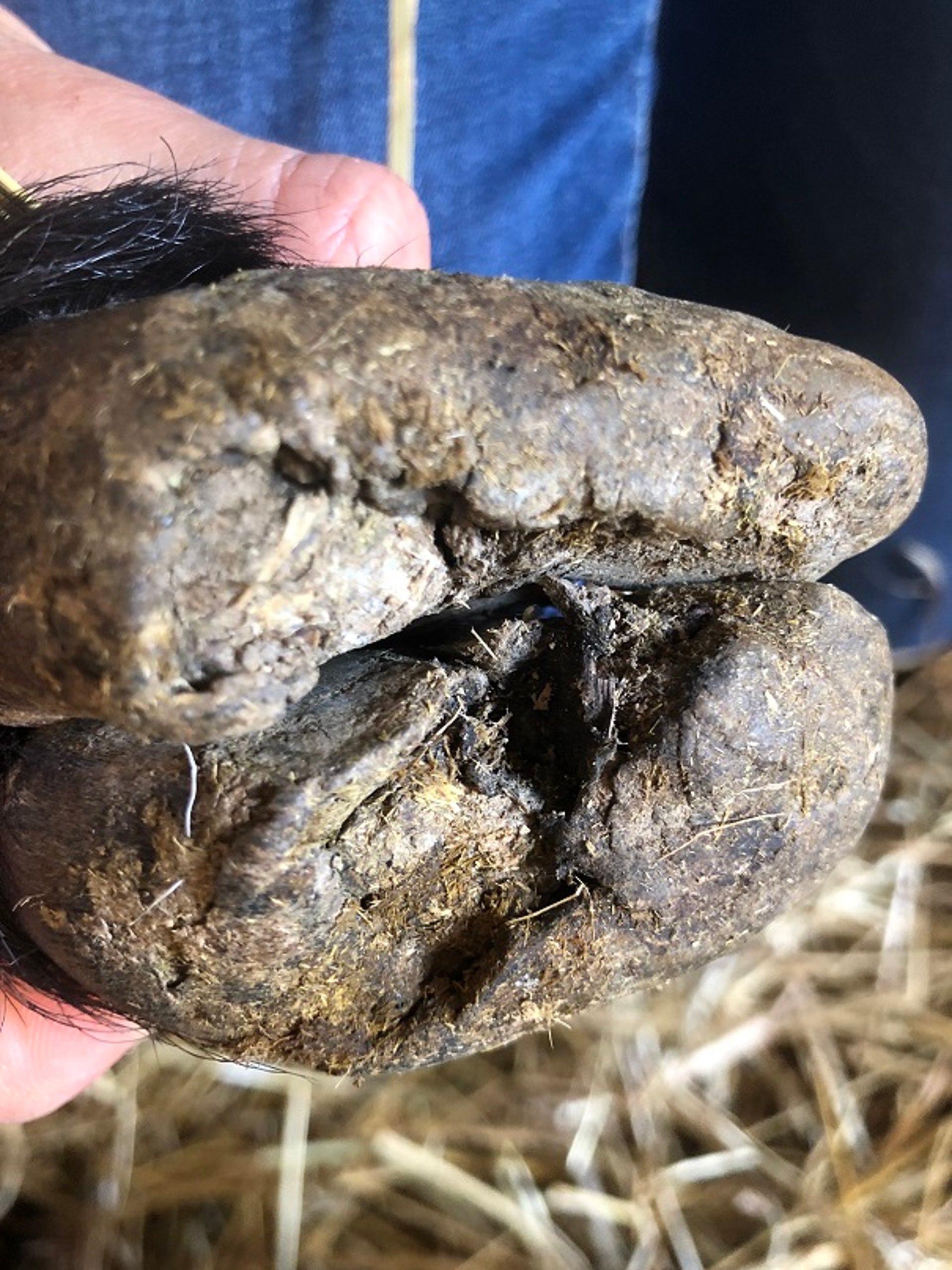
Deformed hoof wall and sole due to footrot infection in a ewe. No hoof trimming was performed during the treatment of this animal.
Courtesy of Dr. Philippa Gibbons.
In early cases, examination generally reveals nothing more than interdigital dermatitis. In slightly more advanced cases, in which the infection has begun to extend into the hoof matrix, there is slight detachment of the hoof wall at the interface of the interdigital skin and hoof. As the disease progresses, the separation of the horn spreads further under the heel and sole. Running one’s thumb between the wall and the underlying tissue in the interdigital space often detaches the wall from the heel and sole, revealing a white, slightly moist (but not purulent), odorous substance. Finally, the outer wall is affected. The horny hoof may eventually be attached only at the toe and the coronet on the lateral side of the foot.
The odor of the necrotic tissue is characteristic and helps to diagnose the disease. Myiasis is a common sequela during fly season; it may extend to the area of the sheep where the affected tissue comes into contact with the body when the sheep is recumbent. Clinical signs of disease persists until the environment dries or the sheep are treated. After apparent healing, D nodosus may remain hidden in small pockets within the foot, where it is detectable only by extensive trimming, becoming active again when moist conditions recur. These sheep are carriers and generally remain infected for life. Immunity to footrot after infection does not seem to occur, because relapses are common.
Diagnosis of Virulent Footrot in Sheep
Compatible clinical signs of lameness, interdigital dermatitis, deformed hoof horn, and foul odor
Culture or PCR assay of individual or pooled samples from interdigital swabs from an animal or herd, differentiating benign and virulent strains

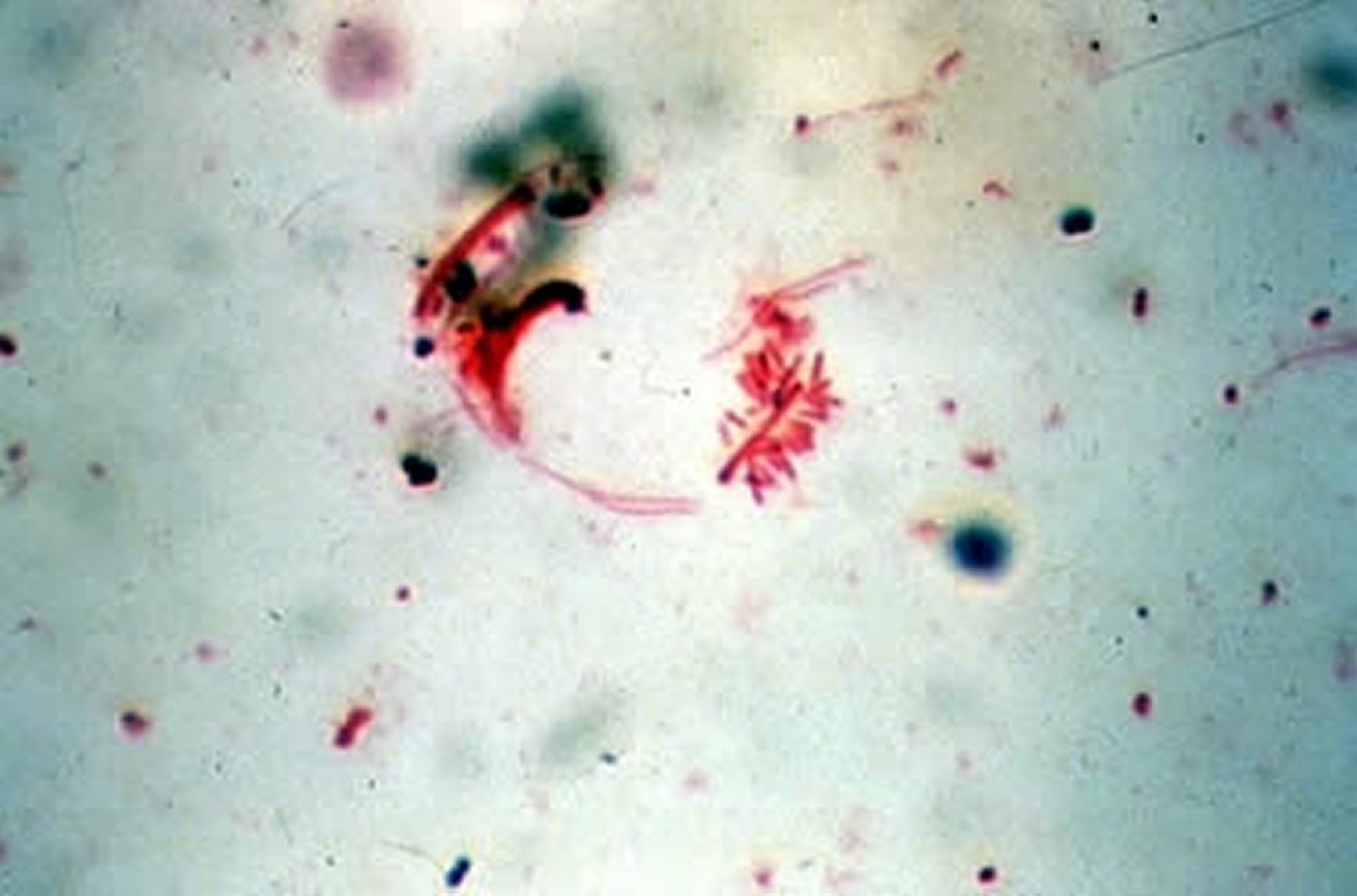
Photomicrograph of a swab smear taken from a footrot lesion in a sheep demonstrating Dichelobacter nodosus organisms. Note classic swollen ends of the organism that is sometimes (as here) characteristically surrounded by adherent, unidentified gram-negative rods. Gram stain; original magnification, 40×.
Courtesy of Dr. John Prescott.
In flocks with virulent footrot, underrunning and separation of the hard horn of the hoof of one or more limbs, complete with the characteristic odor, is diagnostic. If the problem is discovered early, when interdigital dermatitis is the only clinical sign, it should be assumed that the condition is an early stage of contagious footrot, and treatment should be initiated immediately.
Dichelobacter nodosus is a fastidious organism that requires anaerobic conditions for growth and can be challenging to isolate. Prompt plating onto specific media or transportation in appropriate media with anaerobic conditions is essential for success in recovering the organism. Dry or moistened sterile swab samples taken from the interdigital space of all four extremities can be used for culture or pooled for PCR assay and used for herd-level detection or monitoring. Virulence is determined by extracellular proteases and type IV fimbrae, and virulent strains can be identified via thermostability testing of the proteases or via real-time PCR assay for virulence genes. The protease genes aprB2 and aprV2 are associated with benign and virulent strains, respectively. PCR assay testing has been demonstrated to identify D nodosus infection before apparent clinical signs in herds, and it can be a useful eradication tool when available. Diagnostic testing is not available everywhere, and it varies based on region and prioritization of testing and elimination.
Treatment of Virulent Footrot in Sheep
Long-acting parenteral antimicrobials or soaking in a footbath
Excessive trimming of the hoof is not recommended
Treatment efforts may be directed toward temporary control of the disease or total eradication. At certain times, such as during a wet season, temporary control may be the only realistic goal.
Traditionally, treatment consisted of footbaths using antibacterial solutions after careful hoof trimming to remove all dead horn and expose infected tissue and bacteria to air. However, foot soaking for 30–60 minutes has been shown to be more effective even when trimming is not done. In fact, some research has shown that trimming may do more damage than good. A commonly used footbath solution is 10% wt/vol zinc sulfate with 0.2% vol/vol of laundry detergent containing nonionic surfactants such as sodium lauryl sulfate. Formaldehyde at 2%–5% and copper sulfate at 5% are also frequently used with success. Although guidelines for footbaths may vary, they should be at least 6 cm deep, with sheep spending at least 10 minutes in the solution and being allowed to dry in a clean area afterward. Aerosol sprays have been used in lieu of footbaths and include zinc sulfate, tincture of iodine, tetracycline, copper sulfate, formalin, chlorine bleach, and other disinfectants. However, sprays are not as effective as footbaths or soaking.
The advent of long-acting antimicrobials used in combination with topical treatments has improved recovery and reduced carrier animals. Parenteral administration of a long-acting oxytetracycline or a macrolide is an effective systemic treatment. In animals with lameness, NSAIDs for pain management should be considered. Sheep must be placed in a clean area (ie, one in which no sheep have been kept for at least 3 weeks) or in a completely dry lot after they are run through a footbath and administered the antimicrobial. Sheep will become reinfected as soon as the antimicrobial is cleared if returned to a contaminated environment. Treated sheep should be examined once a week to identify those not responding to treatment. Sheep that do not respond should be isolated and preferably culled. Dichelobacter nodosus is difficult to eradicate from animals that have relapsed numerous times. Furthermore, subclinical or relapsing cases take valuable time to handle, identify, and isolate, and they remain a source of infection for other animals.
Prevention and Control of Virulent Footrot in Sheep
Animals from unknown premises or auction houses should not be purchased. Any sheep to be added to the flock should be quarantined for several weeks to prevent the spread of footrot and other chronic diseases. During the quarantine period, the animal’s hoofs should be lightly trimmed and examined closely for pockets and other malformations that suggest a previous D nodosus infection, and testing for presence of D nodosus can be performed if available. Vehicles (eg, trucks or trailers) or facilities in which unknown or infected sheep have been held should be thoroughly cleaned and disinfected before placing uninfected sheep in them. If it is not possible to thoroughly disinfect transport vehicles, zinc sulfate can be liberally scattered over the floor to reduce viable bacteria.
Routine trimming serves as an important opportunity for transmission. When trimming is undertaken, the hoof trimmers should be cleaned with an antiseptic solution between uses, and personnel should change gloves between handling of each sheep.
Because the incubation period of footrot is ~14 days, footbaths at 10-day intervals will control spread of the organism in affected flocks during wet-condition periods. Footrot has been controlled by placing footbaths with 10% wt/vol zinc sulfate solution around water troughs, forcing sheep to walk through them and stand in to drink. Lame sheep should be separated for treatment and not returned to the flock until all evidence of footrot is gone.
Dichelobacter nodosus vaccines accelerate healing in affected sheep and aid in protecting unaffected sheep. They are recommended as an additional tool to control or eradicate the disease. However, their effectiveness depends on the strains causing the infection and those present in the vaccine. No vaccine contains all the various strains of D nodosus, and vaccination without other means of control will most likely select for strains not contained in the vaccine. Alum-precipitated vaccines require two doses 4–6 weeks apart to establish effective immunity, which persists for 2–3 months. Lesions heal within 4–6 weeks if immunity is established. Within 3 weeks after the initial dose, oil-emulsion vaccines induce an immune response that confers protection that may persist for 3–4 months. In endemic areas, revaccination is recommended at intervals of 3–6 months. Reaction to the vaccine is common, resulting in large granulomas and occasional abscesses. Vaccines for F necrophorum have not generally shown much benefit in either treatment or prevention. Availability of the vaccines varies with region, and vaccines are currently unavailable in the US.
Addition of zinc to trace mineral salt, reportedly effective in reducing hoof rot in cattle, has not been shown to be particularly helpful for sheep footrot. However, zinc is important for immunity and skin and hoof health. Providing it in a well-balanced trace mineral mix may be helpful in locations deficient in zinc.
Eradication of Virulent Footrot in Sheep
A successful eradication program requires planning, commitment, and an investment of time and money. Eradication can be achieved only by eliminating all cases of D nodosus infection in a flock and preventing its reintroduction. This may be done by replacing affected animals with footrot-free sheep or by rigorously treating all new infections and culling affected sheep that do not respond readily to treatment. Eradication is easiest when the environment is dry; at other times, treatments should be directed more toward control of transmission.
Affected sheep have historically been identified by close examination of all four extremities; however, PCR assay of interdigital swabs is available in some areas and has been shown to detect carrier animals before clinical signs are evident. Subclinical cases constitute a major problem and cause relapses in seemingly unaffected flocks. Other ruminants, such as goats, deer, or cattle, are potential sources of D nodosus. If they are in contact with the sheep, they should be considered in the eradication program.
In the first step of an eradication program, the hoofs all sheep should be trimmed and carefully examined. The flock should then be divided into affected and unaffected groups. Sheep with no visible lesions are walked through a footbath, isolated, and placed on clean, dry ground (ie, where no sheep have been kept for at least 3 weeks). This group may have some degree of genetic resistance, and identifying them in some way is recommended. Retaining offspring from this group could further help control the disease. This group should be administered an injection of long-acting tetracycline, and any lame sheep should be removed immediately.
The group of sheep with footrot lesions are culled, or after careful hoof trimming, the extremities are soaked for at least 30 minutes and the animals are treated with an antimicrobial and kept separated from the lesion-free group. This second group should undergo foot soaking once a week for a total of three times or be medicated with long-acting oxytetracycline at the time of soaking. At the end of this period, sheep should again be examined and hooves trimmed. Any sheep with an active case of footrot should be culled. This group must be monitored closely during the next wet period to detect any carriers, which are usually the first animals to show lameness. When no relapses have occurred for 1 month or longer, this group may be placed with the lesion-free group. However, returning a single active or subclinical case to the unaffected flock can negate the previous eradication efforts. As testing improves and becomes more available, interdigital swabs may play a more important role in the identification of carrier animals and subclinical cases, which could hasten eradication efforts.
Australia has implemented an effective eradication program involving many flocks. The program has three phases. The control phase is used during periods of active spread or to reduce the number infected. During this phase, vaccination, footbaths, and parenteral antimicrobials can all be used. The eradication phase must take place during the dry season and cannot begin until several weeks after the administration of all medications has been stopped and 10–12 weeks after vaccination. Footbaths and vaccination tend to mask the presence of infection. During this phase, extremities of every sheep are inspected every 3–4 weeks. Infected sheep can be treated with parenteral antimicrobials at the first inspection only. After this, infected sheep are culled at each inspection. This continues until there are two completely negative, consecutive flock examinations. In the surveillance phase, all lame sheep in the flock are examined immediately. If footrot is present, the flock goes back to phase 1 or 2 again.
Key Points
Footrot is a contagious disease that can cause major economic losses and impact animal welfare.
Diagnosis is made based on compatible clinical signs including lameness, interdigital inflammation, and deformed hoof walls.
Treatment involves parenteral antimicrobials, use of footbaths, or both.
Prevention and control depends on strict biosecurity, vaccination when available, and treatment or elimination of infected animals.
For More Information


