


Myotonic muscle disorders share the feature of delayed relaxation of muscle after mechanical stimulation or voluntary contraction due to abnormal muscle membrane conduction. Horses have three known forms of myotonia: myotonia congenita, myotonia dystrophica, and hyperkalemic periodic paralysis (HyPP).
Myotonia Congenita and Dystrophica
The initial clinical signs of myotonia in foals are well-developed musculature and mild pelvic limb stiffness. Bilateral bulging (dimpling) of the thigh and rump muscles is often obvious and gives the impression that the animal is very well developed. Percussion of affected muscles exacerbates the muscle dimpling below a large area of tight contraction that can persist for a minute or more with subsequent slow relaxation. Myotonia congenita usually does not show progression of clinical signs beyond 6–12 months of age, and muscle stiffness may improve with exercise. The cause has not been identified.
Foals with myotonia dystrophica show a progression of clinical signs in the first 1–2 years of life to include areas of muscle atrophy fibrosis and stiffness that worsens with exercise. This results in some areas with excessively large muscle development and other areas of atrophy. Retinal dysplasia, lenticular opacities, and gonadal hypoplasia have been noted in Quarter Horse, Appaloosa, and Italian-bred foals with myotonic dystrophy.

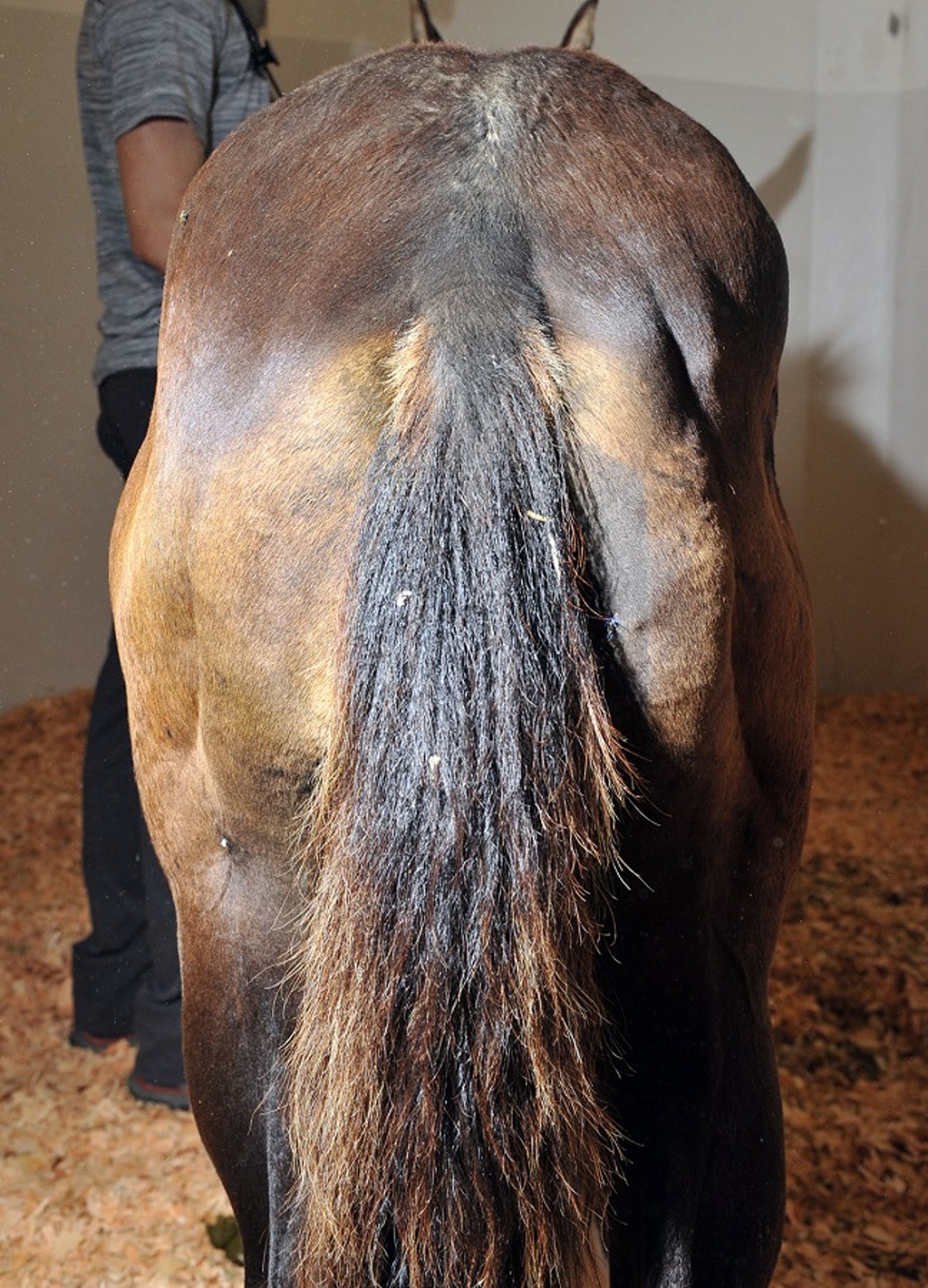
Courtesy of Dr. Stephanie Valberg.
A tentative diagnosis of myotonia can be made on the basis of age and clinical signs of stiff gait, muscle bulging, and prolonged contractions after muscle stimulation. Definitive diagnosis of myotonia requires electromyographic examination. Affected muscle manifests pathognomonic, crescendo-decrescendo, high-frequency repetitive bursts with a characteristic sound, often likened to a dive bomber. Myotonia dystrophica shows dystrophic changes in muscle biopsies not present in myotonia congenita. Dystrophic changes include ringed fibers, numerous centrally displaced myonuclei, sarcoplasmic masses, and an increase in endomysial and perimysial connective tissue. Fiber type grouping and atrophy of both type I and type II muscle fibers may be present.
Horses with myotonia congenita or dystrophica are rarely serviceable, and euthanasia is usually warranted in dystrophic foals because of the severity of stiffness and atrophy that develop over time. Conclusive evidence regarding the genetic basis of this disorder in horses is still not available.
Hyperkalemic Periodic Paralysis
Hyperkalemic periodic paralysis is an autosomal dominant trait affecting Quarter Horses, American Paint horses, Appaloosas, and Quarter Horse crossbreeds worldwide. The point mutation in the voltage-dependent skeletal muscle sodium channel alpha subunit occurs in ~4% of Quarter Horses; however, this percentage is much higher in halter and pleasure horse performance types.
Horses may be subclinically affected. Clinical signs of intermittent muscle fasciculations and weakness are first identified in foals to horses 3 years of age. Homozygous horses are often more severely affected and may be identified at a younger age than heterozygotes. A brief period of myotonia often occurs initially, with some horses showing facial myotonia and prolapse of the third eyelid. Muscle fasciculations, beginning on the flanks, neck, and shoulders may become more generalized. Although most horses remain standing during mild attacks, weakness with swaying, staggering, dogsitting, or recumbency may occur, with severe attacks lasting 15–60 minutes or longer. Heart and respiratory rates may be increased; however, horses remain relatively bright and alert. Respiratory distress occurs in some horses as a result of upper respiratory muscle paralysis. Once episodes subside, horses regain their footing and appear normal with absent or minimal gait abnormalities. Young horses that are homozygous for the HyPP trait may have respiratory stridor and periodic obstruction of the upper respiratory tract that can be fatal.
Common factors that trigger episodes include sudden dietary changes or ingestion of diets rich in potassium (>1.1%), such as those containing alfalfa hay, molasses, electrolyte supplements, and kelp-based supplements. Food withholding, anesthesia or heavy sedation, trailer rides, and stress may also precipitate clinical signs. The onset of clinical signs, however, is often unpredictable. Exercise per se does not appear to stimulate clinical signs; serum CK activity shows no or minimal increases during episodic fasciculations and weakness.
Descent from the stallion Impressive in a horse with episodic muscle tremors is strongly suggestive of HyPP. Hyperkalemia (6–9 mEq/L), hemoconcentration, and hyponatremia occur during clinical episodes; however, a definitive diagnosis requires DNA testing of mane or tail hair. Electromyographic examination of affected horses between attacks reveals abnormal fibrillation potentials and complex repetitive discharges, with occasional myotonic potentials and trains of doublets between episodes. Differential diagnoses for hyperkalemia include delay before serum centrifugation, hemolysis, chronic renal failure, and severe rhabdomyolysis.
Many horses recover spontaneously from HyPP episodes. Owners may abort early mild episodes using low-grade exercise or feeding grain or corn syrup to stimulate insulin-mediated movement of potassium across cell membranes. In severe cases, IV administration of calcium gluconate (0.2–0.4 mL/kg of a 23% solution diluted in 1 L of 5% dextrose) or dextrose (6 mL/kg of a 5% solution), alone or combined with sodium bicarbonate (1–2 mEq/kg), often provides immediate improvement. With severe respiratory obstruction, a tracheostomy may be necessary. Acute death is common, especially in homozygous animals.
Prevention requires decreasing dietary potassium to 0.6%–1.1% total potassium concentration and increasing renal losses of potassium. High-potassium feeds such as alfalfa hay, first cutting hay, brome hay, sugar molasses, and beet molasses should be avoided. Optimally, later cuts of timothy or Bermuda grass hay; grains such as oats, corn, wheat, and barley; and beet pulp should be fed in small meals several times a day. Regular exercise or frequent access to a large paddock or yard are also beneficial. Pasture is ideal for horses with HyPP because the high water content of pasture grass makes it unlikely that horses will consume large amounts of potassium in a short period of time. Complete feeds for horses with HyPP are commercially available.
For horses with recurrent episodes even with dietary alterations, acetazolamide (2–4 mg/kg, PO, every 8–12 hours) or hydrochlorothiazide (0.5–1 mg/kg, PO, every 12 hours) may be helpful. Breed registries and other associations have restrictions on the use of these drugs during competitions. Some horses have both HyPP and polysaccharide storage myopathy (PSSM) ( See also Chronic Exertional Rhabdomyolysis), which may result in an episode of rhabdomyolysis during a hyperkalemic paralytic event with subsequent increased serum CK activity and prolonged recumbency.
There appear to be other idiopathic causes of muscle fasciculations in Quarter Horses that have clinical signs similar to HyPP yet test negative for the HyPP mutation. Reports of elevations in serum potassium during episodes of fasciculations are variably normal or slightly elevated. The cause of these fasciculations is unknown and treatment approaches are currently similar to those for HyPP.
For More Information
Also see pet health content regarding muscle disorders in horses.


