Also see Management of Reproduction: Horses.
The most common noninfectious cause of abortion in horses is twinning. Most abortions related to twinning occur at 8–9 months of gestation and can be preceded by premature lactation. Placental insufficiency ultimately causes the abortion of twins (see ).


Placenta from a mare that aborted twins. Note the avillous area of contact between the two chorioallantoic membranes.
Courtesy of Dr. Ahmed Tibary.
Equids are predisposed to umbilical cord torsion because they have longer umbilical cords (> 85 cm) than other species have. Healthy equine umbilical cords can be twisted several times; thus, umbilical cord twists are not always pathological in horses. Diagnosis of abortion due to cord torsion requires evidence of localized swelling and/or hemorrhage and the presence of more than eight twists (see ).


Umbilical cord torsion with cord edema and hemorrhage in a horse. The umbilicus has more than 12 twists.
Courtesy of Dr. Ahmed Tibary.
Signs of fetal circulatory disturbances, such as subcutaneous edema, a swollen, soft liver, and microscopic mineralization of placental vessels, are also signs of umbilical cord obstruction in horses.
Various congenital fetal abnormalities have been reported in cases of noninfectious equid abortions.
Infectious causes of equid abortion include viral diseases (eg, equine rhinopneumonitis, equine viral arteritis), as well as bacterial and fungal infections.
Fescue Grass Toxicosis as a Cause of Abortion in Horses
Equids that ingest tall fescue grass infected by the endophyte fungus Acremonium coenophialum can experience prolonged gestation, agalactia, edema and premature separation of the placenta, perinatal death, and retained placenta. Abortion in these cases is rare but can occur in the last 2 months of pregnancy as a result of severe edema and premature placental separation.
Diagnosis: In abortions due to fescue grass toxicosis, the placenta is thickened and edematous and does not rupture normally at the cervical star. The chorioallantois precedes the foal through the birth canal instead of remaining attached to the uterus (red bag in the ), resulting in anoxia and death of the fetus. The vasoconstrictive properties of alkaloids produced by A coenophialum appear to be the cause.


Premature placental detachment in a mare with ascending placentitis. Note the velvety aspect of the chorionic surface of the detached placenta.
Courtesy of Dr. Ahmed Tibary.
Treatment and prevention: The way to treat fescue grass toxicosis in horses is prevention. Broodmares should not graze on endophyte-infected fescue pastures. However, if preventing such ingestion is not possible, mares should be treated with domperidone (which prevents ergovaline from inhibiting prolactin release and, hence, prevents agalactia), perphenazine, or reserpine.
Mare Reproductive Loss Syndrome as a Cause of Abortion in Horses
"Mare reproductive loss syndrome" (MRLS) is the term given to equine abortions associated with eastern tent caterpillars (Malacosoma americanum).
In spring of 2001, equine stud farms in Kentucky and neighboring states the US experienced an explosive outbreak of early and late-term abortions, stillbirths, and weak foals that died within a few days (1). Simultaneously, there was a large increase in fibrinous pericarditis and unilateral uveitis in horses of all ages and both sexes (1). Together, these conditions became known as MRLS. Analysis of records showed that MRLS had occurred in the area in earlier years (2).
MRLS has since been diagnosed in other states, including New York and Florida. An abortion storm with similar clinical signs and risk factors has also been reported in Australia (1).
Most equine abortions occur at 40–80 days of gestation; some, as late as 140 days. A few affected mares present colic, fever, and/or purulent vulvar discharge, but most remain clinically normal.
Typically, the first sign of MRLS is abortion or ultrasonographic imaging of a fetus dead in utero. Late-term losses generally occur at 10 months of gestation to term, and the mare usually does not display signs of impending parturition.
Diagnosis: Most fetuses aborted as a result of MRLS are expelled within 2 days to 2 weeks after dying and are autolytic. Neutrophilic placentitis and metritis are usually present. Most mares that are bred again during the same breeding season do not become pregnant; however, conception is usually normal during the next breeding season.
Pathological features of MRLS include the presence of amnionitis and funisitis (inflammation of the umbilical cord involving the umbilical vessels and caused by an intrauterine infection) (see ), with only the amniotic portion of the umbilical cord affected. The placenta and umbilical cord are thickened, edematous, and discolored light brown to yellow. Neutrophilic inflammation of the umbilical cord and placenta is usually present; and neutrophilic funisitis is characteristic of the syndrome.

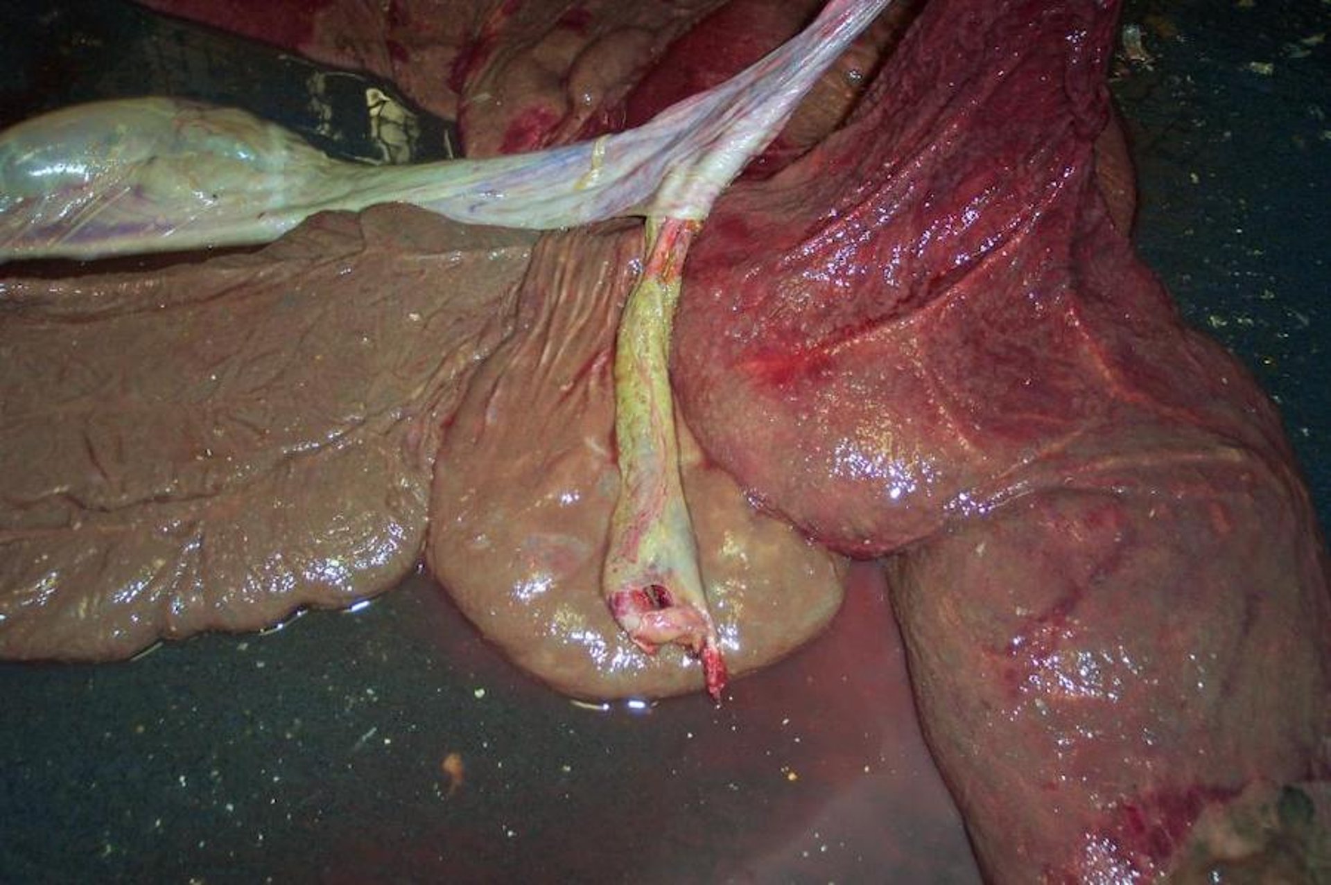
Generalized placentitis and funisitis in a mare. Note the thickened chorionic surface of the placenta with exudate. The umbilical cord is also thickened.
Courtesy of Dr. Ahmed Tibary.
MRLS was initially suspected to have a bacterial etiology; however, no definitive diagnosis emerged. Exposure to eastern tent caterpillars in pastures was considered the main cause.
The setae on the caterpillar's exoskeleton have been proposed as a transporting mechanism of enteric bacteria (3). Setae dislodged from ingested processionary caterpillars migrate through the wall of the cecum and/or colon, pass through the uterine wall, and establish infection via transported gut bacteria.
Another theory suggests that a toxin produced by the caterpillars after they consume black cherry tree leaves causes the inflammatory conditions and abortions that characterize MRLS.
The pathogenesis of MRLS remains unconfirmed. The role of a toxin, rather than setae, fits well with the environmental evidence and the number of cases at any given time.
Prevention and control: MRLS prevention consists of pasture management to control the number of eastern tent caterpillars, along with other procedures to prevent the exposure of pregnant mares to eastern tent caterpillars.
Bacterial Abortion in Horses
Bacterial placentitis is by far the most commonly diagnosed cause of abortion in many horse breeding areas. Placentitis is an important cause of equine late-term abortion, premature delivery, and neonatal death.
Pearls & Pitfalls
|
Except for Leptospira spp and nocardioform infections, most cases of bacterial placentitis are ascending.
Ascending Placentitis in Horses
Ascending placentitis is characterized by premature udder development, increased combined uteroplacental thickness at the level of the cervical star (see ), and, in some cases, mucopurulent vaginal discharge.


Ascending placentitis in a mare. Note the congested and thick cervical star area (arrows).
Courtesy of Dr. Ahmed Tibary.
If placentitis is not treated, placental function is compromised and placental separation ensues, resulting in fetal death and expulsion. In chronic placentitis, the fetus might show delayed development.
Streptococcus equi zooepidemicus, Escherichia coli, Pseudomonas aeruginosa, Enterobacter spp, and Klebsiella pneumoniae are the most frequent bacteria isolated from vaginal discharge, uterus, placenta, and fetal gastric contents in horses.
Other bacteria have also been reported to cause ascending placentitis in mares, including Streptococcus dysgalactiae equisimilis, Enterobacter agglomerans, alpha-hemolytic streptococci, Staphylococcus aureus, and Actinobacillus spp. Examination of the placenta shows an edematous and thickened chorioallantois with fibrinonecrotic exudate at the level of the cervical star.
Diagnosis: After equine abortion or birth, placentitis can be diagnosed by gross inspection and culture of the placenta. To detect placentitis during pregnancy, mares need to be monitored at intervals throughout gestation. Because there are generally no external clinical signs of placentitis, intentional surveillance is required, such as assessing the combined thickness of the uterus and placenta (CTUP) by transrectal ultrasonography.
Treatment: Although the etiological agent of a horse abortion is unlikely to be known during pregnancy, treatment with antimicrobials and altrenogest (a synthetic progestin hormone that mimics natural progesterone) can be undertaken to try to eliminate infection in the placenta and preserve a viable pregnancy.
Prevention and control: Pregnant mares should be kept in clean environments and, in the case of leptospirosis, should not be exposed to the urine of other animals or stagnant water runoff. Some veterinarians perform a Caslick procedure (vulvoplasty) to limit the potential for external contamination of the vagina and mitigate the risk of ascending placentitis.
Leptospiral Placentitis in Horses
Placentitis caused by Leptospira spp is characterized by diffuse lesions secondary to hematogenous spread. Leptospiral placentitis as a cause of abortion seems to be on the rise in Kentucky, Northern Ireland, England, and South America.
Several Leptospira serovars—eg, L interrogans Pomona, Pomona (type kennewicki), and Bratislava; L kirschneri Grippotyphosa; and L borgpetersenii Hardjo (type hardjo-prajitno)—have been isolated from aborted equine fetuses. In North America, the most common isolate is serovar Pomona (type kennewicki), which is carried by several wildlife species, including the striped skunk, raccoon, whitetail deer, and opossum.
Most Leptospira-induced abortions occur between 6 and 9 months of gestation. The placenta is thick, heavy, edematous, hemorrhagic, and occasionally covered with a brown mucoid material on the chorionic surface. Funisitis has also been described in leptospiral abortion. The fetus can have mild to moderate icterus and liver enlargement, and fetal histopathological lesions can include various degrees of nephritis and hepatitis.
Diagnosis: Leptospirosis is diagnosed from results of fluorescent antibody staining of placenta or fetal kidney, liver, or lung and on fetal serological testing.
Prevention: A vaccine is available in the US but has not been shown to protect against abortion.
Leptospirosis is zoonotic; humans can contract the disease from contact with infected urine or tissues from horses.
Treatment, prevention, and control: Treatment of horses with leptospirosis should be initiated promptly, with appropriate antimicrobial therapy and supportive care. Antimicrobials are commonly used to decrease clinical disease and urinary shedding of Leptospira spp; however, evidence regarding the ability of treatment to eliminate renal infection or prevent subsequent reproductive losses is limited.
Mares that have aborted and horses recently infected can shed Leptospira in urine for up to 3 months and should be managed with appropriate biosecurity measures (4).
Nocardioform Placentitis in Horses
Nocardioform placentitis is a distinct type of equine placentitis first described in the US in the mid 1980s (5). Etiological agents implicated in mares with nocardioform placentitis consist of various groups of gram-positive, filamentous, branching bacteria, including Nocardia spp, Rhodococcus rubropertinctus, Amycolatopsis spp, and Crossiella equi.
Nocardioform placentitis can result in abortion, stillbirth, or birth of weak foals at term. Some mares exhibit premature mammary gland development and lactation before abortion.
Infection of the placenta in horses is generally thought to be a sequela of the hematogenous spread of microorganisms from a primary port of entry. The lesion is extensive and severe exudative, mucopurulent, and necrotizing placentitis, frequently located at the base of the uterine horns or at the junction between the body and horns of the placenta. The affected area is thickened, and its chorionic surface is covered with brown, necrotic, mucopurulent exudate and dotted with white or yellow granular structures.
Underneath this mucoid material, the chorionic surface is reddish white, mottled, and roughened. Villous necrosis and adenomatous hyperplasia of the allantoic epithelium, and hyperplasia with or without squamous metaplasia of the chorionic epithelium frequently occur. The fetus is often severely underdeveloped as a result of placental insufficiency and shows no remarkable gross or histopathological lesions.
Placentitis Due to Potomac Horse Fever
Potomac horse fever, caused by Neorickettsia risticii, can be followed by abortion in mid- to late gestation. There is placentitis, and the placenta is often retained.
Diagnosis:N risticii has been isolated from fetal lymphoid tissues after abortion. Histologically, there is fetal colitis. Identification of this colitis provides a presumptive diagnosis.
Prevention: There is a vaccine for Potomac horse fever; however, its efficacy in preventing abortion is unknown.
Equine Mycotic Placentitis as a Cause of Abortion
Mycotic placentitis in horses is due to an ascending infection that causes a thickened chorioallantois with variable exudate. Causative agents include Aspergillus spp, Mucor spp, Candida spp, Histoplasma capsulatum, Coccidoides spp, and Cryptococcus neoformans.
Diagnosis: Fetuses aborted in late gestation as a result of mycotic placentitis might be fresh, with evidence of growth delay. A pale, enlarged liver or dermatitis might be found. Hyphae are found in the placenta, liver, lungs, or gastric contents.
Equine Rhinopneumonitis (Equine Herpesvirus 1 Infection) as a Cause of Abortion
Equine rhinopneumonitis—specifically, equine herpesvirus 1 (EHV-1) infection—is the most important viral cause of abortion in horses; EHV-4 has also been isolated from some cases.
Pearls & Pitfalls
|
The principal mode of EHV transmission from horse to horse is by direct contact through nasal secretions, reproductive tract discharge, placenta, or the aborted fetus. Short-distance airborne transmission of infection is also possible.
Abortion due to EHV-1 infection usually occurs after 7 months of gestation and is not preceded by maternal illness. The placenta can be edematous or normal. Gross fetal lesions include subcutaneous edema, jaundice, increased volume of thoracic fluid, and an enlarged liver with yellow-white lesions approximately 1 mm in diameter. Histologically, these lesions are areas of necrosis containing intranuclear inclusions. Inclusion bodies are also found in necrotic lymphoid tissues. There is often necrotizing bronchiolitis.
Diagnosis: Diagnosis of EHV-1 infection is based on fluorescent antibody staining, PCR assay, or virus isolation from fetal tissues.
Prevention: Prevention of equine rhinopneumonitis is based on vaccinating against EHV-1 at 5, 7, and 9 months of gestation, as well as preventing the exposure of pregnant mares to horses attending shows or other equine events. Abortion can occur despite regular vaccination.
Equine Viral Arteritis as a Cause of Abortion
Abortion can occur 6–29 days after the appearance of initial clinical signs of equine viral arteritis (EVA). Abortion rates can approach 60% in a naive population as the result of direct impairment of placental function and severe fetal infection (6). Arteritis might be found in the fetal myocardium or placenta; usually, however, there are no fetal lesions. Stallions can be persistently infected, and EVA can be transmitted venereally (via natural cover or insemination with shipped cooled or frozen semen) or by aerosol.
Diagnosis: Diagnosis of EVA is based on a history of the disease shortly before abortion, virus isolation or PCR assay of placenta and/or fetal tissues, or seroconversion of the dam.
Prevention: EVA can be prevented by management to minimize viral transmission in breeding populations and to prevent the development of carrier stallions.
In the US, a licensed modified live virus vaccine against EVA is available for use in nonpregnant mares. Antibody titers resulting from vaccination and natural infection cannot be differentiated, and the serological status of horses can affect their import status. Therefore, the serological status of breeding horses should be determined before vaccination, and all subsequent vaccinations should be recorded.
For More Information
Burns SJ, Westerman AG, Harrison LR. Environmental influences on mare reproductive loss syndrome: do they fit with a toxin as the causative agent?J Equine Vet Sci. 2022;114:104001.
Canisso I, Ball BA, Erol E, Squires EL, Troedsson MHT. Comprehensive review on equine placentitis. Proc Ann Conv Am Assoc Equine Practit. 2015;61:490-509.
Sones JL. Placentitis in mares. Equine Reproduction Laboratory, Colorado State University. January 29, 2025. Accessed August 11, 2026.
Also see pet owner content regarding abortion in horses.
References
Sebastian MM, Bernard WV, Riddle TW, Latimer CR, Fitzgerald TD, Harrison LR. 2008. Review paper: mare reproductive loss syndrome. Vet Pathol. 45(5):710-719. doi:10.1354/vp.45-5-710
Powell DG,Troppman A, Tobin T, eds. Proceedings of the first workshop on mare reproductive loss syndrome. Kentucky Agricultural Experiment Station, College of Agriculture, University of Kentucky; 2003. https://publications.mgcafe.uky.edu/files/sr2003-1.pdf
Todhunter KH, Cawdell-Smith AJ, Bryden WL, Perkins NR, Begg AP. Processionary caterpillar setae and equine fetal loss: 1. Histopathology of experimentally exposed pregnant mares. Vet Pathol. 2014;51(6):1117-1130. doi:10.1177/0300985813516638
Divers TJ, DeNotta S. AAEP infectious disease guidelines: leptospirosis. American Association of Equine Practitioners; 2022. https://aaep.org/wp-content/uploads/2024/02/Leptospirosis_DZ_Guidelines_IDC_2021.pdf
Hong CB, Donahue JM, Giles RC Jr, et al. Etiology and pathology of equine placentitis. J Vet Diagn Invest. 1993;5(1):56-63. doi:10.1177/104063879300500113
Léon A, Richard E, Fortier G, et al. Overview of the causes of abortion in horses, their follow-up and management. Reprod Domest Anim. 2023;58(suppl 2):93-101. doi:10.1111/rda.14406