

Acute bovine pulmonary emphysema and edema (ABPEE) is one of the more common causes of acute respiratory distress in adult cattle on pasture. It usually occurs in the fall, 5–10 days after a change to a better, often lush, pasture. It is a disease involving groups of cattle; while a majority of cattle in a given pasture group are often somewhat affected, only a small minority will develop severe respiratory distress. There is no effective treatment; strategic pasture use is important for prevention.
Acute bovine pulmonary emphysema and edema is a respiratory distress syndrome affecting groups of cattle after movement onto lush pasture. Clinical signs result from a pneumotoxin produced by ruminal microorganisms. The clinical course is characterized by self-limiting signs of mild-moderate dyspnea and minimal coughing in most animals, but severe respiratory distress and sudden death can occur in some animals. Once exposed to the offending pasture, moving animals will not prevent the development of clinical signs. Interstitial pneumonia represents a group of respiratory diseases characterized by an acute onset of severe respiratory distress and a combination of lung lesions that include pulmonary edema and congestion, interstitial emphysema, alveolar epithelialization, and hyaline membrane formation.
Etiology of Acute Bovine Pulmonary Emphysema and Edema
Metabolites of the naturally occurring amino acid l-tryptophan probably are responsible for many outbreaks of acute bovine pulmonary emphysema and edema. In the rumen, l-tryptophan is degraded to indoleacetic acid, which can be converted to 3-methylindole by some ruminal microorganisms. Then 3-methylindole is absorbed into the bloodstream and is the source of the pneumotoxicity after metabolism by the mixed function oxidase system, which is very active in the lungs. Apparently, the level of l-tryptophan in crops is most likely to be high in lush, rapidly growing pastures, particularly (but not exclusively) in the fall.
Clinical Findings of Acute Bovine Pulmonary Emphysema and Edema
Acute bovine pulmonary emphysema and edema is most common in adult beef cows but may occur in either sex and in dairy or beef cattle under similar management conditions. Nursing calves are unaffected. Outbreaks usually develop within 5–10 days of a change to better grazing and rarely occur in animals that have been on a field > 3 weeks.
Mild cases may go unnoticed. Cattle are subdued but still alert; there is tachypnea and hyperpnea, but auscultation is usually unrewarding. Such cattle usually recover spontaneously within days. Severely affected cattle show extensive respiratory distress with mouth breathing, extension of the tongue, and drooling. A loud expiratory grunt is common; however, coughing is unusual. In the early stages, auscultation reveals surprisingly soft respiratory sounds. Mild exercise increases dyspnea and may precipitate death.
If death does not occur, the animals improve dramatically and resume eating by the third day. At this stage, auscultation reveals harsh respiratory sounds and, in some animals, dorsal (emphysematous) crackles. Some cattle have subcutaneous emphysema extending along the back from the withers. Full clinical recovery may require 3 weeks.
Lesions on Post-Mortem Examination
In affected cattle that have died or been slaughtered in extremis, the lungs are heavy and do not collapse normally. They are widely affected, with various degrees of firmness; there is extensive edema and emphysema, often with the formation of large, air-filled bullae in interlobular and subpleural regions. Submucosal hemorrhages are often present on the larynx and in the trachea and larger bronchi. Histologically, the lesion is characterized by congestion, alveolar edema, hyaline membrane formation, and areas of early alveolar epithelial hyperplasia of type II pneumocytes; occasionally, areas of bronchiolar necrosis may be found. The emphysema is often dramatic and is limited to interstitial fascia, where it is accompanied by edema.
In animals culled after 3 days of illness, the lungs are still heavy and do not collapse normally. They are pinkish gray and of increased firmness, but edema and emphysema are no longer evident. Histologically, the lungs lesions on Day 3 have transitioned to widespread alveolar epithelial hyperplasia characteristic of a diffuse, acute, proliferative alveolitis is evident.
Diagnosis of Acute Bovine Pulmonary Emphysema and Edema
Recent pasture movement
Acute respiratory distress
Necropsy

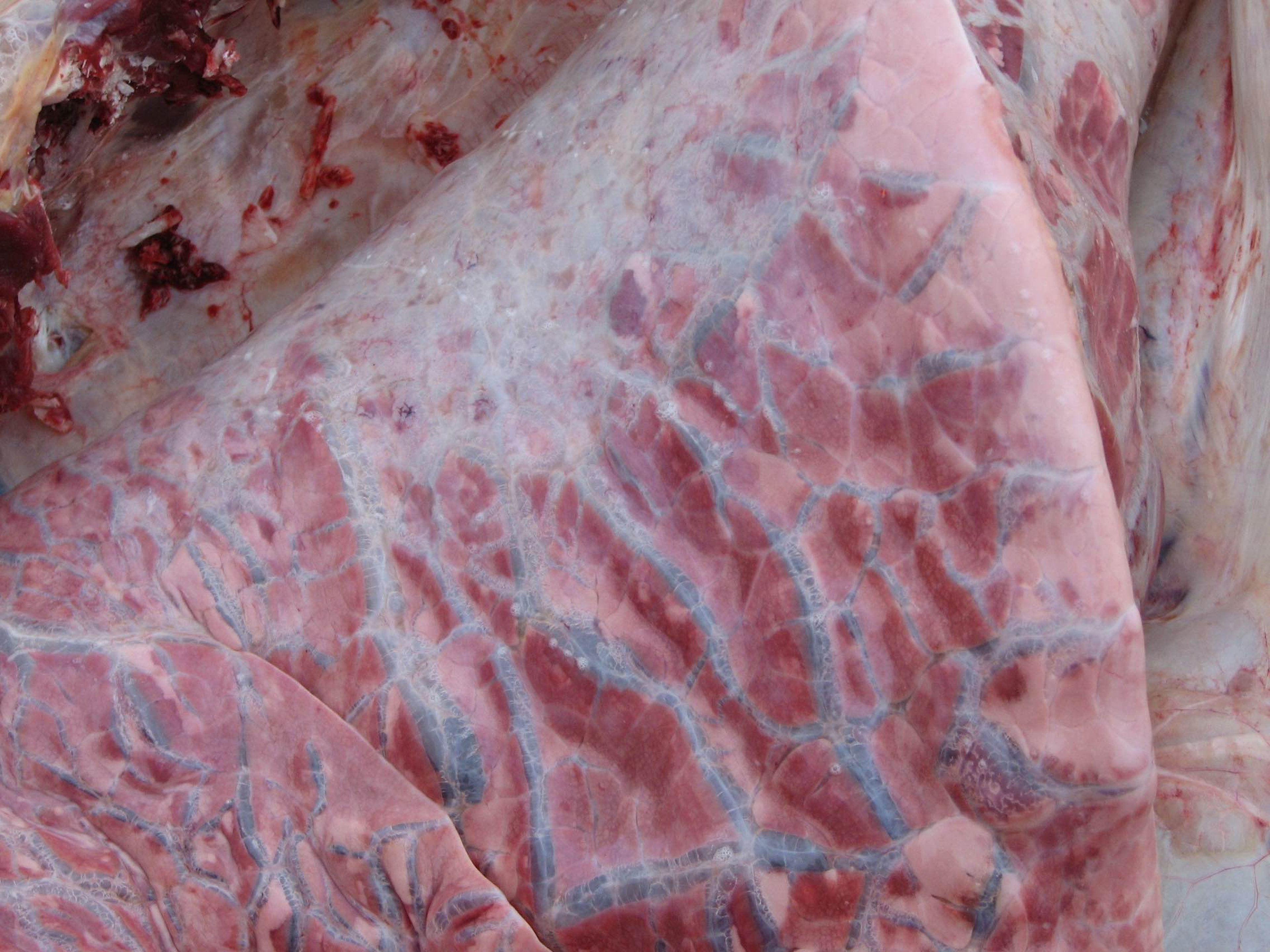
Extensive emphysema and edema associated with acute bovine pulmonary emphysema and edema.
Courtesy of Dr. John Campbell.
Diagnosis of acute bovine pulmonary emphysema and edema is based on history of recent pasture movement for the herd, clinical signs such as acute onset of severe respiratory distress in adult cattle, and pathologic lesions. Because the syndrome is not specific with regard to cause, evidence must be obtained from history of management factors such as change in pasture. Often, cases result in sudden death. Necropsy may reveal lesions of extensive edema. Emphysema may be confirmed histologically.
Treatment of Acute Bovine Pulmonary Emphysema and Edema
No effective treatment
No treatment has been identified for ABPEE. Severely affected animals with acute bovine pulmonary emphysema and edema have so little pulmonary reserve that any driving or handling must be done with caution to prevent immediate death. Moving the cattle is unlikely to prevent the development of clinical signs, once they have already been exposed to the offending pasture.
Control of Acute Bovine Pulmonary Emphysema and Edema
One approach to control of acute bovine pulmonary emphysema and edema is pasture use management, including the following options:
Feeding hay before turnout on pasture and limiting exposure time on suspect pastures
Limiting grazing time and gradually increasing exposure to the pasture over time
Using pastures before they become lush
Delaying use of lush pastures until after a hard frost
Initially grazing pastures with less susceptible stock (cattle < 15 months old or sheep)
Using strip grazing
A medical approach to control involves feeding monensin or lasalocid, which inhibit the bacteria that convert l-tryptophan to 3-methylindole. Treatment with monensin can be started 1 day before introduction to pasture, whereas lasalocid requires a 6-day pretreatment period. These drugs are of no benefit after onset of clinical signs.
Key Points
ABPEE can result in sudden onset of severe respiratory distress or sudden death in adult pastured cattle.
Etiology is associated with movement from dry to lush pasture with high L-tryptophan content; ruminal microorganisms convert this to a pneumotoxic substance (3-methylindole).
No treatment is effective for affected individuals.
Removal from pasture may not prevent new cases.


