

Ovine pulmonary adenocarcinoma is a pulmonary neoplasm due to an infectious virus that causes progressive respiratory distress and weight loss. Diagnosis is based on compatible clinical signs, ultrasonographic evaluation, or necropsy. No treatment is available, so infected sheep should be culled to limit transmission to herdmates.
Ovine pulmonary adenocarcinoma (OPA) is an infectious, viral, neoplastic disease of the lungs of sheep and, rarely, of goats. It has been reported in Europe, Asia, Africa, South America, and North America.
Etiology of Ovine Pulmonary Adenocarcinoma
OPA is an infectious neoplastic lung disease resulting from infection by a beta retrovirus called jaagsiekte sheep retrovirus (JSRV). The virus replicates predominantly in tumor cells, is released into the airways, and is found in respiratory secretions. Transmission of JSRV occurs predominantly through the aerosol route by inhalation of infected respiratory secretions; it may also be transmitted via colostrum and milk.
Clinical Findings of Ovine Pulmonary Adenocarcinoma
The period of incubation for OPA after natural infection extends over months; clinical signs generally become evident when sheep are 2–4 years old. However, disease may occur in lambs 8–12 months old that are progeny of infected dams. The tumors produce clinical signs when they become sufficiently large or numerous enough to interfere with respiration.
Signs in affected sheep include weight loss and increasing respiratory distress and panting. Crackles are audible over a much larger area than the distribution of OPA lesions determined ultrasonographically. Coughing is not prominent, and infected animals are afebrile unless secondary infection develops.
During the advanced stages of disease, the tumor mass may occupy up to 60% of lung parenchyma. Clinical signs end in death after many months; sometimes, however, secondary pasteurellosis causes death within 1–2 days.
Lesions

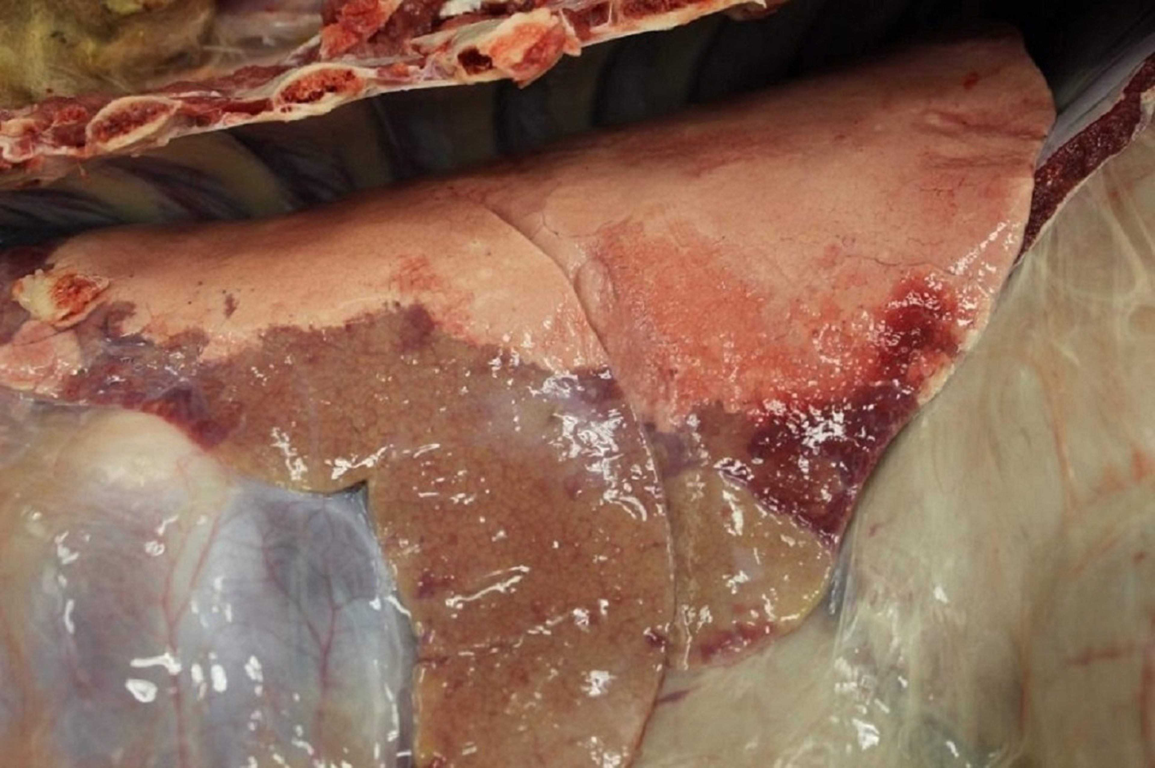
This photograph shows lung tissue affected by ovine pulmonary adenocarcinoma. The lesions are firm, red-brown, flat, and sharply demarcated in the cranioventral lung.
Courtesy of Dr. Philip Scott.
OPA tumors are confined to the lungs and, rarely, the associated lymph nodes. They vary from small nodules to extensive solid areas that involve the ventral parts of one or more lobes and are firm, gray, flat, and sharply demarcated. Copious amounts of white, frothy fluid are present in the airways. Histopathologic changes are due to uncontrolled proliferation of columnar type II pneumocytes and similar cells in the bronchioles (Clara cells).
Diagnosis of Ovine Pulmonary Adenocarcinoma

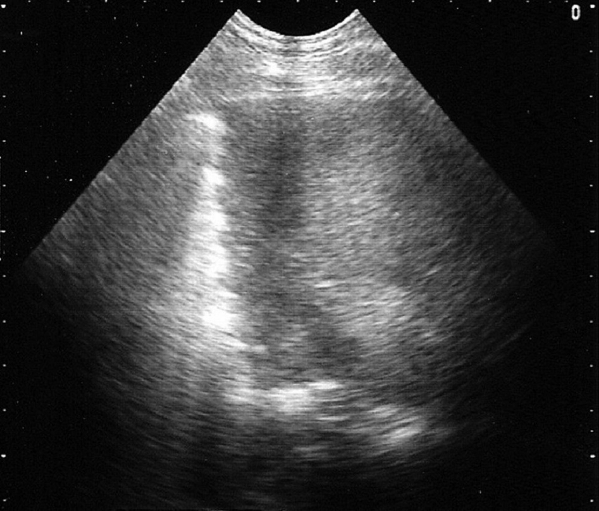
Ultrasonographic image of the lung field of a sheep with OPA. The 5-MHz probe is at the top of the image, dorsal to the left, centimeter markers along the right margin. Abrupt loss of the hyperechoic line represents the visceral pleura replaced by a uniform hypoechoic area that depicts the tumor mass.
Courtesy of Dr. Philip Scott.
Compatible clinical signs and wheelbarrow test
Ultrasonographic evaluation
Chronic weight loss, dyspnea, crackles, and copious amounts of serous nasal discharge from accumulated lung fluid in an adult sheep that is afebrile are highly suggestive clinical signs of OPA.
During the advanced stages of disease, clear, frothy fluid may flow freely from both nostrils when the sheep’s head is lowered during feeding; the quantity of this fluid may exceed 50 mL if the hindquarters are raised when the head is simultaneously lowered (colloquially referred to as the wheelbarrow test). The wheelbarrow test causes affected sheep considerable distress and must be discontinued as soon as some clear fluid appears at the nostrils; euthanasia is warranted immediately after this positive result is obtained. Not all cases of OPA produce this fluid in detectable amounts, even in the advanced stages of disease. Therefore, a negative wheelbarrow test should not be considered conclusive; a positive wheelbarrow test, however, is pathognomonic for OPA.
There is currently no commercial confirmatory serologic test for OPA. The PCR assay has been used in research on OPA. However, although the test is highly sensitive in laboratory assays, it fails to detect JSRV in most infected sheep other than overt clinical cases. The reason is that few infected cells are present in the blood during the early stages of disease progression. Bronchoalveolar lavage has been used on sedated sheep to collect cells from the airways, followed by DNA extraction and PCR assays. Although this method appears to offer better sensitivity than the blood test, the sample collection method does not lend itself to routine on-farm, large-scale testing.
Ultrasonography can be used to differentiate chronic lung diseases and support a diagnosis of OPA, including superficial lung lesions as small as 1–2 cm in diameter. The first indication of changes in the superficial lung parenchyma due to OPA is the abrupt loss of the bright linear echo formed by normal aerated lung tissue (visceral or pulmonary pleura) to be replaced by a hypoechoic area in the ventral margins of the lung lobes at the fifth or sixth intercostal space.
Control of Ovine Pulmonary Adenocarcinoma
Culling of affected sheep
No specific treatment or vaccine for OPA is available. Affected sheep must be culled as soon as clinical suspicions are confirmed by ultrasonographic examination. Treatment with antimicrobials may temporarily improve the clinical appearance of sheep with extensive secondary bacterial infection. Good biosecurity is essential to minimize the risks of introducing OPA to unaffected farms via purchased sheep. At this time, the best recommendation after a diagnosis is confirmed is to remove all animals showing clinical signs suggestive of OPA. However, subclinically infected sheep serve as a reservoir for the virus. Maintaining sheep in single-age groups is the most important management factor to minimize disease.
Key Points
Ovine pulmonary adenocarcinoma is an infectious viral disease that causes weight loss and dyspnea due to lung tumors.
Diagnosis is based on clinical signs, ultrasonographic evaluation, or necropsy.
There is no treatment, and affected sheep should be culled.
For More Information
Center for Food Security and Public Health: Ovine Pulmonary Adenocarcinoma


