

Clitocybe spp and Inocybe spp
Clitocybe are fleshy, small mushrooms with white-tan-gray caps that are convex to flattened and very often have depressed centers. Gills are attached and descend the stalk, and spores are mostly white. The more than 200 species of Clitocybe are widely distributed in North America and usually grow on the ground in open woods, parks, and lawns.
Inocybe are small, brown mushrooms that grow on conifers or broad-leafed trees and oak woods (mycorrhizal). They usually have a conical cap with a knob, ringless stalk, veil-covered immature gills, no remnant of universal veil, and a spore print that is bright rust-colored, orange-brown, or gray-brown.

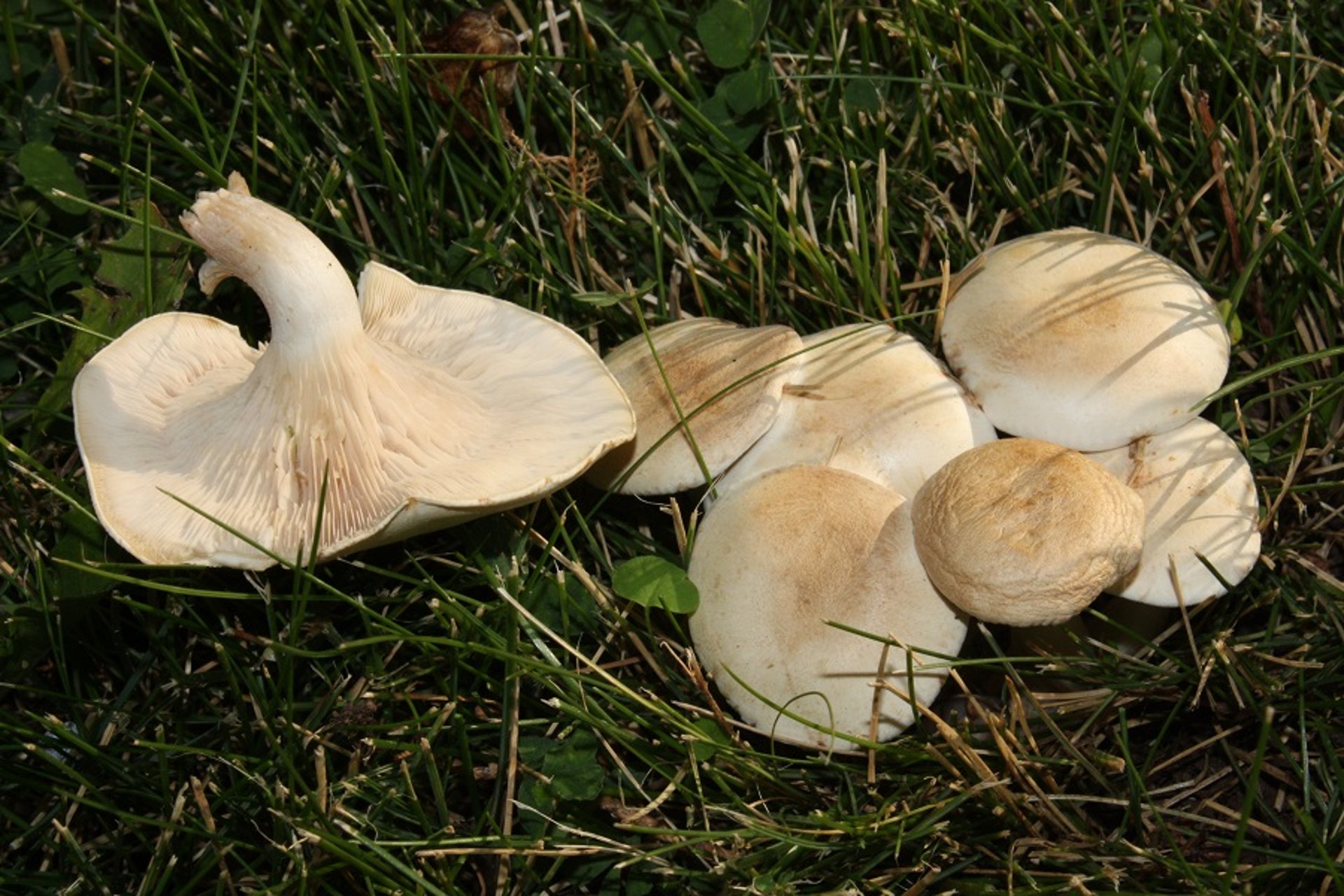
Courtesy of Dr. R. Michael Davis, Plant Pathology, University of California-Davis.
The toxin in these mushrooms is muscarine, a thermostable muscarinic receptor agonist that binds to acetylcholine receptors in the peripheral nervous system. Muscarine is poorly absorbed orally and does not cross the blood-brain barrier. Its cholinergic effects are therefore peripheral. The absorbed portion is quickly distributed throughout the body and undergoes urinary excretion. Muscarine is structurally similar to acetylcholine, a cholinergic neurotransmitter. Unlike acetylcholine, muscarine is not degraded by acetylcholinesterase, and toxicity results from unregulated stimulation at the receptors. Muscarine competes with acetylcholine at cholinergic receptor binding sites, leading to excessive stimulation of postganglionic cholinergic fibers and the subsequent observed clinical signs (cholinergic excess).
Clinical Findings of Muscarine Toxicosis in Animals
Clinical signs can occur rapidly (often within 5 to 30 minutes and generally within 2 hours) after ingestion of mushrooms containing muscarine. Mild to excessive cholinergic stimulation can result in salivation, lacrimation, urination, diarrhea, dyspnea, and emesis (the acronym SLUDDE is often used as a mnemonic for these parts of the cholinergic toxidrome). Dyspnea develops in response to increased bronchial secretions and bronchoconstriction. Bradycardia, miosis, hypotension, shock, and abdominal pain are also possible.
Diagnosis of Muscarine Toxicosis in Animals
Diagnosis is based on a history of mushroom ingestion, identification of suspected mushroom, consistent clinical signs (cholinergic toxidrome), and response to treatment. Muscarine can be detected in urine, and analysis could be considered for confirmation of exposure. Identification of the mushrooms from the environment or vomitus may also be used to support the diagnosis. In severe gastroenteritis cases, tests used to evaluate fluid and electrolyte status, together with a liver profile, may be useful.
Treatment of Muscarine Toxicosis in Animals
The rapid onset of signs (which may include vomiting) after muscarinic mushroom ingestion often makes decontamination unnecessary. Other than offering supportive care (fluid and electrolyte replacement), treatment is not needed in most cases. In life-threatening cases, atropine (beginning dose 0.04 mg/kg, a portion of the calculated dose administered IV, and the remaining portion IM or SC, repeated as necessary) is the treatment of choice. Signs typically respond well to atropine and resolve within 30 minutes of administration. Efficacy of treatment is assessed based on lack of respiratory difficulties and respiratory secretions and not on mydriasis. Excessive atropine treatment can lead to anticholinergic effects (eg, tachycardia, gastrointestinal stasis, behavioral changes, and hyperthermia); therefore, animals should be monitored. Patients can be provided electrolyte supplementation and probiotics orally for up to 3 days after discharge.
Amanita muscaria and A pantherina
Amanita muscaria have a red to orange-red cap with distinctive white or pale yellow warts, gills that are somewhat attached, white spores, ring stalk, and bulbous base (cup). Amanita pantherina have a tan to yellow-brown or dark brown cap with white patches, a ringed stalk with basal bulb, and white spores. Both of these mushrooms are found in coniferous and deciduous forests throughout the United States but are most abundant in the Pacific Northwest.

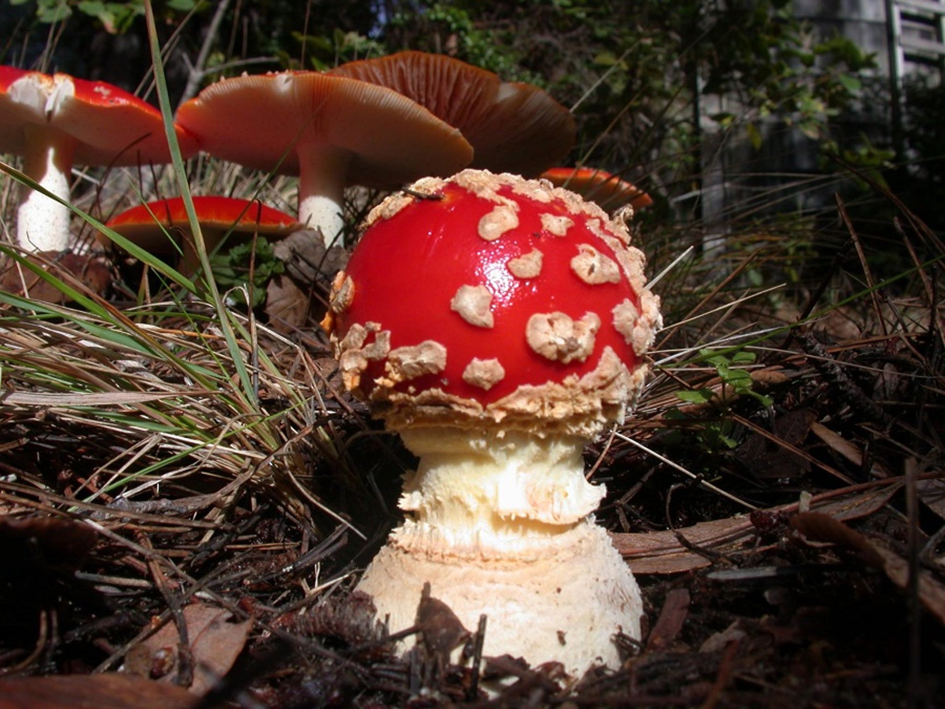
Courtesy of Dr. R. Michael Davis, Plant Pathology, University of California-Davis.
The toxins in these mushrooms are isoxazole derivatives: ibotenic acid, which is structurally similar to the stimulatory neurotransmitter glutamic acid, and its decarboxylated metabolite muscimol, which is stereochemically similar to the inhibitory neurotransmitter gamma-aminobutyric acid (GABA). Ibotenic acid undergoes spontaneous decarboxylation (stomach, liver, and brain), forming muscimol, which is a GABA-receptor agonist. Therefore, muscimol is considered the major toxin responsible for causing clinical signs of toxicosis by increasing the membrane permeability for anions, resulting in a slight, short-lasting hyperpolarization and associated decreased excitability of the receptive neuron. Muscimol also acts on GABAA receptors and has a depressant action. In adult humans, the toxic threshold for ibotenic acid is reported to be 30–60 mg and that of muscimol is 6 mg (ingestion of one average-sized A muscaria). Amanita muscaria is hallucinogenic and has long been used by humans for spiritual and recreational purposes.
Clinical Findings of Muscimol Toxicosis in Animals
The onset of clinical signs occurs within 30–120 minutes after ingestion. Neurologic signs in animals include disorientation, opisthotonus, paresis, seizures, paddling, chewing movements, miosis, vestibular signs (ataxia, head tilt, nystagmus, circling, etc), respiratory depression, and, in severe cases, coma. Duration of clinical signs is ~24 hours.
Diagnosis of Muscimol Toxicosis in Animals
Diagnosis of isoxazole poisoning is based on history of exposure to a mushroom, quick onset of clinical signs (within hours after exposure), type of clinical signs (hallucinations and other CNS effects), and identification of the mushroom. Analysis of urine and stomach contents for ibotenic acid and muscimol by liquid chromatography–mass spectrometry (LC-MS) can confirm poisoning.
Treatment of Muscimol Toxicosis in Animals
Treatment of isoxazole poisoning is supportive, with focus on seizure control (diazepam [0.5 mg/kg, IV, to effect; repeated as needed], phenobarbital [6 mg/kg, IV], or pentobarbital [5–15 mg/kg, IV]). Early decontamination (induction of emesis and administration of activated charcoal) can be tried prior to development of clinical signs. Because of the GABAergic effects of muscimol and ibotenic acid, medications with GABA agonist effects such as diazepam or phenobarbital should be used with caution. Use of these medications in a poisoned animal to control seizures may further aggravate CNS and respiratory depression; if such drugs are used, the animal's respiration should be carefully monitored for need of mechanical ventilation. Muscarine concentrations in A muscaria are very low, and peripheral effects are more anticholinergic than cholinergic. Therefore, atropine treatment is contraindicated.
Chlorophyllum molybdites and Russula emetica
Chlorophyllum molybdites mushrooms have caps that are conical when young, becoming convex to nearly flat. They are initially smooth and brown but become white with brown scales. They have unattached white gills, green or grayish-olive spores, and a ring on the stalk. They are common in the southern and central US. Russula emetica mushrooms have a reddish, slimy cap with short whitish stalk and gills; no veils; and white to yellowish-white spores. They grow singly or in a group and are found throughout the US. Other genera leading to gastrointestinal irritation include Agaricus, Boletus, Entoloma, Gomphus, Hebeloma, Lactarius, Naematoloma, Omphalotus, Ramaria, Rhodophyllus, Scleroderma, and Tricholoma. The toxins in these mushrooms are unknown gastrointestinal irritants that are thought to be high-molecular-weight proteins found throughout the mushroom.
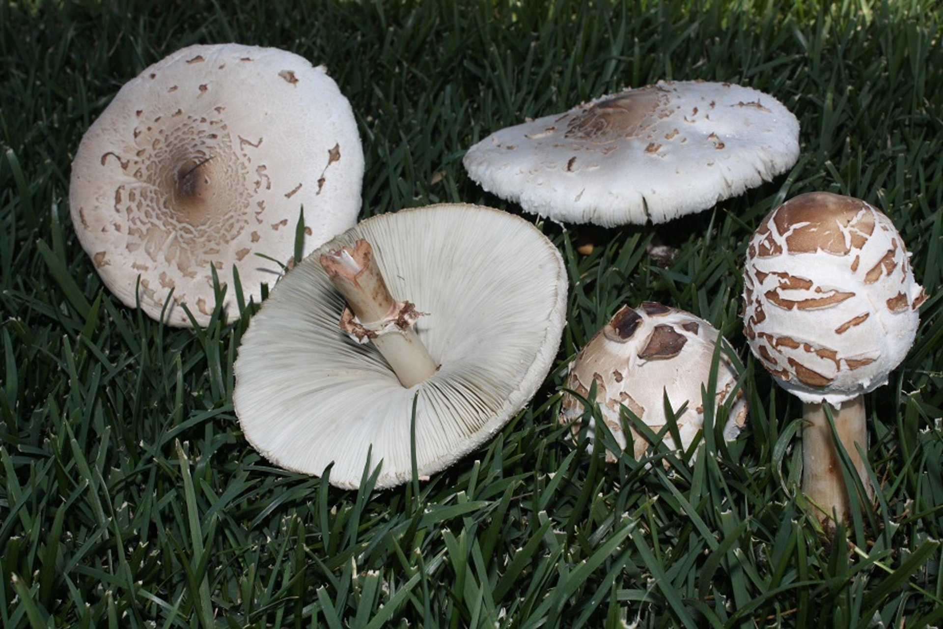
Courtesy of Dr. R. Michael Davis, Plant Pathology, University of California-Davis.
Clinical Findings of Chlorophyllum molybdites and Russula emetica Toxicosis in Animals
Within minutes to hours (15 minutes to 3 hours) after exposure, clinical signs become evident. These include vomiting, bloody diarrhea, abdominal pain, muscle cramps, liver injury, and circulatory disturbances that are self-limiting. Although recovery generally occurs within 6–24 hours, it could be prolonged or rarely become life-threatening in cases of hypovolemic shock, oliguria, or transient increased BUN concentration secondary to dehydration. Death is rare.
Diagnosis of Chlorophyllum molybdites and Russula emetica Toxicosis in Animals
A tentative diagnosis may be based on history and early onset of reported clinical signs (signs of gastroenteritis, such as vomiting and bloody diarrhea). Except for dehydration, there are no systemic clinical signs. The mushrooms should be saved for identification. Gastrointestinal upset is also an initial sign after ingestion of more dangerous hepatotoxic and nephrotoxic mushrooms, although the onset is typically more delayed. A CBC, serum biochemical analysis, and radiography may be useful to assess the clinical picture and help rule out other causes for the clinical signs.
Treatment of Chlorophyllum molybdites and Russula emetica Toxicosis in Animals
The potential rapid onset of vomiting may make decontamination after ingestion of gastrointestinal irritant mushrooms less feasible. There are no specific antidotes. Treatment is supportive and depends on the extent of signs. Intravenous fluid therapy is recommended to maintain hydration. Sucralfate, histamine-2 blockers (famotidine), or proton pump inhibitors (omeprazole) may be used to decrease mucosal irritation. Vomiting should be controlled with antiemetics such as maropitant and metoclopramide. Many cases are self-limiting and resolve without treatment. The severity of clinical signs depends on the type of mushroom, amount ingested, and individual sensitivity. In most cases, the prognosis is good, and full recovery is expected within a few hours to days.
Psilocybe cubensis, Conocybe cyanopus, Gymnopilus spectabilis, and Panaeolus subbalteatus
Psilocybe cubensis are small, brown, slender-stalked mushrooms. They commonly grow in piles of dung and fertilized grasses in moist areas throughout the US, especially in the southeast and northwest. The mushrooms develop a blue-green color in handled and injured areas. They have yellowish, sticky to moist caps with brown gills; a persistent ring on the stalk; and a purple-brown spore print. Conocybe cyanopus are small and fragile, with brown caps, brown gills, and a large ring midway down the long, thin stalk. The spore print is cinnamon brown. They are widely distributed in North America. Gymnopilus spectabilis are large yellow-orange mushrooms with an orange to rust-orange spore print. They are found in clusters on wood, stumps, or the ground over buried wood. Panaeolus subbalteatus have broadly conical to flat caps with a dark belt around margin, brown gills, hairy reddish stalks, and a blackish spore print. They are widely distributed in North America.

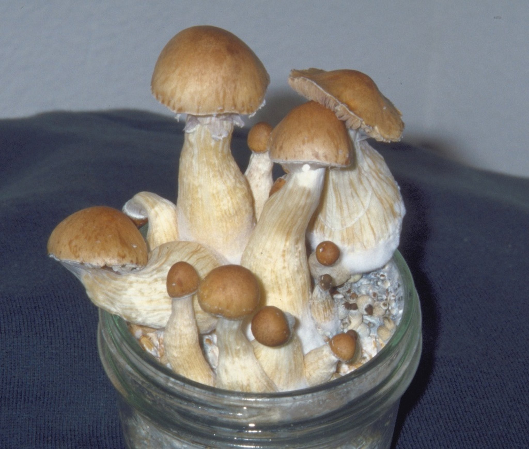
Courtesy of Dr. R. Michael Davis, Plant Pathology, University of California-Davis.
These hallucinogenic mushrooms contain psilocybin and psilocin, which are indole alkaloids similar to lysergic acid diethylamide (LSD) and are classified as controlled substances. Psilocybin undergoes rapid dephosphorylation (plasma, kidneys, and liver), forming psilocin, which is structurally similar to serotonin (stimulatory to serotonin receptors in the CNS and peripheral nervous system). Psilocybin and psilocin cross the blood-brain barrier and concentrate in brain tissue. Growth conditions, geographic location, storage conditions, and mushroom species are factors that influence toxin concentrations. Ingestion of five or six dried mushroom caps is reported to be toxic.
Clinical Findings of Psilocybin and Psilocin Toxicosis in Animals
Clinical signs occur within 0.5–1 hours; rarely, they are delayed up to 3 hours after ingestion. Signs include aggression, ataxia, vocalization, nystagmus, seizures, and increased body temperature, with recovery within 6 hours of observed clinical signs.
Diagnosis of Psilocybin and Psilocin Toxicosis in Animals
A history of mushroom ingestion, identification of suspected mushrooms, consistent clinical signs, and response to supportive care are critical for diagnosis. Exposure can be confirmed by detection of psilocin and psilocybin in urine by select veterinary diagnostic laboratories.
Treatment of Psilocybin and Psilocin Toxicosis in Animals
Because of the short-lasting effects, mild cases may resolve without treatment. Supportive treatment may be necessary when severe clinical signs are present. Seizures can be controlled with diazepam (0.5–1 mg/kg, IV or per rectum, with incremental dose increase of 5–10 mg, to effect) or phenobarbital (6 mg/kg, IV, IM, or PO, every 6 hours to effect up to a maximum of 24 mg/kg total over 24 hours). Body temperature should be monitored. In some cases, leaving the animal untreated in a quiet, dark environment might be all that is necessary.


