In addition to designing and implementing preventive-care protocols for all animals in the collection, zoo veterinarians must formulate clinical care programs to respond to the needs of collection animals. At a minimum, clinical care programs should include immediate notification of any reportable diseases to relevant local, state, or federal authorities; daily observation of each animal in the collection by a competent team member (often a member of the husbandry staff); immediate notification of any health problems to the veterinary team; appropriate diagnostic tests to rule out infectious diseases; and prompt implementation of treatment as indicated by results of examination or testing.
Clinical care programs for zoo animals include all the pillars of preventive care, including the implementation of quarantines on the basis of risk analysis, routine fecal examinations (at least once a year) to identify the presence of parasites, physical examination, collection of biological samples (taking advantage of the physical or chemical restraint of an animal for any other reason), vaccination against relevant diseases, routine necropsies in the event of any death, and maintenance of detailed medical records.
Animal care staff are usually the first to notice and report clinical signs of disease, which are often subtle or easily missed by the untrained observer. Diagnosis and treatment of zoo animals are similar to domestic species, although the methods of drug administration and restraint can differ. Additionally, attention must be paid to both individual and population health as well as understanding any disruptions in social hierarchies should an animal require hospitalization or removal from the exhibit for treatment.
A comparative medical approach to preventive care is generally most successful and involves application of medical or surgical information about diseases affecting free-ranging animals, related domestic animals, or humans. Frequently, other veterinary experts or human medical or dental specialists are consulted for advice or assistance with complicated medical or surgical cases. Knowledge of taxonomy and comparative anatomy, physiology, behavior, nutrition, and pathology is useful.
Behavioral Training of Zoo Animals
The zoo should develop a behavioral husbandry program that promotes behavioral enrichment (ie, providing opportunities for animals to express species-specific behaviors) and monitors and improves animal welfare. Health, or freedom from sickness and disease, is one of the five pillars, or domains, of animal welfare. Veterinarians must therefore participate in the zoo’s animal welfare program, which includes routine holistic welfare assessments.
Animal welfare programs can also facilitate objective quality-of-life monitoring, which can help evaluate treatment outcomes for chronic conditions and identify indicators for humane euthanasia. Clinical care programs should also integrate with behavioral husbandry programs to improve monitoring of animal health and decrease risks associated with examination and immobilization. For more information on zoo animal welfare, see the American Association of Zoo Veterinarians Animal Welfare Resources.
The principles of operant conditioning with positive reinforcement can be applied to all species, and training zoo animals to perform behaviors on command facilitates various management and medical procedures.
Behavioral restraint refers to animals remaining still voluntarily to allow for a procedure without the use of physical restraint or chemical immobilization. It is particularly useful for medical procedures, including injections by hand, venipuncture, ultrasonographic examination, hoof care, palpation, and physical examination. Other useful behaviors include stepping onto a scale or perch to be weighed, entering a restraint device (eg, squeeze cage or transport crate), and moving from one enclosure to another (“shifting”) on demand.
For species that need routine medical procedures or husbandry work, positive-reinforcement training can provide safety for personnel, who can deliver care on the opposite side of a protective barrier, and give the animal some scope of choice.
Physical Restraint of Zoo Animals
The difficulties involved in physical examinations and medical interventions with uncooperative patients are magnified when working with nondomesticated species. Animals pose a direct risk to those handling them because of their teeth, claws, beaks, venom, tails, spines, and many other defense mechanisms.
Inadequate restraint increases the risk of escape and thus endangers zoo staff, the public, other animals in the collection, and the animal itself, as lethal restraint can be necessary. Furthermore, it is impossible to perform a thorough physical examination, collect samples, or administer treatments without proper restraint.
Analgesics should be included in restraint protocols if the patient might be in pain before the physical examination or if the planned intervention could cause more than just discomfort or momentary pain. When behavioral restraint is not possible, physical restraint can be considered on the basis of the animal’s history and temperament, the experience of animal care staff, and the available equipment.
Physical restraint can be performed by hand or with the aid of specific devices. For most zoo animals, physical restraint is only appropriate for administering parenteral sedatives or anesthetics or for short, nonpainful procedures. Whenever possible, animals should be trained to voluntarily enter the restraint device. In some cases, the animal can be enticed, baited, or herded into a restraint; however, physical force should be avoided.
Ideally, restraint devices should be designed as part of the animal’s regular housing and located in an area where the animal is normally shifted as part of the daily routine. Exhibits should contain nest boxes or restraint pens equipped with doors that operate remotely to confine the animal. From these areas, the animal can be transferred to a restraint device, anesthetic chamber, or shipping crate.
Ungulates can be restrained in funnel-shaped chutes that gradually narrow until the animal cannot turn around. Among the restraint tools for small- to medium-size ungulates are chutes with removable bottoms, which suspend the animal. Squeeze cages of appropriate size are used to contain carnivores and primates (see ). One wall of the cage can be moved to narrow the interior space, allowing the animal to be temporarily immobilized at the other end to enable the administration of drugs by manual injection.

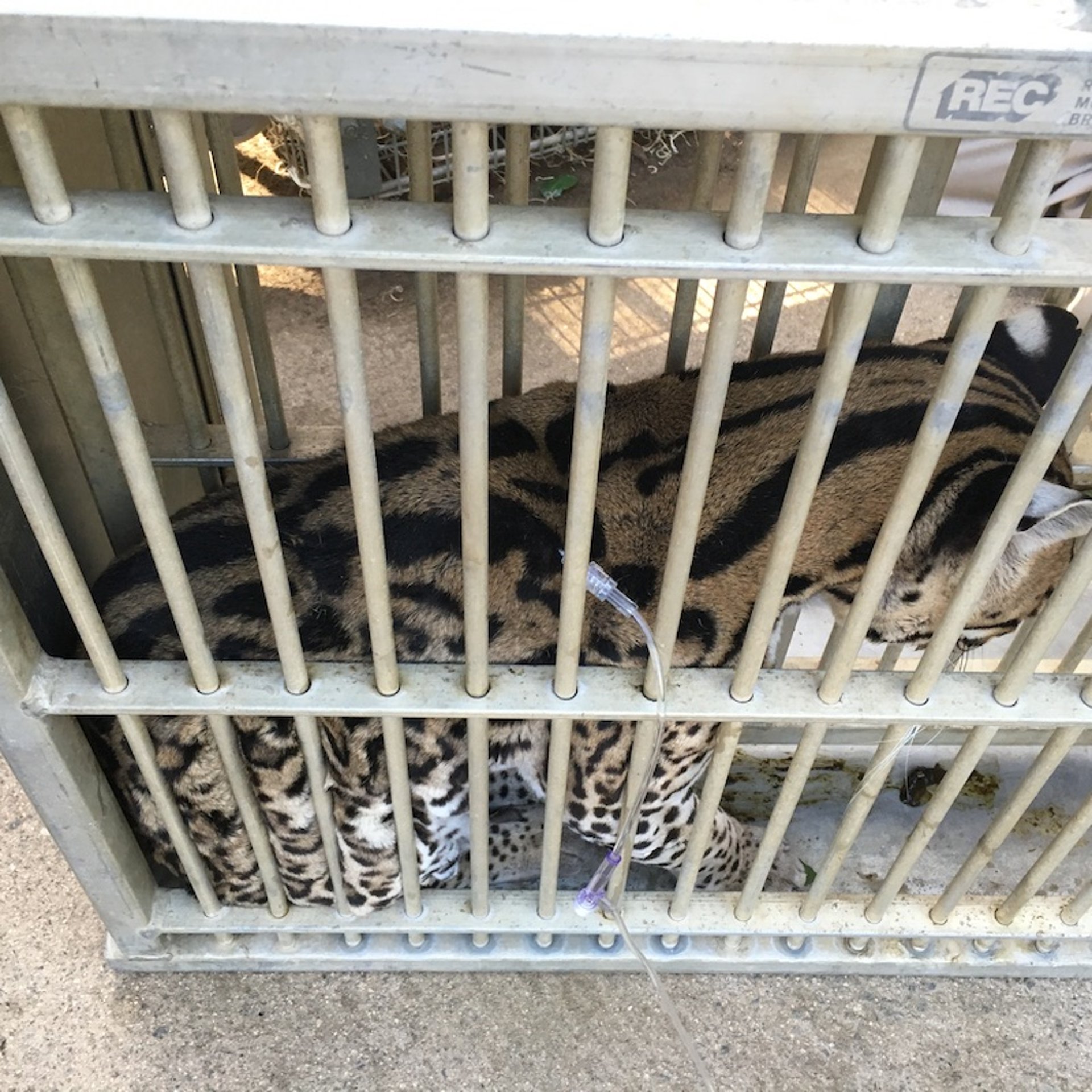
An ocelot being restrained in a squeeze cage. The ocelot voluntarily enters the cage, and the sliding wall squeezes down to allow for minimal movement and therefore administration of parenteral injections, including the subcutaneous fluid administration shown here.
Courtesy of Dr. Meredith M. Clancy.
Nets and burlap sacks are helpful for the brief restraint of small mammals. For larger animals, harnesses, ropes, and chains can be used in conjunction with sedation. Small mammals and birds can be caught and restrained in long-handled hoop nets. These nets must be deep enough that the animal can be confined in the blind end, with the upper part of the net twisted to prevent escape. Often, minor, noninvasive procedures that do not warrant the time and risk of anesthesia can be performed on small mammals and birds using such physical restraint alone.
Poles and baffle boards can be used to prevent direct contact between animals and handlers. Personal protective equipment, including protective eyewear and gloves, should be used as needed. Although heavy gloves can limit dexterity, they can protect handlers from bites, scratches, and punctures during manual restraint. Veterinarians should consult with animal care staff before restraining an animal to select the most appropriate method on the basis of the animal's temperament and training level, the medical and nonmedical teams’ experience, the available equipment, and the design of the holding facilities and areas.
Diagnostic Techniques for Zoo Animals
The fundamental diagnostic technique is obtaining a thorough history, doing a medical record review, and performing thorough, systems-based visual and physical examinations (often requiring sedation or general anesthesia). Because many zoo animals must be immobilized for clinical evaluation, it is often preferred to collect as much diagnostic information as possible at once, rather than across serial examinations. For this reason, blood work, diagnostic imaging, and collection of additional biological samples (eg, biopsies, swabs, or serum for banking) can be performed opportunistically and not only when strictly indicated by the observed clinical signs.
Choice of laboratory testing is often driven by the ease of sample collection, clinical suspicion, and the availability of validated assays, much of which is dependent on the species of the patient. Available diagnostics common in veterinary practice include hematology, serum/plasma biochemistry, serological testing, cytology, fecal examination, urinalysis, and aerobic, anaerobic, fungal, or viral cultures. Special attention, however, must be given to the type of test and the species in which it is applied.
While many assays can be used across taxa, many are also species-specific and can have altered sensitivity and specificity, leading to false-positive or negative results or results that are frustratingly ambiguous or impossible to interpret. When in doubt, clinicians should contact diagnostic laboratories for advice, work with laboratories that specialize in the species in question, or consult the literature prior to choosing a diagnostic test and when interpreting its result.
Molecular diagnostics are useful in zoo practice. For the detection of specific pathogens, PCR assay and sequencing are generally preferred, as next-generation sequencing, though available, is difficult to interpret in a clinical setting. Diagnostic imaging, including radiographic and ultrasonographic evaluation, has become routine in zoo practice, and advanced imaging such as CT is often used for zoo animals. Endoscopy and laparoscopy allow for minimally invasive diagnostic and surgical access, although technical expertise is required. Virtually any technique used for other species can be modified for use in zoo species.
Drug Administration in Zoo Animals
Few drugs are labeled for use in zoo species; however, extralabel use laws allow for the legal use of many drugs in species for which they are not licensed. Evidence-based prescription of drugs, including antimicrobials and anesthetic agents, is possible by consulting the ever-increasing species-specific pharmacokinetic and pharmacodynamic studies in zoo animals.
When species-specific pharmacokinetic data are unavailable, prescription can be on the basis of extrapolation, deductive reasoning, and experience. Metabolic scaling has been used historically; however, it has important limitations across taxonomy in zoo species. Dosing, therapeutic benefit, and potential adverse effects, including contraindications and toxicity, are often not known and can vary even among closely related species and drugs, so caution is advised.
Drug administration can be challenging in zoo animals. Oral medication administration is facilitated by hiding it in food items, including invertebrate prey (bioencapsulation). Working with compounding pharmacies can improve compliance by modifying dosage volumes, formulations, and flavoring. Risks of oral administration can include inadvertent consumption by nontarget individuals for patients that are housed in social groups and poor compliance in Old World primates that might hide medications in cheek pouches. Enteral antimicrobials should be used with caution in foregut fermenters, such as ruminants, macropods, and Old World folivorous monkeys, because of the risk of GI dysbiosis.
Parenteral administration is preferred for many medications, especially sedatives and anesthetics. Parenteral administration can also be preferable if long-acting or depot formulations of medications are available. With behavioral training, many patients will voluntarily allow the administration of subcutaneous or intramuscular injections.
Remote delivery of parenteral medication is commonly used in zoo practice, especially for anesthetics. Several types of remote delivery systems are available, and choice depends on the size of the patient, the distance at which the drug must be delivered, and the appropriate selection of drug concentrations to minimize dosage volume. Veterinarians must understand species-specific anatomy to target the largest muscle bodies to decrease risk of trauma or other complications from poorly aimed injections.
Pole syringes are basically canes to which a loaded syringe with a protective cover is attached. They can be used at a distance of 1–2 m (3.28–6.56 feet). The simplest ones can be made of metal or PVC pipes that act as an extension of the plunger. Once the needle has been inserted into the animal's body, the pole must be pushed to depress the plunger.
“Jab sticks” are a more advanced type of pole syringe that have a chamber with a spring that is activated when the needle is inserted into the animal, so the plunger advances without the operator having to push the stick. The benefit of the jab stick is that it allows for more rapid drug administration. The drawback is that it injects forcefully, with greater risk of trauma, especially in smaller patients.
For greater distances, pneumatic or gunpowder darts can be used. Pneumatic darts have two chambers separated by a rubber piston. The front chamber contains the drug to be administered, while the rear chamber contains pressurized air, which is introduced using an empty syringe and a one-way valve. A pointed needle with apertures on the sides, rather than the tip, is attached to the front and the apertures covered with a lubricated rubber stopper to maintain pressure as the rear chamber is filled with air. When the needle enters the animal, the rubber stopper does not, allowing the pressurized air to push the piston forward, injecting the drug into the patient. In species with thick skin (eg, bears, megavertebrates), barbed needles help the needle to be retained in the patient until the full volume of drug has been injected. Pneumatic darts can be sterilized and reused. However, over time and with repeated use, they can bend and cause unpredictable trajectories.
Gunpowder darts have a single chamber into which the drug is loaded. The rear end contains a cartridge with a spring that is activated upon impact, allowing the components to mix and react rapidly, generating gas that pushes an internal piston within the chamber and causes the contents to be released through the needle. Gunpowder darts are for single use only. Darts should be loaded immediately prior to use, as some drugs can lose potency over time when exposed to plastic or aluminum. All darts are decorated with brightly colored fletching (the fin-shaped device at the rear) that improves aerodynamic stability and facilitates their identification and recovery after firing (see images of medication delivery via dart in , , and ).

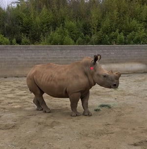
Appropriate placement of a dart with medication on a southern white rhinoceros (Ceratotherium simum simum).
Appropriate placement of a dart with medication on a southern white rhinoceros (Ceratotherium simum simum).
Courtesy of Dr. Meredith M. Clancy.

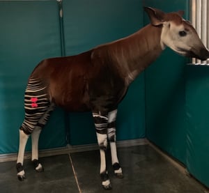
Appropriate placement of a dart with medication in a captive okapi (Okapia johnstonii)
Appropriate placement of a dart with medication in a captive okapi (Okapia johnstonii)
Courtesy of Dr. Meredith M. Clancy.

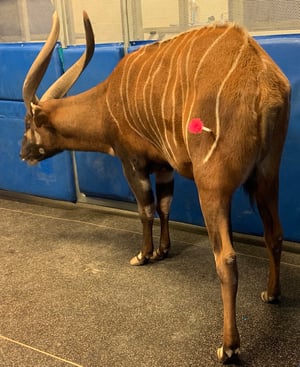
Appropriate placement of a dart with medication in a captive Eastern bongo (Tragelaphus eurycerus isaaci).
Appropriate placement of a dart with medication in a captive Eastern bongo (Tragelaphus eurycerus isaaci).
Courtesy of Dr. Meredith M. Clancy.
Appropriate placement of a dart with medication on a southern white rhinoceros (Ceratotherium simum simum).
Appropriate placement of a dart with medication on a southern white rhinoceros (Ceratotherium simum simum).
Courtesy of Dr. Meredith M. Clancy.
Appropriate placement of a dart with medication in a captive okapi (Okapia johnstonii)
Appropriate placement of a dart with medication in a captive okapi (Okapia johnstonii)
Courtesy of Dr. Meredith M. Clancy.
Appropriate placement of a dart with medication in a captive Eastern bongo (Tragelaphus eurycerus isaaci).
Appropriate placement of a dart with medication in a captive Eastern bongo (Tragelaphus eurycerus isaaci).
Courtesy of Dr. Meredith M. Clancy.
Darts can be fired with blowguns, air guns, or powder rifles. Blowguns are straight, hollow tubes into which a pneumatic dart is loaded. The operator seals their lips around the end of the tube, aims, and with a single, strong exhalation propels the dart toward the target. Air guns, either pistols or rifles, fire darts using compressed carbon dioxide. A pressure regulator allows the operator to determine how much force is applied to each shot. Powder rifles are similar to air guns. However, they use a powder cartridge to fire each dart, rather than compressed air.
Dart rifles can include optical and laser sights. Firing darts can be tricky, because the trajectory depends on the dart's weight and shape, the volume of the drug, and the force with which it is fired. Zoo animal veterinarians must practice with their equipment at regular intervals to maintain proficiency.
In zoo practice, IV access often requires adequate tranquilization or sedation or containment in restraint devices or small enclosures during treatment. As in domestic animals, indwelling IV catheters and IV treatments are reserved for cases requiring the most intensive treatment, as it can be labor intensive to maintain IV access in zoo species. IV access also allows for total IV anesthetic administration in some species (see ).

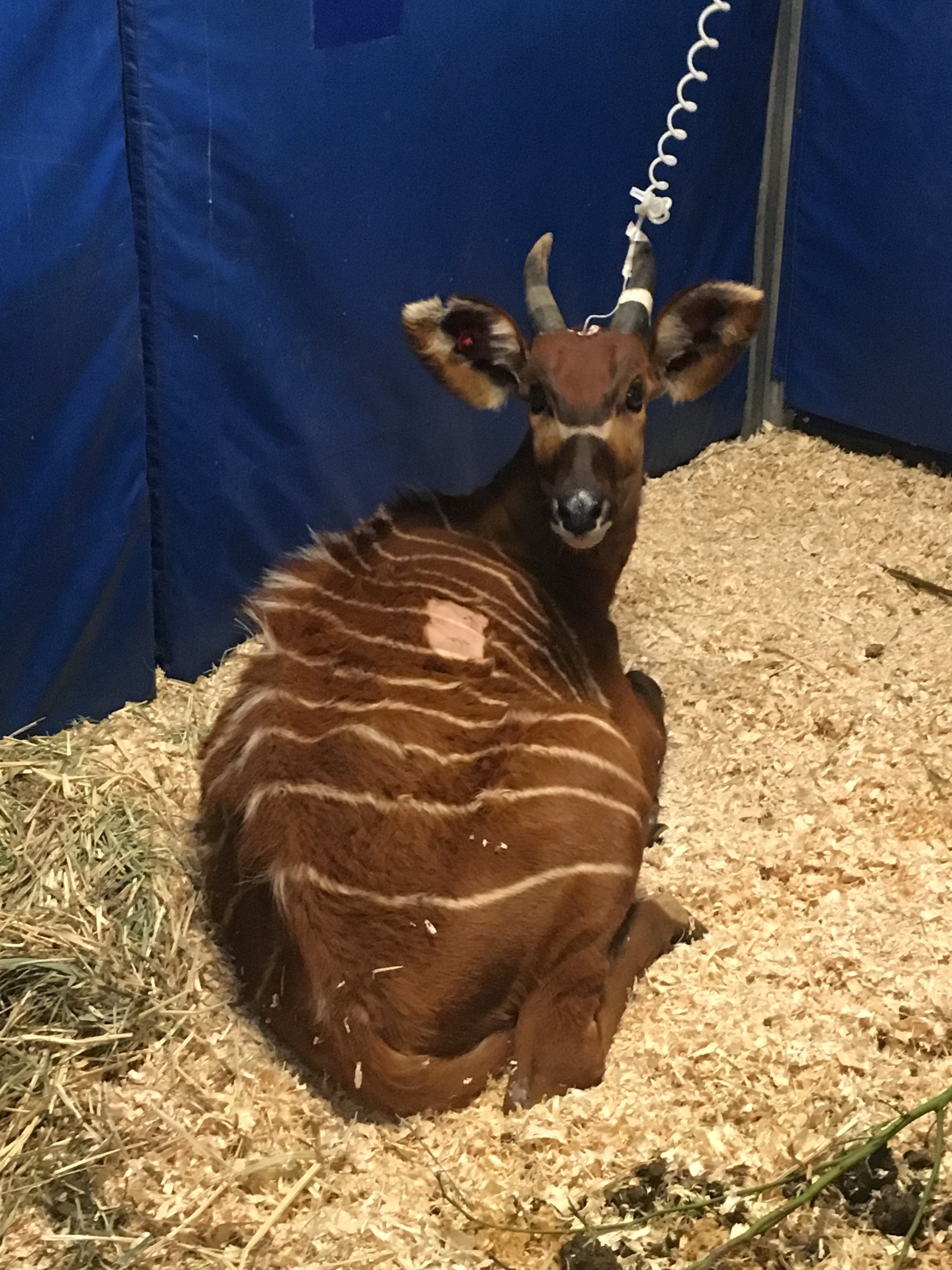
Use of a jugular venous catheter to facilitate administration of intravenous fluids, antimicrobials, and parenteral nutrition in a hospitalized Eastern bongo (Tragelaphus eurycerus).
Courtesy of Dr. Meredith M. Clancy.
Anesthesia Considerations for Zoo Animals
Chemical restraint or immobilization refers to the use of sedatives and anesthetics to limit the voluntary movement of patients. In zoo animals, many procedures routinely accomplished on domestic animals with minimal restraint require anesthesia to ensure the welfare and safety of both animals and personnel.
In zoo animal medicine, the same general principles of sedation and anesthesia apply as in domestic animal practice, with some additional considerations. It is imperative that the restraint team be trained before performing any procedure and that each member be assigned a specific role, with the veterinarian generally designated as ultimately responsible for the procedure.
Before immobilization, the veterinarian should be familiar with the species and anesthetic agents. Anesthesia records for the individual, other conspecifics in the collection, or published references for the species should be reviewed. Response to anesthetic agents can vary even among closely related species, so consultation with experienced colleagues is advised.
An anesthetic plan should be developed for each anesthetic event and should include the anesthetic drugs and doses to be used, other needed pharmaceuticals (eg, emergency drugs, analgesics, anthelmintics, vaccines), monitoring equipment, and any other special equipment needed to perform the procedure at hand.
The location in which an animal is anesthetized is an important consideration and can influence drug selection. Removing water sources and trip or entanglement hazards can decrease risks. However, it is recommend to always plan for availability of adequate staff and access points to the animal in case early intervention is required, especially if anesthesia is performed in a herd situation where other animals are present.
Zoo animals have acute sensory perception and often become nervous or restless when their routine is disrupted or changed. Before drugs are administered and take effect, noise, activity, and the number of personnel should be decreased as much as possible. Once the animal is sedated, its eyes should be covered with an opaque cloth and ear canals can be lightly plugged with cotton to decrease visual and auditory perception. Oxygen can be supplemented by face mask using standard veterinary products or custom-made masks using plastic bottles and adhesive tape, depending on the facial anatomy and the position of the nostrils of the species.
Care must be taken not to occlude the nasal passages of obligate nasal breathers (such as ungulates, swine, rabbits, and rodents). It is recommended to pad the area where large patients are handled to prevent radial nerve paralysis, other peripheral neuropathies, and perfusion problems or pressure necrosis. When possible, an IV catheter should be placed as soon as possible, except in the case of shorter and less invasive procedures.
Anesthetic monitoring is performed in the same manner as with domestic animals, using standard equipment (eg, pulse oximetry, electrocardiography, direct or oscillometric blood pressure monitoring, capnography). However, this equipment cannot replace direct observation and assessment of the patient (eg, respiratory and heart rate by observation and use of stethoscopes, appearance of mucous membranes, capillary refill time, assessment of reflexes). Anesthetic monitoring should continue through to recovery, as long as it is safe to do so.
All zoos must have standard operating procedures (SOPs) for animal escapes, however unlikely. These SOPs must be reviewed regularly and compliance enforced, including regularly scheduled tabletop and live-action drills that involve staff across all the zoo’s departments.
When a dangerous animal escapes, an incident command system is established, often with the veterinarian (or curator or public safety officer) serving as the designated authority. Emergency chemical immobilization can be used if an animal cannot be safely returned to containment using recall training and if staff and public safety can be ensured without the use of lethal force.
SOPs regarding chemical immobilization for emergency response must, at a minimum, specify who is authorized to administer anesthetic drugs, define the situations in which they will be used, identify their location, and describe emergency procedures in case of accidental human exposure.
During emergency immobilization, the use of rapid-action protocols with short induction times is indicated. Limiting the number of drugs in an emergency protocol can decrease the preparation time of a dart for remote administration. Charts of emergency immobilization drugs and doses for various animals in the collection should be available and updated regularly.
For carnivores and primates, protocols based on tiletamine-zolazepam are often used, generally with the addition of an alpha-2 agonist. The butorphanol-azaperone-medetomidine combination is used in many herbivores, although the use of ultrapotent opioids can be indicated for large species.
Aside from the urgency required to respond to an escape situation, the anesthesia of zoo animals follows the same general principles as for their domestic relatives. Whenever possible, clinicians should tailor an anesthetic protocol to each patient to minimize risks, both for the patient and for zoo staff.
The cyclohexamine ketamine is one of the few anesthetic agents licensed for use in zoo animals and is often used in combination with an alpha-2 adrenergic receptor agonist and/or benzodiazepine. Combination of ketamine with other drugs decreases the required dose and results in smoother induction and recovery than if ketamine is used alone. Highly concentrated ketamine acquired from compounding pharmacies decreases dose volume, which is especially important for remote delivery.
Tiletamine-zolazepam, a dissociative anesthetic-tranquilizer combination, is relatively safe in most species, has a rapid induction, and can be concentrated to 200 mg/mL, allowing for small dose volumes. Because no complete antagonist exists, recoveries can be longer than with combinations that can be completely reversed.
Alpha-2-adrenergic receptor agonists such as xylazine, detomidine, medetomidine, or dexmedetomidine, have sedative and analgesic effects and can be used as sole agents or in combination protocols. When combined with ketamine, alpha-2 agonists can provide a surgical plane of anesthesia in many species and a safe anesthesia in dangerous animals. Combined with an ultrapotent opioid, alpha-2 agonists can also provide the basis for anesthesia in many species of hoofstock or megavertebrates.
Dosage-dependent peripheral vasoconstriction caused by alpha-2 adrenergic receptors can lead to alterations in blood pressure, which can interfere with pulse oximetry monitoring or make venipuncture or catheterization more difficult. Smoother and more rapid recovery can be achieved by use of reversal agents (eg, atipamezole, tolazoline, or yohimbine). Atipamezole is dosed at 5 mg/mg medetomidine and 10 mg/mg dexmedetomidine (1, 2, 3). Resedation has been observed in some species in which higher doses of alpha-2 agonists were administered.
Species sensitivity to alpha-2 agonists can vary greatly, as does the dosage required, depending on the combination protocol selected. Alpha-2 agonists should not be administered as the sole anesthetic agent for dangerous carnivores, because these animals can appear sedated but can respond aggressively when stimulated.
Pearls & Pitfalls
|
The neuroactive steroid alfaxalone has been growing in use in zoo animals, with studies demonstrating safety and efficacy in species ranging from marine invertebrates to fish to reptiles to zoo mammals (4, 5, 6, 7, 8, 9). Dose-dependent anesthesia with smooth recovery has been observed in multiple species. Volume can be a limiting factor, with high volumes required for relatively small species. Addition of an analgesic is required for painful procedures, because alfaxalone has no known analgesic properties.
The rapid onset and short duration of propofol make it useful as a supplemental agent if IV access is possible. Isoflurane or sevoflurane can be used to induce anesthesia in small mammals, birds, and reptiles or as a maintenance agent for longer anesthetic events. Other agents used in combination anesthesia or sedation include the benzodiazepine anxiolytic midazolam, the phenothiazine tranquilizer acepromazine, and the butyrophenone tranquilizer azaperone.
Ultrapotent opioids, such as etorphine and thiafentanil, are often indicated for the chemical restraint of megavertebrates. Safety is paramount when working with these drugs, as even inadvertent casual skin exposure can cause respiratory and cardiac depression and death in humans. When working with ultrapotent opioids, veterinarians should, at a minimum, work in pairs; inform all safety and first-responder personnel; use appropriate personal protective equipment, including two pairs of disposable gloves, eye and/or face shields, and long sleeves; draw up reversals first; dispose of contaminated needles and syringes in rigid, sealed containers; and wash contaminated surfaces and the patient's injection site thoroughly with water. When handling an ultrapotent opioid, carry a copy of the package insert in a pocket so that emergency first responders can easily access it in case of accidental exposure.
Ultrapotent opioids are used in combination with other agents (eg, alpha-2 agonists, azaperone, acepromazine), as use as sole agents can cause respiratory depression and muscle rigidity. Etorphine is commonly included in anesthetic protocols for large ungulates, such as large equids and rhinoceroses. It can cause tachycardia, hypertension, hypoventilation, and increased oxygen demand. Thiafentanil oxalate is a synthetic derivative of fentanyl and a potent opioid. Compared to etorphine, thiafentanil has a shorter induction time. However, it is equally efficacious. Because of thiafentanil's rapid absorption and metabolism, patients do not become renarcotized after reversal, and ungulates usually recover without the need for drug reversal. Adverse effects of thiafentanil can include changes in respiratory rate, heart rate, blood pressure, and body temperature; cyanosis; and reaction to auditory stimuli.
Naltrexone is the antidote of choice for ultrapotent opioids. Reported dosing varies with respect to the opioid being reversed and the species in question. It should be administered intramuscularly, except in emergency situations, in which 25% of the total dose can be administered intravenously (4). When administered intravenously, naltrexone can produce a rapid and abrupt return to consciousness and mobility and, in some cases, induce extrapyramidal signs. Renarcotization is always a risk, and repeated dosing might be required.
In many large carnivores, especially big cats, blood potassium concentrations can increase over time during anesthesia, so serial monitoring is indicated for prolonged procedures. Hyperkalemia causes an increase in the amplitude of QRS complexes and peaked T waves on electrocardiogram in addition to bradyarrhythmia and cardiac arrest. Treatment involves IV insulin administered with dextrose along with reversal of anesthesia as soon as it safe to do so.
Capture myopathy (also called exertional myopathy) is a form of exertional rhabdomyolysis that can occur in zoo animals during extreme stress associated with handling or transport. It occurs as part of four described syndromes:
Hyperacute or capture shock syndrome is rare and characterized by weakness, muscle rigidity, ataxia, and acidosis, with death occurring < 6 hours after the capture event.
Acute or ataxic-myoglobinuric syndrome is the most common capture myopathy. It is characterized by acute necrosis of skeletal and cardiac muscle resulting in weakness, ataxia, and myoglobinuria that causes kidney failure and death within hours or days after capture.
Subacute syndrome is similar to acute syndrome; however, it involves rupture and hemorrhage of the appendicular muscles. It can occur immediately or up to 48 hours after capture, and humane euthanasia is often indicated.
Delayed, chronic, or peracute syndrome is when affected animals recover without complications at the time of capture and die days or weeks later after exposure to further stress, such as a second capture attempt.
Risk factors associated with capture myopathy include environmental factors (eg, high temperatures), factors associated with capture (eg, prolonged chase before or after darting, the type of drug used), and factors associated with the patient (eg, concomitant diseases, body condition, age, sex, or reproductive status).
The pathogenesis of capture myopathy includes the perception of fear, activation of the sympathetic and adrenal nervous systems resulting in an endogenous catecholamine response, and muscle activity. Species that have a strong flight response, including prey species such as ruminants or some birds (eg, cranes and flamingos), are considered particularly susceptible. Risk can be mitigated by scheduling immobilizations during the cooler times of the year and day, minimizing visual and auditory stimuli before and immediately after darting to decrease stress, confining animals in a dimly lit stable or enclosure to prevent prolonged chase, and preventing or correcting hyperthermia using cooled IV fluids, cold-water enemas, or ice or alcohol applied to the inguinal region or extremities.
Parenteral administration of vitamin E and selenium during anesthesia can decrease the risk of capture myopathy during recovery. Intravenous crystalloid fluid administration treats acidosis and hyperkalemia, improves blood pressure and perfusion by increasing intravascular volume, and improves renal perfusion and diuresis, decreasing the risk of myoglobinuric syndrome. The administration of benzodiazepines can decrease the endogenous catecholamine-mediated stress response and induce muscle relaxation. The use of sodium bicarbonate to treat metabolic acidosis has been discussed (10), although it is not routinely used. Corticosteroids can be used for analgesia and to decrease inflammation without compromising renal perfusion. Animals that survive the recovery period can benefit from physical therapy and analgesia.
Anesthetic induction can be achieved in most carnivores following the same principles as for domesticated companion-animal carnivores. In big cats, ketamine is frequently used in conjunction with an alpha-2 agonist, such as medetomidine or xylazine (11). The addition of midazolam can smooth induction and recovery and decrease the risk of seizures. Ketamine and midazolam combinations are recommended for cats with neurological or cardiovascular disease. Medetomidine-butorphanol-midazolam and ketamine-medetomidine combinations, with or without midazolam, have been used successfully in cheetahs (Acinonyx jubatus) (12, 13, 14).
Historically, the combination of tiletamine and zolazepam has been associated with seizures, ataxia, and death up to 4 days after the anesthetic event in many feline species, especially tigers (Panthera tigris); however, this has since been largely refuted (15, 16). Anesthesia can be maintained by inhalation of isoflurane or sevoflurane administered by mask or, preferably, by endotracheal intubation. Reversal agents can shorten recovery times.
Brown bears (Ursus arctos) can be effectively anesthetized with combinations of tiletamine-zolazepam and xylazine or medetomidine. The most commonly used protocols for spectacled bears (Tremarctos ornatus) and sloth bears (Melursus ursinus) include tiletamine-zolazepam with ketamine-medetomidine, and for giant pandas (Ailuropoda melanoleuca), ketamine-xylazine (17, 18, 19, 20).
Small carnivores, such as viverrids, herpestids, and procyonids, can be induced solely by inhalational anesthesia, although this can be contraindicated because of the stress experienced by the animal during physical restraint for induction.
In sea otters (Enhydra lutris), ketamine is considered to have a narrow margin of safety, and alpha-2 agonists alone, such as dexmedetomidine, do not reliably induce sedation. Therefore, the use of opioid-based protocols combined with benzodiazepines is preferred (21, 22).
For ungulates, detomidine-butorphanol can be used to perform standing sedation in a wide variety of species, including various equids (eg, zebra [Equus quagga], wild horse [Equus przewalskii]), Asian elephants (Elephas maximus), rhinoceroses, and various artiodactyls (23). Wild asses (Equus africanus) tend to be particularly aggressive and can require higher doses than other equids and longer times before they can be safely approached.
Prior to approaching the animal, external stimuli (eg, noise, light) should be minimized, two accessible escape routes identified, and the entire team informed of the plan. Lowered head, drooped eyelids and upper lip, ataxia, absence of tail twitching and ear movements, and unresponsiveness to auditory, tactile, and visual stimuli are indications that it can be safe to approach the animal. Potential adverse effects include second-degree atrioventricular block and bradycardia in equids, tachycardia in Indian rhinoceroses (Rhinoceros unicornis), and bradypnea and bradycardia in white rhinoceroses (Ceratotherium simum).
Standing sedation of giraffes should be performed in a corridor or chute with rigid walls and nonslip floors for safety. Tapirs (Tapirus spp) can be premedicated with butorphanol and xylazine (24). Induction is with an alpha-2 agonist, such as detomidine (preferred), although ultrapotent opioids can be used. Intubation is performed blindly.
The shoulder or neck muscles are the easiest to inject in rhinoceroses. Compared to other ungulates, rhinoceroses are disproportionately susceptible to the effects of opioids relative to their size. Anesthetic protocols can include etorphine and butorphanol with midazolam alone, midazolam and medetomidine, or azaperone. Butorphanol or another mixed opioid agonist-antagonist (eg, diprenorphine, nalbuphine, or nalorphine) are included in these protocols to improve oxygenation. Other ways to support respiration include nasal or intratracheal insufflation with a nasotracheal tube, endotracheal intubation (facilitated by IV propofol induction), and intermittent positive-pressure ventilation. Rhinoceroses should be kept in lateral recumbency during anesthesia and sternal recumbency with the head elevated for recovery.
Common anesthetic agents used for cervids include butorphanol-azaperone-medetomidine (commercially available as a premixed product) or azaperone-medetomidine with or without alfaxalone (25, 26, 27). Hypoventilation and hypoxia are the most common anesthetic complications, so oxygen supplementation is recommended. Atipamezole can be used for recovery. Okapi (Okapia johnstoni) have been anesthetized with intramuscular butorphanol-medetomidine followed by ketamine-thiafentanil (28).
Protocols for the effective chemical restraint of giraffes (Giraffa spp) include combinations of xylazine, azaperone, detomidine, or butorphanol or medetomidine-ketamine with or without an ultrapotent opioid (29, 30, 31). Their long necks make giraffes challenging anesthetic candidates, as cervical malposition can obstruct the airways or cause focal myopathies. Anesthetic induction with a controlled transition from standing to sternal recumbency should be done. When in lateral recumbency, the neck should be supported by a board or ladder and the head kept above the rumen and tilted downward to decrease the risk of regurgitation and aspiration, respectively. The neck angle should be changed every 10–15 minutes to decrease the risk of muscle spasms. As with all megavertebrates, with giraffes, plenty of padding must be placed under the animal's shoulder and radius to prevent radial nerve paralysis.
Etorphine-acepromazine or medetomidine-ketamine can be used in small antelopes like impalas (Aepyceros melampus). Medetomidine-ketamine and medetomidine-alfaxalone-azaperone have been used for the immobilization of Canadian mouflon (Ovis canadensis); however, it can cause severe hypoxemia. Thiafentanil is the anesthetic of choice for pronghorn antelopes (Antilocapra americana). Other acceptable protocols for large ungulates include butorphanol-medetomidine with either azaperone or midazolam (32, 33, 34). Nalbuphine-medetomidine-azaperone protocols can be sometimes be preferable, especially for field procedures, because controlled drugs are avoided. Naltrexone can be used to reverse nalbuphine and atipamezole to reverse medetomidine.
Peccaries (Tayassuidae) and nondomestic swine, like warthogs (Phacochoerus spp) and red river hogs (Potamochoerus porcus), can be immobilized with protocols similar to domestic pigs. Unlike some domestic breeds, other suids are not prone to malignant hyperthermia; however, they can become hyperthermic because of their low body surface area to volume ratio, large amounts of subcutaneous fat, and poor capacity to dissipate heat. All suids and peccaries are obligate nasal breathers and tend to hypoventilate under general anesthesia. Therefore, endotracheal intubation and intermittent positive-pressure ventilation (IPPV) are recommended. Intranasal insufflation of oxygen can be used; however, IPPV by nasal mask is contraindicated because of the risk of gastric distention and cranial displacement of the diaphragm.
Regulatory Issues Regarding Zoo Animals
Veterinary practice in zoos is subject to the same local, state, and federal laws that govern and regulate practice in other contexts. Additionally, veterinarians must understand those state, federal, and international laws that pertain to transboundary animal transport and diseases, animal welfare, and management of endangered, migratory, dangerous, and marine species.
In the US, zoos must obey the Animal Welfare Act and are subject to periodic audits by the USDA. Other countries have adopted laws with a similar purpose, such as the Zoo Licensing Act 1981 in the UK and the Zoological Parks Authority Act 2001 in Australia.
The Drug Enforcement Agency governs the purchase, storage, and use of controlled substances in the US, and additional regulations apply to the use of ultrapotent opioids, which is somewhat specific to zoo practice. The American Veterinary Medical Association (AVMA) sets standards for veterinary practice in the US, including guidelines for humane euthanasia, that apply to zoo practice.
Many species in zoos are managed as a metapopulation, so the movement of animals or biological samples across state and national borders is common. In institutions accredited by the Association of Zoos and Aquariums (AZA), these metapopulations can be managed by a Species Survival Plan Program (SSP) or the European equivalent, an European Association of Zoos and Aquariums (EAZA) Ex situ Program (EEP).
Collection, transport, and exhibition of wildlife require compliance with local, state, and federal laws. Zoo veterinarians must be accredited by the USDA in order to sign health certificates for interstate and international transport and comply with any necessary infectious disease screening. In the EU, the UK, Norway, Liechtenstein, and Switzerland, the Balai Regulation 92/65 (Council Directive 92/65/EEC) governs the transnational movement of animals (including semen, ova, and embryos) and stipulates the terms of quarantine of animals between zoological institutions in member states. Additional regulations can be mandated by the National Oceanographic and Atmospheric Administration, by the National Marine Fisheries Service, and under the Migratory Bird Treaty Act of 1918 and the Marine Mammal Protection Act, depending on the species and activity in question.
In the US, additional regulations overseeing the importation, quarantine, and keeping of animals such as nonhuman primates and bats are mandated by the Centers for Disease Control and Prevention (CDC). Specific regulations can dictate management of certain species, including robust requirements for tuberculosis testing in captive elephants of 3 negative trunk wash cultures collected within a 7-day period performed at a frequency dependent on herd status. These can change according to recommendations from the US Animal Health Association to federal and state agencies. Familiarity with these regulations and relationships with regulatory veterinarians are essential for any practicing zoo veterinarian to ensure compliance.
Veterinary import permits aim to protect national food animal industries from potential foreign diseases on the basis of recommendations made by the World Organisation for Animal Health (WOAH). These permits are issued by the Animal and Plant Health Inspection Service (APHIS) of the USDA and by the Consumers, Health, Agriculture, and Food Executive Agency (CHAFEA) in the EU.
The Convention on International Trade in Endangered Species of Wild Fauna and Flora (CITES) is a multilateral treaty drafted by the International Union for Conservation of Nature (IUCN) and in force since 1975. CITES regulates the international trade of endangered animals. The species listed in Appendix I are those that are threatened and for which a permit is required before transport. Species listed in Appendix II might or might not be threatened; however, their international trade is regulated, and an export permit is required before transport.
There can also be national requirements regulated by the Fish and Wildlife Service in the US and by the European Environment Agency in the EU. Additionally, the packaging and shipment of animals and biological samples must comply with the requirements of the International Air Transport Association (IATA) and, for international transport specifically, the Committee of Experts on the Transport of Dangerous Goods of the Economic and Social Council (ECOSOC) of the United Nations.
Many zoological institutions are accredited by independent third parties, such as the AZA (North America), the EAZA (Europe and parts of Asia), the Latin American Association of Zoos and Aquariums (Central and South America), the Southeast Asian Association of Zoos and Aquariums (Southeast Asia), the Pan-African Association of Zoos and Aquariums (Africa), and the Australasian Zoo and Aquarium Association (Australia, Oceania, and the South Pacific). Participation is voluntary, and although not legally binding, accreditation is contingent upon adherence to guidelines that are generally stricter and more granular than those imposed by the state and federal governments, thereby raising the standards of care for the living collection.
Of particular interest to zoo veterinarians are the animal care manuals and best practice guidelines published by the AZA and EAZA Taxon Advisory Groups (TAGs), respectively. TAGs are composed of a team of zoo professionals, including veterinarians with experience and expertise in a specific taxon, working under the auspices of the AZA or EAZA. They help determine the standards of care for their respective taxa, help establish in situ and ex situ conservation goals, and assist in the management of these populations in member institutions. Their manuals and guidelines are freely available and detail husbandry and veterinary care, including recommendations and standards for preventive medicine, reproduction and contraception, and necropsy protocols, for various taxa kept under human care in member institutions.
Although not legally binding, zoo animal health programs of member institutions are expected to follow these manuals and guidelines for the relevant taxa whenever possible, in addition to meeting all relevant accreditation standards.
Zoonoses of Zoo Animals
Zoonoses are an important consideration at zoos for the health of the animals, staff, and visiting public. While humans are susceptible to many infectious diseases of animals, the reverse is also true. An occupational health program should be developed for personnel who come into contact with collection animals. This should include biosecurity training (eg, recognizing clinical signs of common zoonotic diseases, hygiene [including handwashing], avoiding contact with animals and their waste) and provision of personal protective equipment.
Humans and collection animals can also be susceptible to diseases carried by free-ranging wildlife, such as rabies transmitted by raccoons, foxes, or bats. In countries where rabies is endemic, rabies prophylaxis (ie, vaccination) is often recommended for staff who work or might work with susceptible species.
Mycobacterial infections have a wide variety of zoonotic potential. Atypical mycobacteriosis can occur as a secondary infection of open wounds and is a particular risk in aquaculture. Tuberculosis can be transmissible among humans, nonhuman primates, elephants, and other mammals. Routine tuberculosis testing should be arranged in conjunction with the zoo veterinarian, the institution's occupational health specialist, and/or the government public health department for those employees in contact with susceptible species.
Birds infected with Chlamydia psittaci can develop clinical signs or can be nonclinically affected carriers. This intracellular bacterium can cause disease in humans, especially those who are immunocompromised. Reptiles can intermittently shed Salmonella enterica serovars in their feces, as can other carnivorous bird and mammal species. These and other enteric pathogens—both bacterial and parasitic—underscore the importance of appropriate personal protective equipment, such as gloves, and hygiene, such as frequent handwashing.
Nonhuman primates are particularly susceptible to diseases carried by humans, including measles. Infection with human herpes simplex virus can be fatal in some species of New World primates, such as callitrichids. Severe acute respiratory coronavirus 2 (SARS-CoV-2), associated with COVID-19, can cause severe disease in some zoo species, including gorillas (Gorilla gorilla) and big cats, in particular lions (Panthera leo), tigers (Panthera tigris), and snow leopards (Panthera uncia).
Influenza viruses can be transmitted among various taxa, including humans, great apes, pigs, marine mammals, and birds. Further information on zoonotic (and nonzoonotic) infectious diseases of zoo animals can be found in the Transmissible Diseases Handbook published by the European Association of Zoo and Wildlife Veterinarians and the Infectious Disease Manual published by the American Association of Zoo Veterinarians. (Also see Zoonoses.)
For More Information
Transmissible Diseases Handbook. European Association of Zoo and Wildlife Veterinarians.
Infectious Disease Manual. American Association of Zoo Veterinarians.
West G, Heard D, Caulkett N, eds. Zoo Animal and Wildlife Immobilization and Anesthesia. 2nd ed. Wiley-Blackwell; 2014.
References
US Food and Drug Administration. [atipamezole hydrochloride] sterile injectable solution, dogs. Freedom of Information Summary, ANADA 200-753. Approved July 11, 2023. https://animaldrugsatfda.fda.gov/adafda/app/search/public/document/downloadFoi/14244
US Food and Drug Administration. [dexmedetomidine hydrochloride] sterile injectable solution for dogs and cats. Freedom of Information Summary, ANADA 200-824. Approved September 19, 2025. https://animaldrugsatfda.fda.gov/adafda/app/search/public/document/downloadFoi/17446
US Food and Drug Administration. [medetomidine hydrochloride] sterile injectable solution, dogs. Freedom of Information Summary, ANADA 200-610. Approved May 25, 2017. https://animaldrugsatfda.fda.gov/adafda/app/search/public/document/downloadFoi/1461
US Food and Drug Administration. [naltrexone hydrochloride], captive non-food-producing minor species hoof stock. Freedom of Information Summary, MIF 900-003. Approved November 3, 2016. https://www.fda.gov/media/101557/download
O'Dell JH, Kock MD, Thompson PN, Meyer LCR. Minimum effective naltrexone dose to antagonise etorphine immobilisation and prevent the complications of renarcotisation in domestic goats. Vet Rec. 2017;181(18):481. doi: 10.1136/vr.104435
Taylor WP. Wildlife Capture and Chemical Restraint Manual. Alaska Department of Fish and Game; 2000. https://www.adfg.alaska.gov/static/home/library/pdfs/wildlife/research_pdfs/wildlife_capture_chemical_restraint_manual.pdf
Ølberg R-A, Kovacs KM, Bertelsen MF, Semenova V, Lydersen C. Short duration immobilization of Atlantic walrus (Odobenus rosmarus rosmarus) with etorphine, and reversal with naltrexone. J Zoo Wildl Med. 2017;48(4):972-978. doi:10.1638/2016-0232R.1
Wiedner E. Proboscidea. In Miller RE, Fowler MD, eds. Fowler's Zoo and Wild Animal Medicine. Volume 8. Miller RE, Fowler ME (eds). Saunders; 2015:517-532. doi:10.1016/b978-1-4557-7397-8.00053-0
Fowler ME, Mikota SK, Steffey EP. In Fowler ME, Mikota SK, eds. Chemical restraint and general anesthesia. Biology, Medicine, and Surgery of Elephants. Blackwell Publishing; 2006:91-118. doi:10.1002/9780470344484.ch9
Hoover L, Oyama MA, Reineke EL. Clinical use of sodium bicarbonate in small animals: indications, electrolyte changes, and survival outcome. J Small Anim Pract. 2026. doi:10.1111/jsap.70174
Cushing A. Felids. In West G, Heard D, Caulkett N, eds. Zoo Animal and Wildlife Immobilization and Anesthesia. 3rd ed. Wiley; 2025:821-832. doi:10.1002/9781119539278.ch39
Stagegaard J, Hørlyck A, Hydeskov HB, Bertelsen MF. Ketamine-medetomidine and ketamine-medetomidine-midazolam anesthesia in captive cheetahs (Acinonyx jubatus)—comparison of blood pressure and kidney blood flow. J Zoo Wildl Med. 2017;48(2):363-370. doi:10.1638/2016-0128R.1
Colburn AMW, Murray S, Hayek L-AC, Marker L, Sanchez CR. Cardiorespiratory effects of dexmedetomidine-butorphanol-midazolam (dbm): a fully reversible anesthetic protocol in captive and semi-free-ranging cheetahs (Acinonyx jubatus). J Zoo Wildl Med. 2017;48(1):40-47. doi:10.1638/2016-0042.1
Lafortune Marin M, Gunkel C, Valverde A, Klein L, Citino SB. Reversible anesthesic combination using medetomidine‐butorphanol‐midazolam (MBMZ) in cheetahs (Acinonyx jubatus). Proceedings AAZV. 2005. https://www.researchgate.net/publication/303749282
Kreeger TJ, Armstrong DL. Tigers and Telazol: the unintended evolution of caution to contraindication. J Wildl Manage. 2010;74(6):1183-1185. doi:10.2193/2009-186
Norman A, Simpson-Brown E, Patterson S. A retrospective study comparing the safety of ketamine-medetomidine and tiletamine-zolazepam-medetomidine for chemical restraint of captive Panthera spp. J Zoo Wildl Med. 2025;56(1):16-24. doi:10.1638/2024-0018
Caulkett N, Arnemo JM. Bears. In West G, Heard D, Caulkett N, eds. Zoo Animal and Wildlife Immobilization and Anesthesia. 3rd ed. Wiley; 2025:775-784. doi:10.1002/9781119539278.ch35
McEntire MS, Hope KL, Hayek L-C, Siegal-Willott JL. Review of anesthetic protocols in Andean bears (Tremarctos ornatus), sloth bears (Melursus ursinus), and giant pandas (Ailuropoda melanoleuca) at the Smithsonian Institution's National Zoological Park, 1995-2016. J Zoo Wildl Med. 2020;51(1):67-79. doi:10.1638/2018-0165
Evans AL, Sahlén V, Støen O-G, et al. Capture, anesthesia, and disturbance of free-ranging brown bears (Ursus arctos) during hibernation. PLoS One. 2012;7(7):e40520. doi:10.1371/journal.pone.0040520
Esteruelas NF, Cattet M, Zedrosser A, et al. A double-blinded, randomized comparison of medetomidine-tiletamine-zolazepam and dexmedetomidine-tiletamine-zolazepam anesthesia in free-ranging brown bears (Ursus arctos). PLoS One. 2017; 2(1):e0170764. doi:10.1371/journal.pone.0170764
Monson DH, McCormick C, Ballachey BE. Chemical anesthesia of northern sea otters (Enhydra lutris): results of past field studies. J Zoo Wildl Med. 2001;32(2):181-189. doi:10.1638/1042-7260(2001)032[0181:caonso]2.0.co;2
Ready ZC, Adams L, Herrick K, et al. Retrospective review of dexmedetomidine-butorphanol-midazolam or medetomidine-butorphanol-midazolam for anesthesia of sea otters (Enhydra lutris) under professional care. J Zoo Wildl Med. 2025;56(3):497-506. doi:10.1638/2024-0141
Bouts T, Dodds J, Berry K, et al. Detomidine and butorphanol for standing sedation in a range of zoo-kept ungulate species. J Zoo Wildl Med. 2017;48(3):616-626. doi:10.1638/2016-0047.1
Bush M, Citino SB, Lance WR. The use of butorphanol in anesthesia protocols for zoo and wild mammals. In Miller RE, Fowler ME, eds. Fowler's Zoo and Wild Animal Medicine: Current Therapy. Volume 7. Elsevier Saunders; 2012:596-603. doi:10.1016/b978-1-4377-1986-4.00077-9
Miller BF, Osborn DA, Lance WR, Howze MB, Warren RJ, Miller KV. Butorphanol-azaperone-medetomidine for immobilization of captive white-tailed deer. J Wildl Dis. 2009;45(2):457-467. doi:10.7589/0090-3558-45.2.457
Mathieu A, Caulkett N, Stent PM, Schwantje HM. Capture of free-ranging mule deer (Odocoileus hemionus) with a combination of medetomidine, azaperone, and alfaxalone. J Wildl Dis. 2017;53(2):296-303. doi:10.7589/2016-09-210
Pon K, Caulkett N, Woodbury M. Efficacy and safety of a medetomidine-azaperone-alfaxalone combination in captive white-tailed deer (Odocoileus virginnianus). J Zoo Wildl Med. 2016;47(1):29-37. doi:10.1638/2015-0121.1
McFarland A, Mama KR, Kinney ME, et al. Repeated use of a thiafentanil-based anesthesia protocol in an okapi (Okapia johnstoni). J Zoo Wildl Med. 2020;50(4):993-996. doi:10.1638/2019-0037
Citino SB, Bush M. Giraffidae. In West G, Heard D, Caulkett N, eds. Zoo Animal and Wildlife Immobilization and Anesthesia. 2nd ed. Wiley-Blackwell; 2014:809-821. doi:10.1002/9781118792919.ch58
Roeder MM, Bapodra-Villaverde P, Sadler R, et al. Evaluation of thiafentanil-medetomidine-ketamine combinations for anesthesia without the use of a restraint device in zoo-housed giraffes (Giraffa camelopardalis). J Zoo Wildl Med. 2025;56(3):483-496. doi:10.1638/2024-0111
Delk KW, Mama KR, Rao S, Radcliffe RW, Lamberski N. Comparison of anesthesia of adult giraffe (Giraffa camelopardalis) using medetomdine-ketamine with and without a potent opioid. J Zoo Wildl Med. 2019;50(2):457-460. doi:10.1638/2018-0119
Perrin KL, Denwood MJ, Grøndahl C, Nissen P, Bertelsen MF. Comparison of etorphine-acepromazine and medetomidine-ketamine anesthesia in captive impala (Aepyceros melampus). J Zoo Wildl Med. 2015;46(4):870-879. doi:10.1638/2015-0114.1
Williams M, Caulkett N, Neuhaus P, Ruckstuhl K, Boysen S, Fahlman A. Comparison of the efficacy and safety of medetomidine-ketamine versus medetomidine-azaperone-alfaxalone combination in free-ranging Rocky Mountain bighorn sheep (Ovis canadensis). J Zoo Wildl Med. 2018;49(3):662-670. doi:10.1638/2017-0161.1
Kreeger TJ, Cook WE, Piché CA, Smith T. Anesthesia of pronghorns using thiafentanil or thiafentanil plus xylazine. J Wildl Manage. 2001;65(1):25-28. doi:10.2307/3803272