

For discussion of ocular squamous cell carcinoma, see Overview of Neoplasia of the Eye and Associated Structures.
The eyelids consist of four parts: 1) the outer, very thin and mobile skin; 2) the strong and encircling orbicularis oculi muscle anchored at the medial canthus; 3) the thin and poorly developed fibrous tarsus, which contains the sebaceous meibomian glands and attaches the eyelid to the bony orbital rim; and 4) the thin and flexible palpebral conjunctiva, which continues to the conjunctival fornix or conjunctival cul-de-sac. Cilia (eyelashes) may or may not be present, depending on the species. Eyelid disorders may be associated with facial and orbital abnormalities, specific breeds, and adjunct skin diseases, as well as with many systemic diseases.
Conformational Abnormalities

Courtesy of Dr. Sameeh M. Abutarbush.
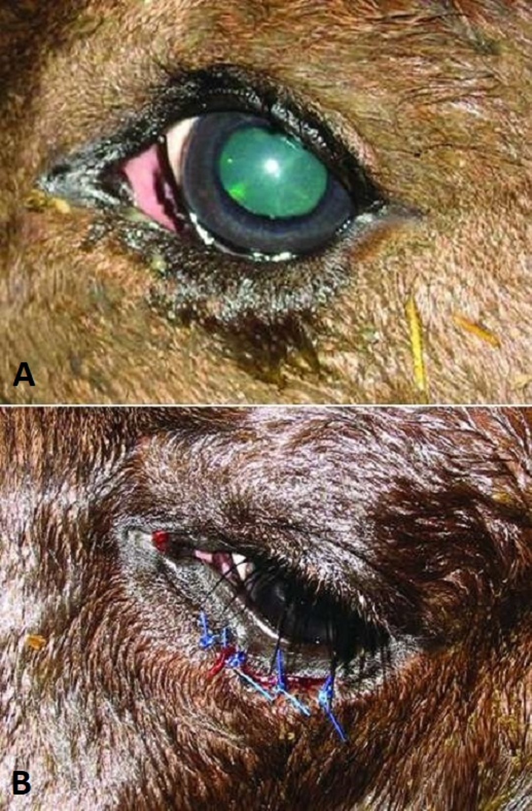
Entropion is an inversion of all or part of the eyelid margins that may involve one or both eyelids and the canthi. It is the most frequent inherited eyelid defect in many canine and ovine breeds. It may also follow cicatrix formation or severe blepharospasm due to ocular or periocular pain. Inversion of the cilia (eyelashes) or facial hairs causes further discomfort, conjunctival and corneal irritation, and if protracted, corneal scarring, pigmentation, and corneal ulceration. Early spastic entropion may be reversed if the inciting cause is quickly removed or if pain is alleviated by everting the eyelid hairs away from the globe with temporary tacking sutures in the eyelid margin Temporary eyelid-tacking sutures or surgical staples left in place for 2–3 weeks may be used to treat entropion in puppies, foals, and sheep. In young dogs, tacking sutures may need to be replaced multiple times because surgical correction provides the best result once the dog has reached its more mature head size, at about 5–6 months old. Established entropion usually requires surgical correction that, in breeds with heavy upper facial folds (such as the Chinese Shar-Pei), may need to include tacking of the dorsal facial folds to achieve optimal results.

Courtesy of K. Gelatt.
In foals and lambs, entropion is secondary to enophthalmos from mild dehydration and/or corneal ulceration. These cases should be treated with temporary eyelid-tacking sutures and treatment for corneal ulceration, if present. Primary entropion surgery should not be performed in foals or lambs, because it will result in appreciable cicatricial ectropion as the patient reaches adult size. Similarly, dogs and cats of any age with spastic entropion are best treated with temporary eyelid-tacking sutures until the reason for their spastic entropion (corneal ulceration or other keratitis) is resolved. After their keratitis is resolved, entropion surgery may no longer be indicated.
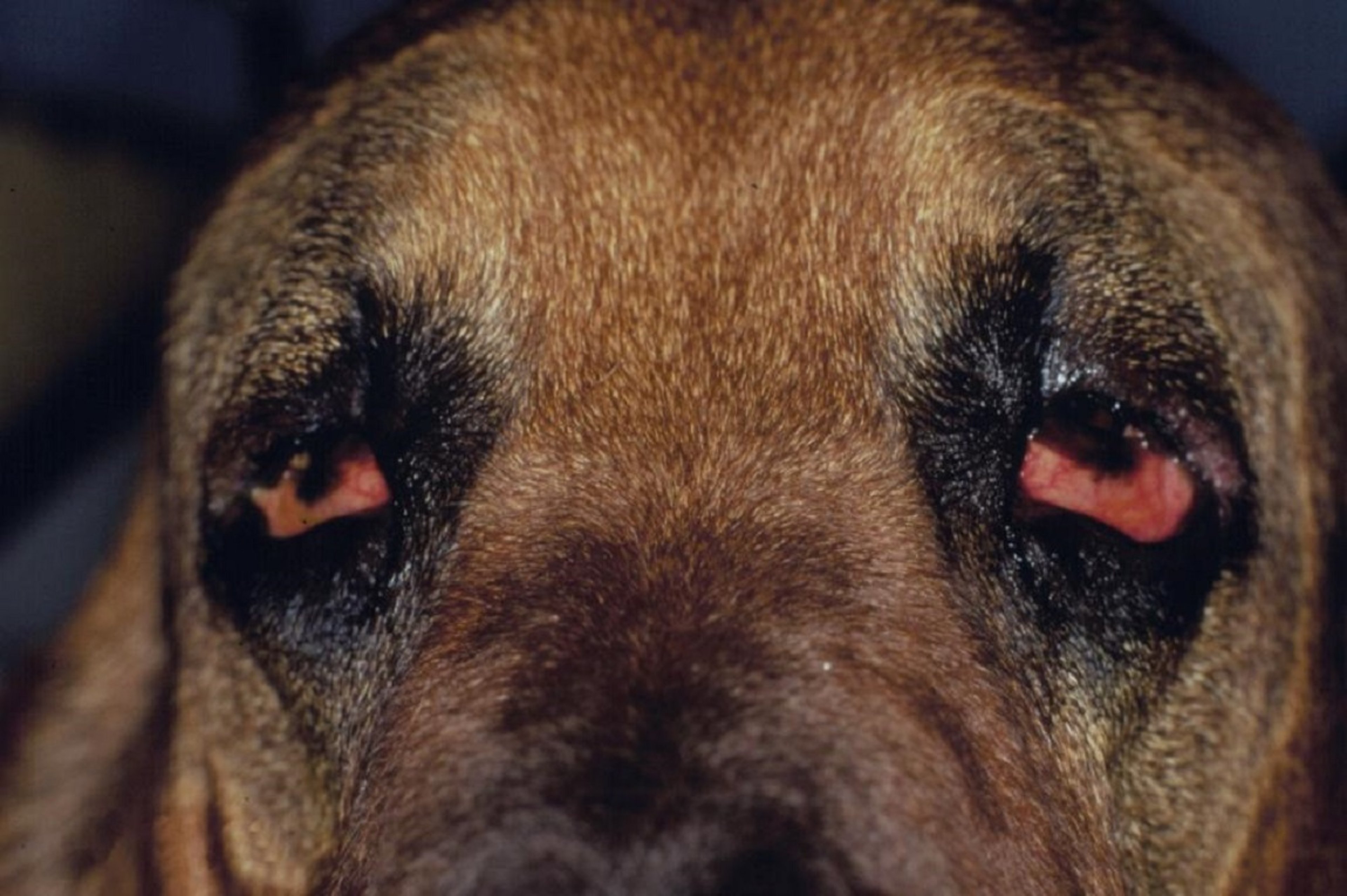
Ectropion is a slack, everted eyelid margin, usually with a large palpebral fissure and elongated eyelids. It occurs mainly in larger dog breeds and is usually associated with excessively long eyelids. It is a common bilateral conformational abnormality in a number of dog breeds, including the Bloodhound, Bullmastiff, Great Dane, Newfoundland, Saint Bernard, and several Spaniel breeds. Contracture of the eyelid due to scarring or facial nerve paralysis may produce unilateral ectropion in any species. Conjunctival exposure to environmental irritants and secondary bacterial infection can result in chronic or recurrent conjunctivitis. Topical anti-inflammatory medications may temporarily control mild or intermittent inflammation. Mild cases may also be controlled by repeated, periodic lavage with eyewash and chronic lubrication with topical gel or ointment. Generally, surgical eyelid-shortening procedures are indicated to provide more normal eyelid function and more appropriate protection of the globe.

Courtesy of K. Gelatt.
Lagophthalmos is an inability to fully close the eyelids and protect the cornea from drying and from trauma. It may result from extremely shallow orbits (in brachycephalic breeds), exophthalmos due to a space-occupying orbital lesion, or facial nerve paralysis. Corneal scarring, pigmentation, and ulceration usually result. Unless an inciting cause can be corrected, the treatment is frequent topical lubrication with gels or ointments and surgical blepharoplasty to decrease the eyelid opening. Depending on the breed or the cause, shortening or closure of the medial or lateral canthus may be indicated. Excessive nasal skin folds and facial hair may aggravate the damage caused by lagophthalmos.
Abnormalities of the cilia include extra eyelashes (distichiasis) or misdirected eyelashes on the eyelid margin. Anomalies of the cilia are common and probably inherited in some dog breeds, but they are rare in other animal species. Epiphora, corneal vascularization, corneal ulceration and scarring may result.
Distichiasis is a condition in which additional eyelashes grow out of the meibomian gland opening along the eyelid margin. It may be difficult to detect because the extra eyelashes are often the same color as the normal eyelid hair. In some instances, the extra eyelashes are very fine and result in neither clinical signs nor damage. Distichiasis is not treated unless corneal and/or conjunctival disease results. The presence of numerous and/or rigid extra eyelashes more often causes clinical signs and requires treatment. Successful removal of extra eyelashes requires destruction of the follicular base of the eyelids while not injuring the eyelid margin. The most popular method is cryotherapy applied at the base of the meibomian gland on the palpebral conjunctiva. The eyelid margin may lose its pigment after cryotherapy; however, pigment is usually regained in the subsequent months. Inadequate cryotherapy can result in the recurrence of distichiasis.
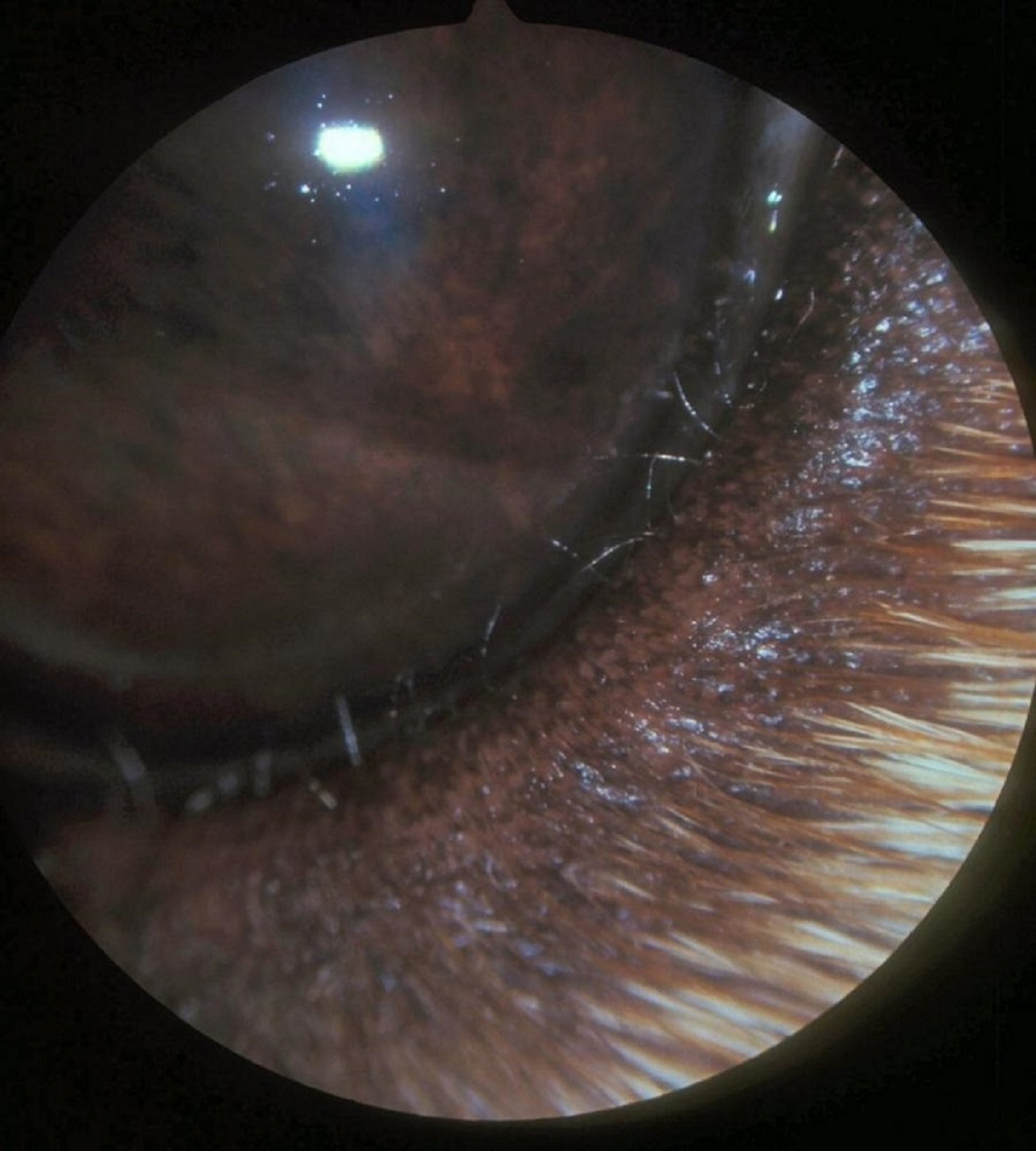
Ectopic cilia also grow from the base of the meibomian gland, but they protrude through the conjunctival surface and irritate the cornea. Ectopic cilia protruding through the palpebral conjunctiva can cause profound pain and corneal ulceration. Conjunctival swelling can make the identification of ectopic cilia challenging. If the extra cilia cause corneal damage, the cilia should be treated by excision, cautery, or cryotherapy.
Inflammation
Blepharitis (inflammation of the eyelids) can result from eyelid disease, extension of a generalized dermatitis, local glandular infections, or irritants such as plant oils or solar exposure. The eyelids can be the original site of involvement for agents that lead to a generalized dermatitis. In pyogranulomatous blepharitis, both the upper and lower eyelids can be involved. Dermatophytes (all species), Demodex canis (dogs), D cati or D gatoi (cats), and bacteria such as staphylococci often are involved. The mucocutaneous junction of the skin and conjunctiva can be the site of lesions of immune-mediated diseases such as pemphigus. Skin scrapings, cultures, and/or biopsies may be required for an accurate diagnosis as well as to direct specific treatment. Localized glandular infections may be acute or chronic (stye [glands of Zeis and Moll glands] and chalazion [meibomian glands]). Meibomianitis can affect the upper and lower eyelids; Staphylococcus and Streptococcus spp are usually involved.

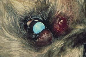
Two large abscesses in the lower and nasal eyelid margins of a Miniature Poodle's eye.
Two large abscesses in the lower and nasal eyelid margins of a Miniature Poodle's eye.
Courtesy of K. Gelatt.

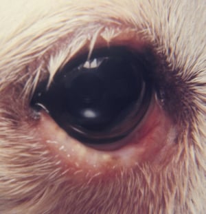
Multiple small abscesses are visible at the openings of the meibomian glands on the lower eyelid margin.
Multiple small abscesses are visible at the openings of the meibomian glands on the lower eyelid margin.
Courtesy of K. Gelatt.

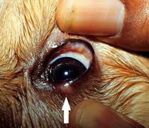
Multiple small abscesses of the meibomian glands of the upper eyelid. A small lesion on the lower eyelid margin (arrow) demonstrates what these small abscesses look like from the skin surface.
Multiple small abscesses of the meibomian glands of the upper eyelid. A small lesion on the lower eyelid margin (arrow)
Courtesy of Dr. Ralph Hamor.
Two large abscesses in the lower and nasal eyelid margins of a Miniature Poodle's eye.
Two large abscesses in the lower and nasal eyelid margins of a Miniature Poodle's eye.
Courtesy of K. Gelatt.
Multiple small abscesses are visible at the openings of the meibomian glands on the lower eyelid margin.
Multiple small abscesses are visible at the openings of the meibomian glands on the lower eyelid margin.
Courtesy of K. Gelatt.
Multiple small abscesses of the meibomian glands of the upper eyelid. A small lesion on the lower eyelid margin (arrow) demonstrates what these small abscesses look like from the skin surface.
Multiple small abscesses of the meibomian glands of the upper eyelid. A small lesion on the lower eyelid margin (arrow)
Courtesy of Dr. Ralph Hamor.
For blepharitis, systemic treatment (antimicrobial and anti-inflammatory) is often indicated in addition to topical antimicrobial and anti-inflammatory treatment. In more severe or chronic cases, this treatment may need to be continued for weeks. Supportive care of hot packing and frequent cleansing is indicated in acute cases. Non-ophthalmic topical preparations should be avoided, to prevent corneal and conjunctival contact and possible irritation. Immune-mediated causes of blepharitis may require longterm treatment to control the clinical signs.


