


Optic nerve diseases are often difficult to detect and diagnose. Of the optic nerve diseases, inflammation of the optic nerve (optic neuritis) is most frequent.

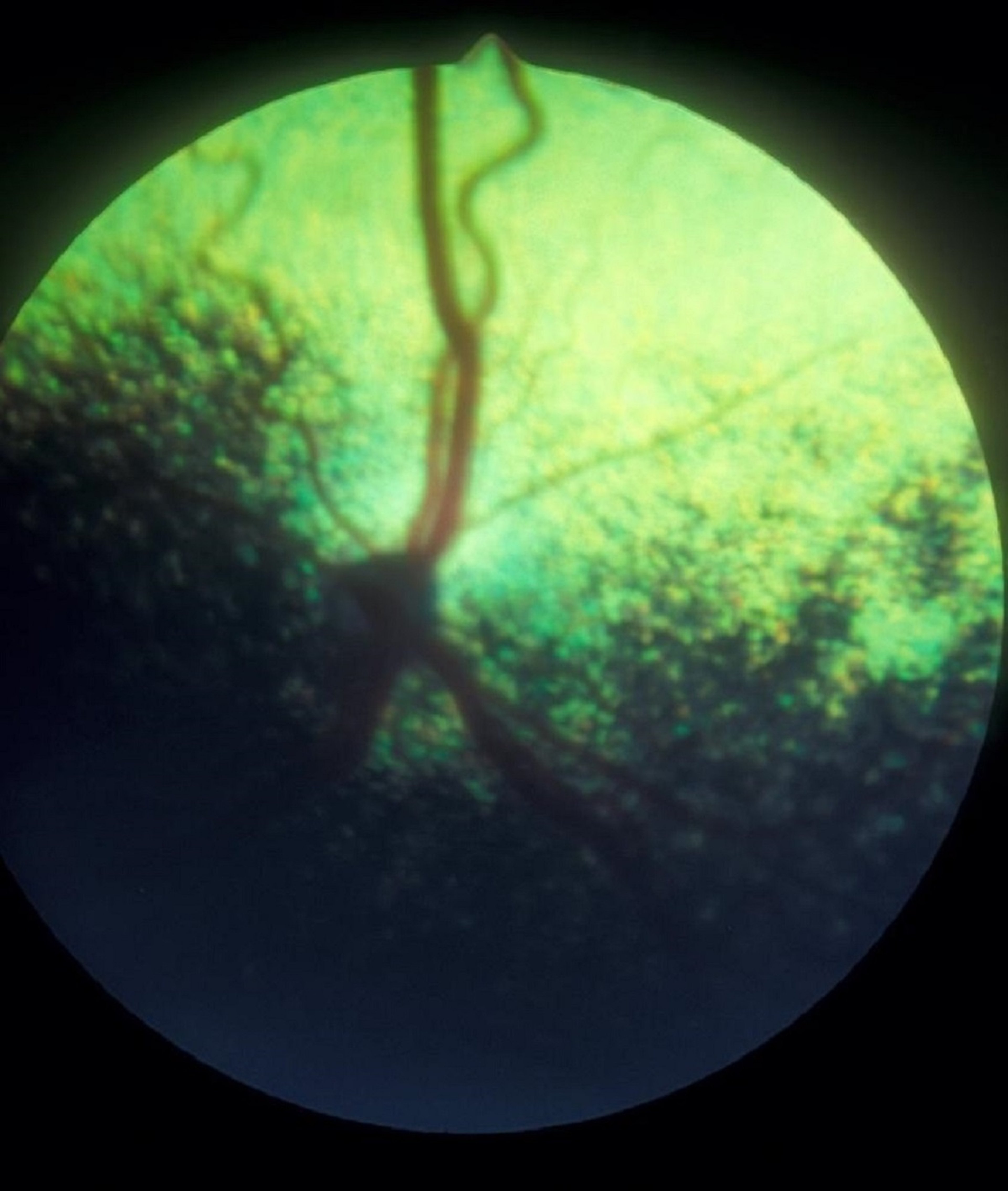
Optic nerve hypoplasia in the eye of a dog. Note the small, dark optic disc.
Courtesy of K Gelatt.
Optic nerve hypoplasia may be inherited in Miniature Poodles; in kittens and calves, it may result from in utero infections with feline panleukopenia and bovine viral diarrhea, respectively. In calves, the cause may be maternal avitaminosis A. The condition may be unilateral or bilateral, and it can occur with or without other ocular anomalies. The number of retinal blood vessels varies and is often normal. The visual deficit depends on the amount of hypoplasia present. Bilateral involvement is manifested as blindness in the neonate. Unilateral involvement is often an incidental finding later in life or becomes manifest if the other eye acquires a blinding disease.

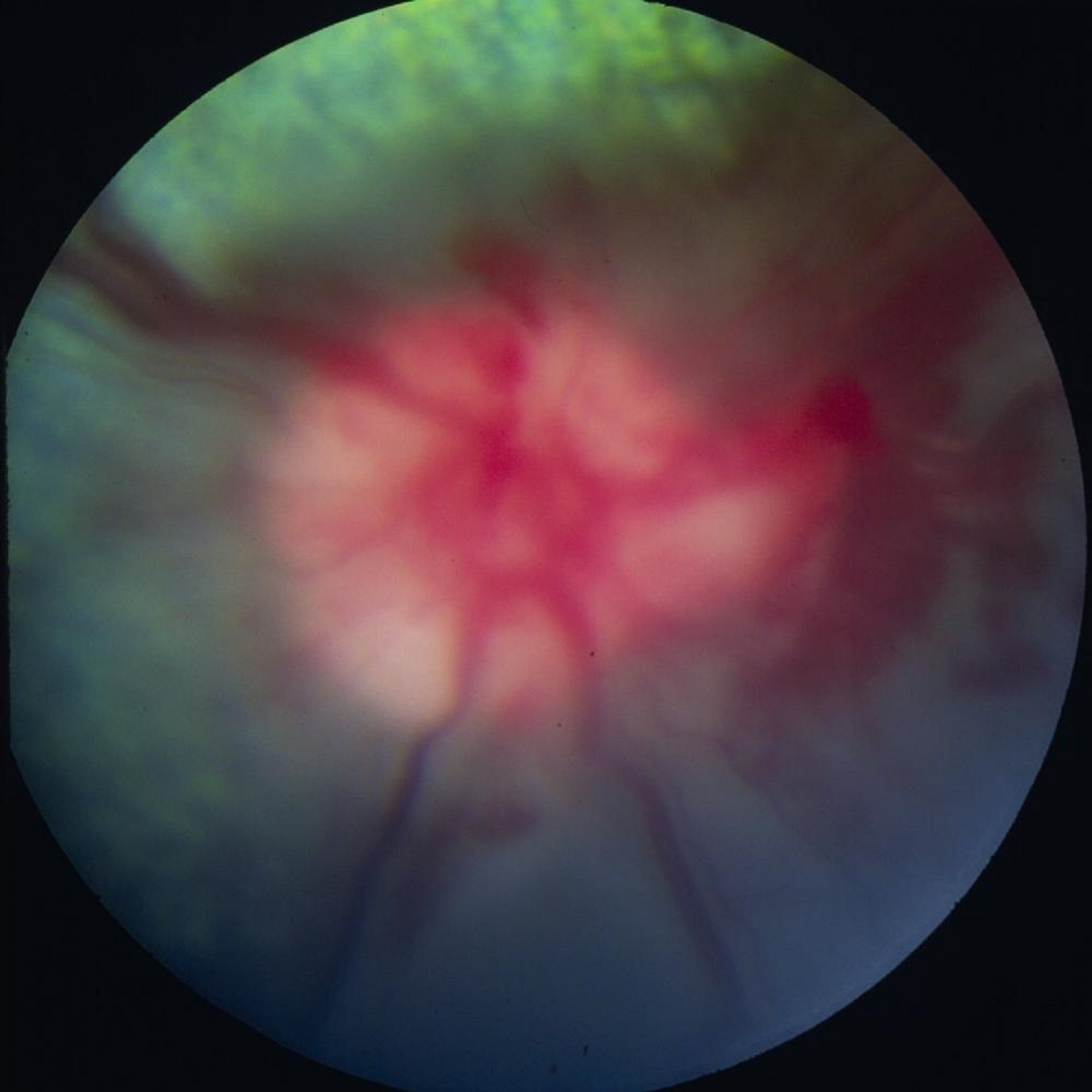
Optic neuritis in the eye of a dog with meningoencephalitis. Note the raised optic disc, with blurred margins, and the areas of hemorrhage.
Courtesy of K. Gelatt.
Bilateral optic neuritis produces acute blindness and dilated and fixed pupils. If the optic disc is affected, it is raised and edematous, with blurred margins and peripapillary hemorrhages, venous congestion, and often inflammatory cells in the adjacent retina and vitreous. If only the retrobulbar optic nerve is affected, the optic disc may appear normal on ophthalmoscopy. Causes of optic neuritis may vary by the species affected and include viral, mycotic, protozoan, and parasitic infections; trauma; reticulosis; toxins; and other causes. The most common cause is meningoencephalitis; physical examination, MRI and CSF tap are usually necessary to establish the possible diagnoses. Specific treatments are directed toward the cause, and systemic corticosteroids are important to decrease inflammation of and damage to the optic nerve.

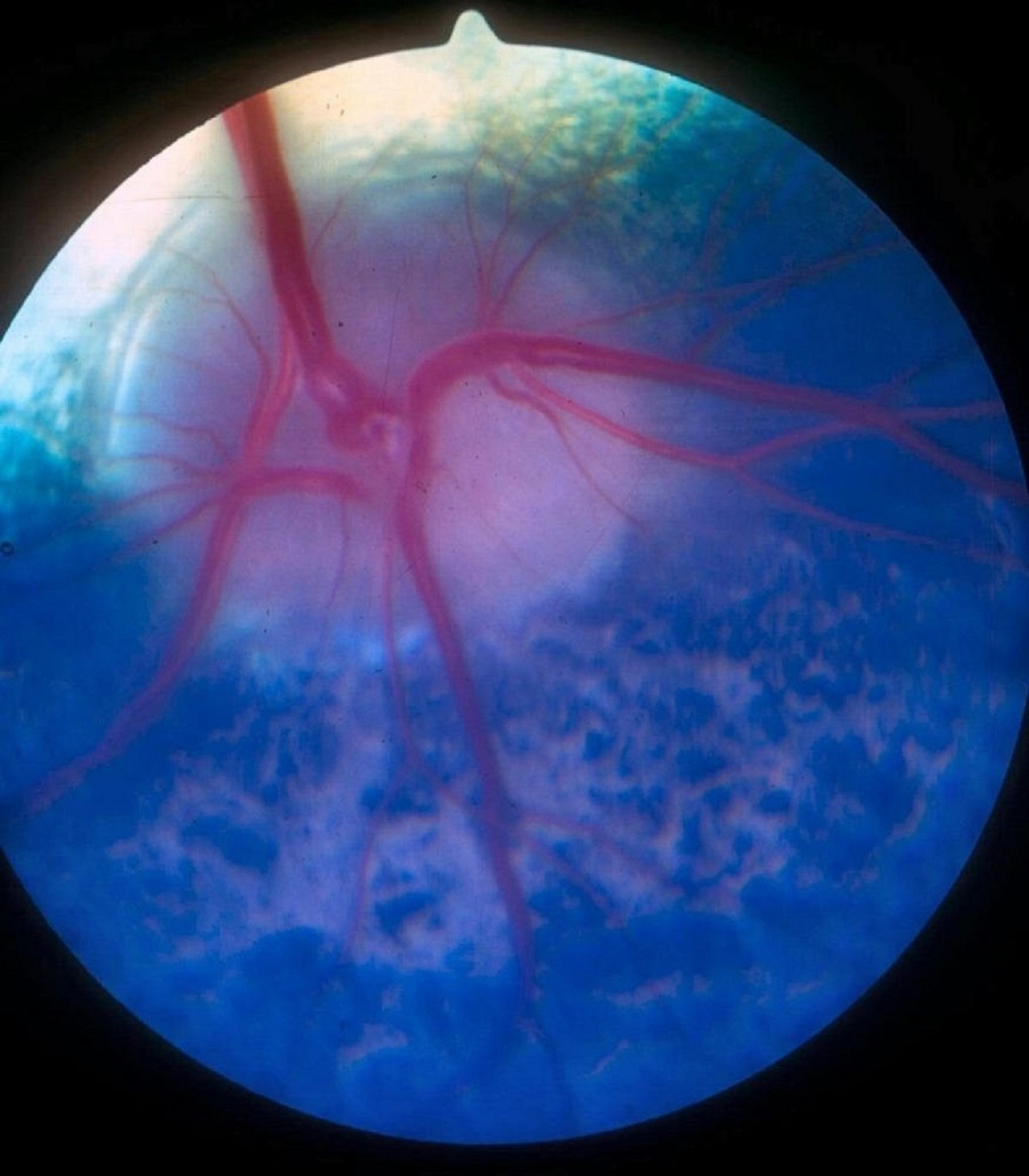
Papilledema resulting from increased CSF pressure in a growing heifer. The optic disc margins appear fuzzy and the surrounding fundus has a translucent appearance.
Courtesy of K. Gelatt.
Papilledema, swelling of the optic disc, is associated with increased intracranial pressure. The optic disc appears raised above the surface of the adjacent retina, and venous congestion is present. Vision and the light pupillary reflexes are not usually affected, unless optic atrophy develops. In young calves with avitaminosis or hypovitaminosis A, optic nerve hypoplasia occurs. In young growing cattle, papilledema secondary to increased CSF pressure occurs.

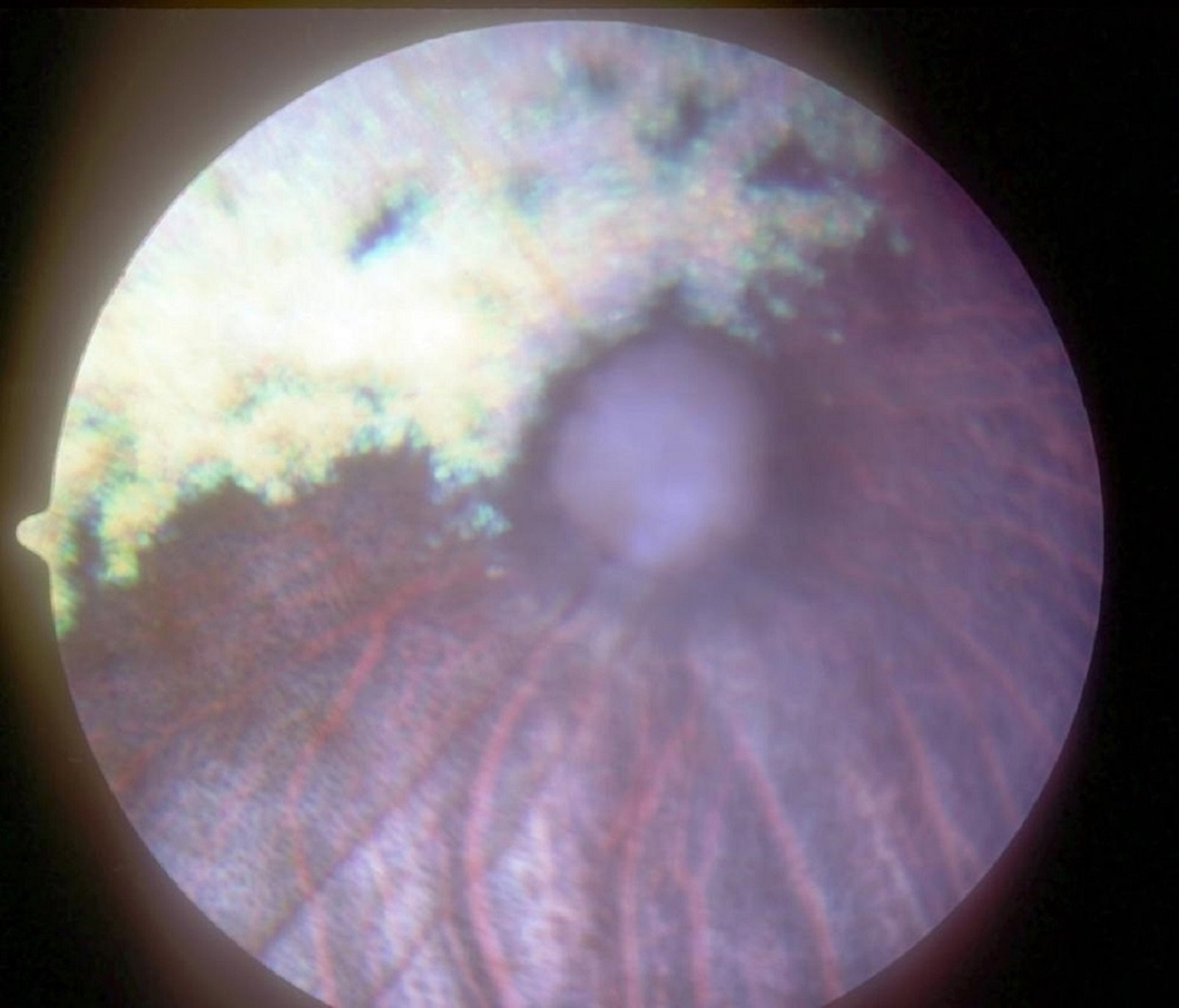
Advanced retinal atrophy and optic nerve atrophy in the eye of a dog. The optic disc is atrophied and gray. Almost no retinal vessels are visible radiating from the optic disc. The tapetum is diffusely hyperreflective because of diffuse retinal atrophy.
Courtesy of K. Gelatt.
Optic nerve atrophy may develop after glaucoma, trauma, advanced retinal degeneration, prolonged ocular hypotension, or inflammation. The optic disc appears depressed, as well as smaller and darker than normal, with a marked decrease in the retinal vasculature. Both direct pupillary reflex and vision are absent. There is no treatment.


