


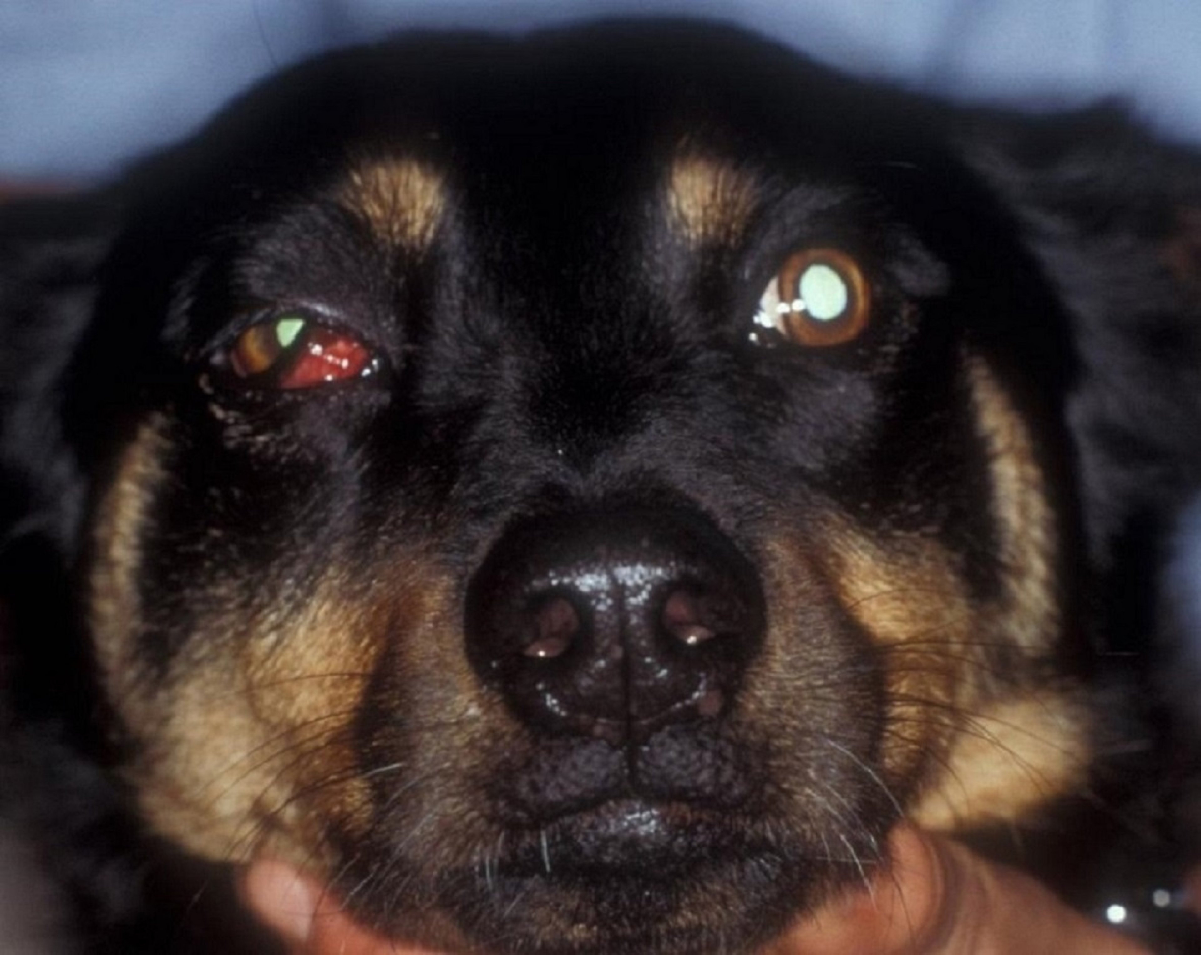
The dog in this photograph is showing acute onset of orbital cellulitis. Exophthalmos and elevation of the third eyelid are evident.
Courtesy of K. Gelatt.
Clinical signs of orbital cellulitis are acute pain on opening the mouth, eyelid swelling, unilateral prolapse of the nictitating membrane, forward displacement of the globe (exophthalmos), and conjunctivitis. Keratitis may develop from lagophthalmos. Clinical signs are directly related to the increased size of the orbital tissues. In dogs, expansion of the orbital tissues pushes the globe and nictitating membrane forward and impairs the blink reflex. The infection is often distributed through the orbital tissues. Inflammation of any of the numerous tissues of the orbit or periorbital tissues may cause orbital cellulitis. Tooth root abscesses may erode through bone to cause orbital cellulitis. Foreign bodies (eg, migrating grass awns) and zygomatic sialadenitis are additional causes. Orbital hemorrhage and neoplasia may mimic cellulitis, except there is usually no pain on opening the mouth. In acute cases, systemic broad-spectrum antimicrobials and systemic anti-inflammatory agents are generally curative, but if swelling behind the last molar is present, drainage of this area is indicated. Warm compresses and topical lubricants to protect the cornea are also indicated. Relapses may occur, and orbital imaging (CT, radiography, and/or ultrasonography) of the adjacent teeth, sinuses, and nasal cavity are recommended. If exposure keratitis is present, a temporary tarsorrhaphy is indicated to protect the globe while treatment is implemented.


