


Hemorrhagic septicemia, a disease of ungulates (primarily water buffalo and cattle in tropical regions), is caused by Pasteurella multocida serotypes B and E. Subclinical, pneumonic, and disseminated (endotoxemic) forms occur, often with peracute distress and high mortality rates. Antimicrobial treatment very early in disease (at the onset of fever) can decrease mortality rate; however, vaccination is a more effective method of controlling outbreaks. Definitive diagnosis requires the isolation of P multocida serotypes B:2 or E:2 or detection by molecular capsular and somatic serotype assays.
Hemorrhagic septicemia (HS) is an acute, highly fatal form of pasteurellosis that affects mainly water buffalo (Bubalus bubalis), cattle (Bos taurus and Bos indicus), and bison (Bison bison and Bison bonasus). It is a WOAH-listed animal disease and is considered the most economically important bacterial disease of water buffalo and cattle in tropical areas of Asia, especially India (particularly Karnataka, Andhra Pradesh, and other southern states), Pakistan, and other parts of southeast Asia (Bangladesh, Myanmar, Cambodia, Thailand, Malaysia, Laos, Vietnam, Indonesia, and the Philippines), where water buffalo populations are high. Disease has the greatest impact on small-scale livestock production operations, where husbandry and preventive practices are commonly poor and free-range management is common.
HS is also an important disease in Africa (Central, Southern, and northeastern) and portions of Central Asia and the Middle East (Afghanistan, Iraq, Syria, Iran, and countries in the Caucasus region), with infrequent occurrences in pigs and cattle in southern Europe (southern Spain, Germany, Austria).
Confirmed outbreaks of HS in the Americas have been reported, beginning in the 1920s, in bison, cattle, and wild cervids; most recently, isolated cases were reported in young calves in 2020 and 2022 (1).
Natural HS occurs infrequently in pigs, sheep, and goats and has been reported in camels, elephants, rhinoceros, horses, donkeys, yaks, and various species of deer and other wild ruminants.
Etiology and Pathogenesis of Hemorrhagic Septicemia
Hemorrhagic septicemia, as defined by WOAH, is caused by Pasteurella multocida serotypes B:2 and E:2 under the Carter-Heddleston classification system, which correspond to serotypes 6:B and 6:E in the Namioka-Carter classification system. Various other P multocida serotypes can cause HS-like disease in cattle and water buffalo, which must be differentiated from HS as defined.
Serotype B:2 has been identified in most areas where the disease is endemic (Asia), whereas serotype E:2 has been found only in Africa. Molecular characterization of the recent cases of serotype B HS in calves in North America demonstrated that those strains are different than the endemic strains circulating globally. Novel serotype B strains could be responsible for sporadic reemergence of cases of HS in North America and Europe.
Infection begins in the tonsil and adjacent nasopharyngeal tissues. Subsequently, bacteremia leads to dissemination and rapid growth of bacteria in various locations, tissue injury, a host cytokine response, and release of lipopolysaccharides that results in rapidly progressing endotoxemia.
Septicemic pasteurellosis that is clinically similar to HS is caused by a wide variety of other P multocida serotypes (predominately serotype A). Septicemic pasteurellosis has been reported most commonly in wild ruminants and occurs in geographic areas where HS is not endemic, such as the US, Europe, the UK, Australia, and Canada.
P multocida is notably involved in several important animal diseases, including fowl cholera in birds, atrophic rhinitis and pasteurellosis in pigs, and snuffles and other diseases in rabbits. Although P multocida can cause zoonotic infections in humans, these infections are generally associated with animal bites, scratches, or exposure to infected animal secretions. The HS syndrome does not naturally transmit from infected livestock to humans and therefore is not itself considered zoonotic.
Epidemiology of Hemorrhagic Septicemia
Up to 5% of healthy water buffalo and cattle are estimated to be colonized by small numbers of P multocida serotype B:2 or E:2, which can be shed during periods of stress (2). Common stressors associated with outbreaks of hemorrhagic septicemia include high temperature and humidity, concurrent infection (blood parasites or foot and mouth disease), poor nutrition, or work stress.
Although outbreaks can occur at any time, disease is most prevalent during the rainy season. Increased outbreaks associated with high rainfall are most likely due to the multiple stressors present during this time and the moist conditions, which prolong the survival time of the organism in the environment.
Infection occurs by contact with infected oral or nasal secretions from either healthy carrier animals or animals with clinical signs of disease, or by ingestion of contaminated feed or water.
In endemic areas, HS affects older calves and young adults, and morbidity and mortality rates are variable. In nonendemic areas, epizootics can occur with high morbidity and mortality rates that can reach 100%. Water buffalo tend to have higher morbidity rates with more severe disease than cattle.
Recovery can stimulate acquired immunity to homologous and often heterologous strains of P multocida, and some affected animals become healthy carriers that can be a source of infection for future outbreaks.
Clinical Findings of Hemorrhagic Septicemia
In cases of hemorrhagic septicemia in water buffalo and cattle, clinical signs can appear 1–3 days after infection, and death can occur within 8–24 hours after the first signs develop. In peracute cases of hemorrhagic septicemia that result in death within 8–24 hours, animals often have fever, hypersalivation, nasal discharge, and labored respiration; however, because of the short duration of disease, these clinical signs can easily be overlooked.
Acute disease generally persists for up to 3 days, with a longer duration of up to 5 days occurring less frequently. In acute cases, HS is characterized by the following clinical signs:
fever of 40–41.1°C (104–106°F)
lethargy or restlessness and reluctance to move
hypersalivation
lacrimation
nasal discharge that begins as serous and progresses to mucopurulent
Subcutaneous swelling in the pharyngeal region that extends to the ventral neck and brisket (and sometimes the forelimbs), progressive respiratory distress, cyanosis, terminal recumbency, and sometimes abdominal pain with diarrhea also occur. See image.

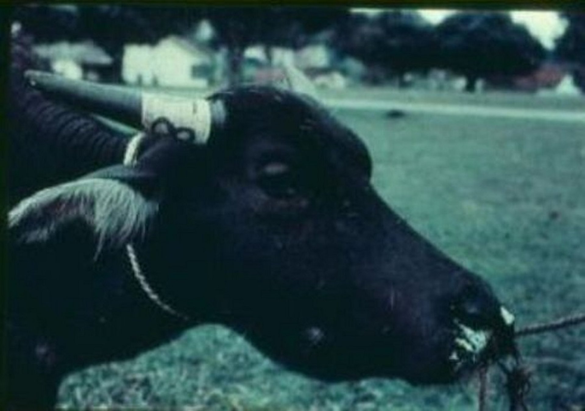
Hemorrhagic septicemia in a water buffalo, showing edematous swelling of the neck and mandibular region.
Courtesy of George Joseph.
Lesions
The characteristic lesion of hemorrhagic septicemia is swelling of the subcutis and muscle of the submandibular region, neck, and brisket by clear to blood-tinged edema fluid. Serous to serofibrinous fluid might also be present in the thorax, pericardium, and abdominal cavity. Widespread congestion typically occurs, with petechiae and ecchymoses in tissues and on serosal surfaces, particularly in the respiratory, GI, and urinary systems. Hemorrhages are often most prominent in the pharyngeal and cervical lymph nodes. Pulmonary congestion and edema (sometimes with interstitial pneumonia) and gastroenteritis occur in some cases.
Diagnosis of Hemorrhagic Septicemia
History, clinical signs, and characteristic lesions
Culture, blood tests, and molecular analysis
Clinical diagnosis of hemorrhagic septicemia in endemic areas is based on history, lapses in vaccination, environmental conditions, and the characteristic clinical signs and lesions of disease. Although typical outbreaks of HS are readily recognized in endemic regions, serotype A septicemic pasteurellosis, acute salmonellosis, anthrax, and noninfectious toxicoses should also be considered as differential diagnoses.
Sporadic cases are more difficult to diagnose clinically and could be confused with blackleg, lightning strike, or snakebite.
A definitive diagnosis of HS is based on cultural isolation and biochemical characterization of P multocida serotype B:2 or E:2 (or other less common serotypes recognized by WOAH as causing HS) from the blood and tissues of a patient with typical clinical signs. Various other P multocida serotypes can cause HS-like disease in cattle and water buffalo, which must be differentiated from HS.
Various serotyping methods have been used, such as indirect hemagglutination, coagglutination, counter immunoelectrophoresis, and immunodiffusion tests. Molecular techniques, including pulsed-field gel electrophoresis, Southern blots, loop-mediated isothermal amplification, PCR-based protocols, and whole genome sequence analysis, have been used to differentiate between capsular and somatic serotypes and strains. PCR array techniques are most feasible for use in endemic areas and can be used with various samples, including blood, tissues, or bacteria from broth or plate cultures.
Treatment and Prevention of Hemorrhagic Septicemia
Antimicrobial therapy
Vaccination
For treatment of hemorrhagic septicemia in water buffalo and cattle, antimicrobials are effective if administered very early in the disease, typically before or at the onset of pyrexia. However, because HS progresses rapidly, treatment is often unsuccessful. During outbreaks, any patient with a fever should be treated with antimicrobials as soon as possible to quickly obtain therapeutic systemic antimicrobial concentrations (3).
Pearls & Pitfalls
|
Consistently efficacious antimicrobials include fluoroquinolones (such as enrofloxacin and ciprofloxacin), cephalosporins (such as ceftiofur), florfenicol, and tetracyclines. Various sulfonamides, penicillin, gentamicin, kanamycin, tilmicosin, and chloramphenicol have been used effectively in some HS outbreaks (3). However, plasmid- and chromosomal-mediated multidrug resistance seems to be increasing for some strains of P multocida, and resistance to tetracyclines and penicillin has been reported for serotype B:2.
Killed vaccines are most commonly used for prevention and include bacterins, alum-precipitated and aluminum hydroxide gel vaccines, and oil-adjuvant vaccines prepared according to WOAH standards. In animals > 3 years old, an initial two doses, 1–3 months apart, are recommended, followed by booster vaccinations once or twice yearly.
The oil-adjuvant vaccine provides protection for 9–12 months and is administered annually. It is most effective when administered 1 month before the monsoon or rainy season. Although it provides the strongest immunity, it is unpopular in the field because of its viscosity and the difficulty of administration. Oil-based vaccines combined with polysorbate 80 or saponin have also been used in attempts to increase the ease of administration or immune protection.
The commonly used alum-precipitated and aluminum hydroxide gel vaccines have shorter durations of immunity (approximately 4–5 months with variable protective efficacy), and twice-yearly booster vaccinations are recommended.
For maximal effectiveness, vaccines should be made from the strains of P multocida circulating in the regions of intended use.
Maternal immunity can interfere with vaccine efficacy in calves.
Various attenuated or modified live vaccines and subunit vaccines made from either purified or recombinant bacterial components have been investigated experimentally, in some cases with good efficacy. However, these products are not generally commercially available.
Failure of vaccination to control HS is common because of inadequate vaccine coverage, which is mostly due to variable management systems that make annual vaccination difficult. In many cases, vaccination is practiced only in the face of an outbreak using a bacterin (broth or gel) vaccine for rapid coverage in the affected area, followed by ring vaccination using the oil-adjuvant vaccine.
WOAH has recommended the establishment of disease-free zones (absence of disease for 3 years), so that vaccination can be focused on areas where infection is common.
Zoonotic Risk of Hemorrhagic Septicemia
The P multocida serotypes that cause hemorrhagic septicemia have not been recovered from infections in humans. However, because many serotypes of P multocida have the potential to infect humans, appropriate precautions should be taken when dealing with suspected cases of HS or HS-like disease.
Key Points
Hemorrhagic septicemia is predominantly a disease of water buffalo and cattle in tropical areas and is caused by Pasteurella multocida serotypes B:2 and E:2.
HS can occur in subclinical, pneumonic, or disseminated forms; endotoxemia associated with disseminated (bacteremic) disease is often associated with high mortality rates.
Vaccines are available with variable efficacy in field outbreaks and for disease management.
Appropriate antimicrobial treatment soon after the onset of disease can decrease mortality rate.
For More Information
Haemorrhagic septicaemia. Manual of Diagnostic Tests and Vaccines for Terrestrial Animals (Terrestrial Manual) of the World Organisation for Animal Health 2024.
Haemorrhagic septicemia. World Organisation for Animal Health. Terrestrial Animal Health Code. 2024.
Committee of Foreign and Emerging Diseases. Hemorrhagic septicemia. In: Brown C, Torres A, eds. Foreign Animal Diseases. US Animal Health Association; 2008:297-300.
Spickler AR. Hemorrhagic septicemia. Center for Food Security and Public Health. Iowa State University. 2019.
Almoheer R, Wahid MEA, Zakaria HA, et al. Spatial, temporal, and demographic patterns in the prevalence of hemorrhagic septicaemia in 41 countries in 2005-2019: a systematic analysis with special focus on the potential development of a new-generation vaccine. Vaccines. 2022;10(2):315.
Annas S, Zamri-Saad M, Jesse FFA, Zunita Z. Comparative clinicopathological changes in buffalo and cattle following infection by Pasteurella multocida B:2. Microb Pathog. 2015;88:94-102.
References
Maddock KJ, Stenger BLS, Pecoraro HL, Roberts JC, Loy JD, Webb BT. Hemorrhagic septicemia in the United States: molecular characterization of isolates and comparison to a global collection. J Vet Diagn Invest. 2025;37(5):771-778. doi:10.1177/10406387251342528
Farahmand-Azar S, Tukmechi A, Ownagh A. Molecular typing and phylogenetic analysis of Pasteurella multocida isolates from cattle and buffaloes of West Azerbaijan, Iran. BMC Vet Res. 2025;22(1):76. doi:10.1186/s12917-025-05185-x
Lestari TD, Khairullah AR, Damayanti R, et al. Hemorrhagic septicemia: a major threat to livestock health. Open Vet J. 2025;15(2):519-532. doi:10.5455/OVJ.2025.v15.i2.3



