

Vesicular stomatitis is a viral disease of primarily horses and cattle, transmitted mainly by biting flies and midges. The disease results in characteristic vesicular lesions that can occur on the muzzle, lips, tongue, ears, sheath, udder, ventral abdomen, and/or coronary bands. The disease is generally self-limiting, with no specific treatment, but some patients may require supportive care. Vesicular stomatitis is a reportable disease in most countries, including the US, with diagnostic confirmation by complement fixation test, PCR assay, and/or virus isolation at an approved reference laboratory.
Vesicular stomatitis is a viral disease caused by two distinct serotypes of vesicular stomatitis virus—New Jersey and Indiana. Vesiculation, ulceration, and erosion of the oral and nasal mucosa and epithelial surface of the tongue, coronary bands, and teats are typically observed in clinical cases, along with crusting lesions of the muzzle, ventral abdomen, ears, and sheath. Clinical signs of vesicular stomatitis have been observed primarily in horses and cattle, and occasionally in pigs, sheep, goats, llamas, and alpacas. Serologic evidence of exposure has been found in many species, including cervids, nonhuman primates, rodents, birds, dogs, antelope, and bats.
In ruminants and swine, the disease is clinically indistinguishable from foot-and-mouth disease, swine vesicular disease, and vesicular exanthema of swine, which must be ruled out first on diagnostic testing. Vesicular stomatitis virus is zoonotic and can be transmitted to humans through direct contact with lesioned animals, so personal protective measures should be used when handling infected animals.
Etiology of Vesicular Stomatitis in Large Animals
Vesicular stomatitis viruses are members of the family Rhabdoviridae and genus Vesiculovirus. They are the prototypes of the Vesiculovirus genus. The viruses are bullet shaped and generally 180 nm long and 75 nm wide. The genomic structure is a single strand of negative-sense RNA composed of five genes (N, P, M, G, and L, representing the nucleocapsid protein, phosphoprotein, matrix protein, glycoprotein, and the large protein, which is a component of the viral RNA polymerase).
Although there are many members of the Vesiculovirus genus, the New Jersey and Indiana serotypes are of particular interest in the Western hemisphere. These two viruses are similar in size and morphology but generate distinct neutralizing antibodies in infected animals. Both were isolated in outbreaks during the 2000s in the US.
Epidemiology and Transmission of Vesicular Stomatitis in Large Animals
Vesicular stomatitis occurs sporadically in the US. Historically, outbreaks occurred in all regions of the country. However, since the 1980s, they have been limited mostly to western states and occur seasonally, usually May through October, with some outbreaks overwintering and continuing into a subsequent year or years. Outbreaks occurred in the US in 1995, 1997 to 1998, 2004 to 2006, 2009, 2010, 2012, 2014 to 2015, 2019 to 2020, and 2023. The largest outbreak in the past decade occurred in 2019 and resulted in 1,144 affected premises in 8 states.
Vesicular stomatitis viruses are endemic in South America, Central America, and parts of Mexico but have not been observed naturally outside the Western hemisphere.
The virus can be transmitted through direct contact with infected animals with clinical signs of disease (lesions) or by biting insects. Black flies (Simuliidae), sand flies (Lutzomyia), and biting midges (Culicoides spp) have been shown to be competent vectors; however, other insects may act as mechanical vectors as well.
Exposure to insects that carry the virus is often associated with nearby moving water sources, such as creeks and rivers or irrigation of pastures, or standing water sources, such as ponds.
Experimental studies have shown that feeding of infected insects on mucosal surfaces and nonhaired areas of the body were associated with development of lesions at those sites, whereas feeding of infected insects on haired areas of the body resulted in antibody production without development of lesions.
The prevalence of clinically affected animals in a herd is generally low (10%–20%), but seroprevalence within the herd may approach 100%. Viremia has not been detected in species that exhibit clinical signs of vesicular stomatitis, although experimental studies have shown transmission of virus, presumably via lymphatics, between cofeeding black flies on cattle.
Virus is routinely isolated from active lesions in affected animals, and these lesions serve as a source of virus transmission by direct contact and contamination of shared feed and water stations.
Many vertebrate species have serologic evidence of exposure; however, no definitive reservoir or amplifying host of vesicular stomatitis viruses has been identified.
Clinical Findings of Vesicular Stomatitis in Large Animals
The incubation period for vesicular stomatitis is 2–8 days and is typically followed by a fever. By the time animals develop other clinical signs and are examined, however, they are rarely febrile.
Ptyalism is often the first clinical sign of disease. Vesicles in the oral cavity are rarely observed in naturally occurring cases because of rupture soon after formation; therefore, ulcers are the most common lesion observed during initial examination. Ulcers and erosions of the oral mucosa, sloughing of the epithelium of the tongue, and lesions at the mucocutaneous junctions of the lips are commonly present in cattle and horses (see ).

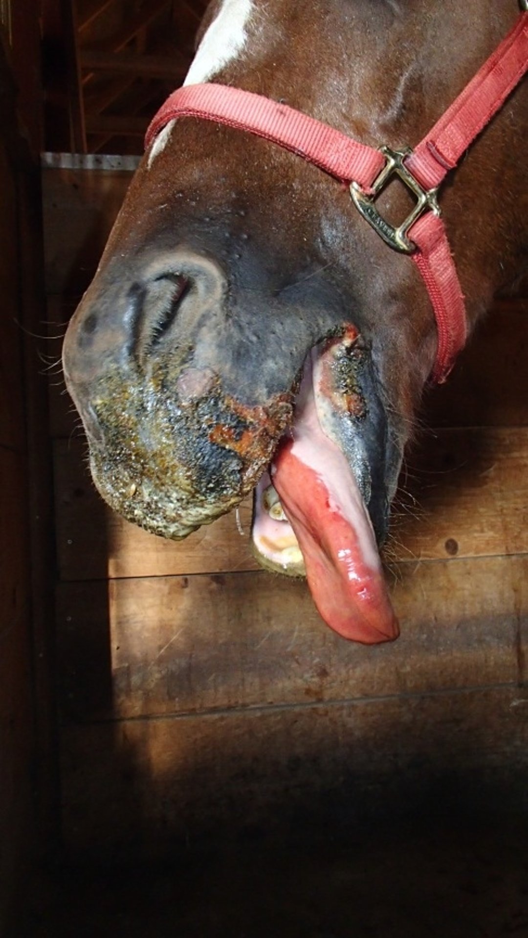
Ruptured vesicular lesions and sloughed epithelium on lips, muzzle, and tongue of a horse, caused by vesicular stomatitis virus.
Courtesy of Dr. Jason Lombard.
Ulcers and erosions on the teats are not uncommon in cattle and may result in secondary cases of mastitis in dairy cows. Coronitis with erosions at the coronary band occurs in some cattle, horses, and pigs, with subsequent development of lameness. Crusting lesions of the muzzle, ventral abdomen, ears, sheath, and udder of horses are typical during outbreaks in the western US. Loss of appetite due to oral lesions and lameness due to foot lesions are normally of short duration.
The disease is generally self-limiting and resolves completely within 10–14 days. Virus-neutralizing antibodies against either serotype persist and have been documented for 10–12 years after an outbreak in individual horses that had previous clinical signs of vesicular stomatitis; nonetheless, reinfection can occur after a second exposure.
Diagnosis of Vesicular Stomatitis in Large Animals
Clinical signs with serologic or antigen detection
In most areas, including the US, vesicular stomatitis is a reportable disease. Samples for diagnostic purposes are generally taken by a foreign animal disease diagnostician or other regulatory veterinarian and are tested by officially designated government laboratories.
Diagnosis is based on the presence of typical clinical signs and either antibody detection through serologic tests, viral detection through isolation, or detection of viral genetic material by molecular techniques. Samples for viral isolation or molecular detection may include vesicular fluid, epithelial tags from lesions, or swabs of lesions. Vesicular stomatitis viruses are easily propagated in cell culture.
Three commonly used serologic tests are competitive ELISA, virus neutralization, and complement fixation. Whereas competitive ELISA (cELISA) and virus neutralization titers persist for many years after an outbreak, complement fixation titers are relatively short-lived and provide the best serologic evidence of recent infection. Antigen detection assays to confirm diagnosis include PCR tests and virus isolation.
Of primary concern in diagnosis is differentiation of vesicular stomatitis from clinically indistinguishable but much more devastating viral diseases, including foot-and-mouth disease in ruminants and swine, swine vesicular disease, and vesicular exanthema of swine. Horses are not susceptible to foot-and-mouth disease. Both noninfectious and infectious causes of oral lesions must be considered.
Treatment, Control, and Prevention of Vesicular Stomatitis in Large Animals
Supportive care
Vector mitigation
Isolation of lesioned animals
Vesicular stomatitis is self-limiting, with no specific treatment other than supportive care. Reluctance to eat due to oral pain can be mitigated by providing softened feeds. Cleansing lesions with mild antiseptics may help avoid secondary bacterial infections. Aged patients or those with underlying medical conditions may require administration of IV fluids if oral lesions result in a reluctance to drink.
Management factors suggested to decrease risk of exposure to the virus include the following:
limiting time on pasture during insect season
providing shelters or barns during insect feeding times
implementing other procedures that decrease animal contact with insects, such as application of insecticides
If animals need to be kept on pasture during outbreaks of vesicular stomatitis, then keeping them pastured away from moving surface water (such as streams, irrigation canals, and rivers) may decrease the risk of exposure to vectors carrying vesicular stomatitis virus.
Affected animals should be isolated, and movement of other animals from the affected premises restricted. Vesicular stomatitis is a reportable disease in most areas, including the US, so state and federal animal health officials must be notified when it is suspected. In the US, affected premises are placed under state quarantine for a period of at least 14 days from the onset of lesions in the last affected animal. Commercially produced vaccines are not available in the US; however, vaccines are available in some Latin American countries.
Veterinarians act as a part of the surveillance network as they examine animals involved in shows, exhibitions, races, and interstate or international movement before writing a health certificate (ie, certificate of veterinary inspection). When veterinarians observe suspect cases of vesicular stomatitis, they should report to both their state and federal animal health officials.
Reporting suspected disease will prompt a regulatory investigation. Lesion swab and serum samples from suspected animals are submitted for testing to approved veterinary diagnostic laboratories. During outbreak years, data regarding laboratory-confirmed cases of vesicular stomatitis, along with the number of premises with cases, are posted on the Animal and Plant Health Inspection Service of the USDA website.
Zoonotic Risk of Vesicular Stomatitis in Large Animals
The vesicular stomatitis viruses are zoonotic and may cause self-limiting influenza-like disease (headache, fever, myalgia, and weakness) lasting 3–5 days in people working in close contact with the virus (eg, laboratory exposure, direct contact with lesions in infected animals). Rarely, humans can develop vesicles on the buccal and pharyngeal mucosa, lips, and nose. More severe signs, including encephalitis, are rare. Personal protective equipment should be used when handling lesioned animals to avoid contact with the virus present in the lesions.
Key Points
Vesicular stomatitis is a viral disease of primarily horses and cattle, transmitted by biting insect vectors and direct contact with lesioned animals.
The disease produces vesicular lesions on the muzzle, lips, tongue, udder, sheath, ears, and/or coronary bands, which is clinically indistinguishable from foot-and-mouth disease, swine vesicular disease, and vesicular exanthema of swine. Diagnostic testing at an approved regulatory laboratory must be conducted for definitive diagnosis.
Vesicular stomatitis is reportable to state and federal animal health officials in the US, and affected premises will be quarantined to limit disease spread.
Vesicular stomatitis is potentially zoonotic, and personal protective equipment should be used when handling lesioned animals.
For More Information


