

Photosensitivity is a skin condition that occurs when photoactive substances interact with ultraviolet (UV) light to produce free radical and reactive oxygen species locally, resulting in skin damage. Lesions are most common on unpigmented skin with frequent UV exposure. Photosensitization may be due to primary intoxication with a photosensitizing agent or to the accumulation of plant-derived photosensitizing agents secondary to altered hepatic metabolism. Thus, hepatic function should always be evaluated in patients presenting with suspected photosensitization. Affected patients are photophobic and develop skin bullae, ulcers, and necrosis. Diagnosis is based on signalment and clinical signs, along with measurement of porphyrins in blood, urine, and, in some cases, feces. Treatment consists primarily of supportive and palliative care.
Photosensitization occurs when skin (especially areas exposed to light and lacking substantial protective hair, wool, or pigmentation) becomes more susceptible to ultraviolet (UV) light because of the presence of photodynamic agents. Unlike sunburn and photodermatitis, photosensitization does not result in pathological skin changes in the absence of a photodynamic agent.
In photosensitization, unstable, high-energy molecules are formed when photons react with a photodynamic agent. These high-energy molecules initiate reactions with substrate molecules of the skin, leading to the release of free radicals that, in turn, result in increased permeability of cell and lysosomal membranes.
Damage to a cell's outer membrane enables leakage of cellular potassium and cytoplasmic extrusion. Damage to lysosomal membranes within a cell releases lytic enzymes into the cell, potentially leading to skin ulceration, necrosis, and edema (see ).

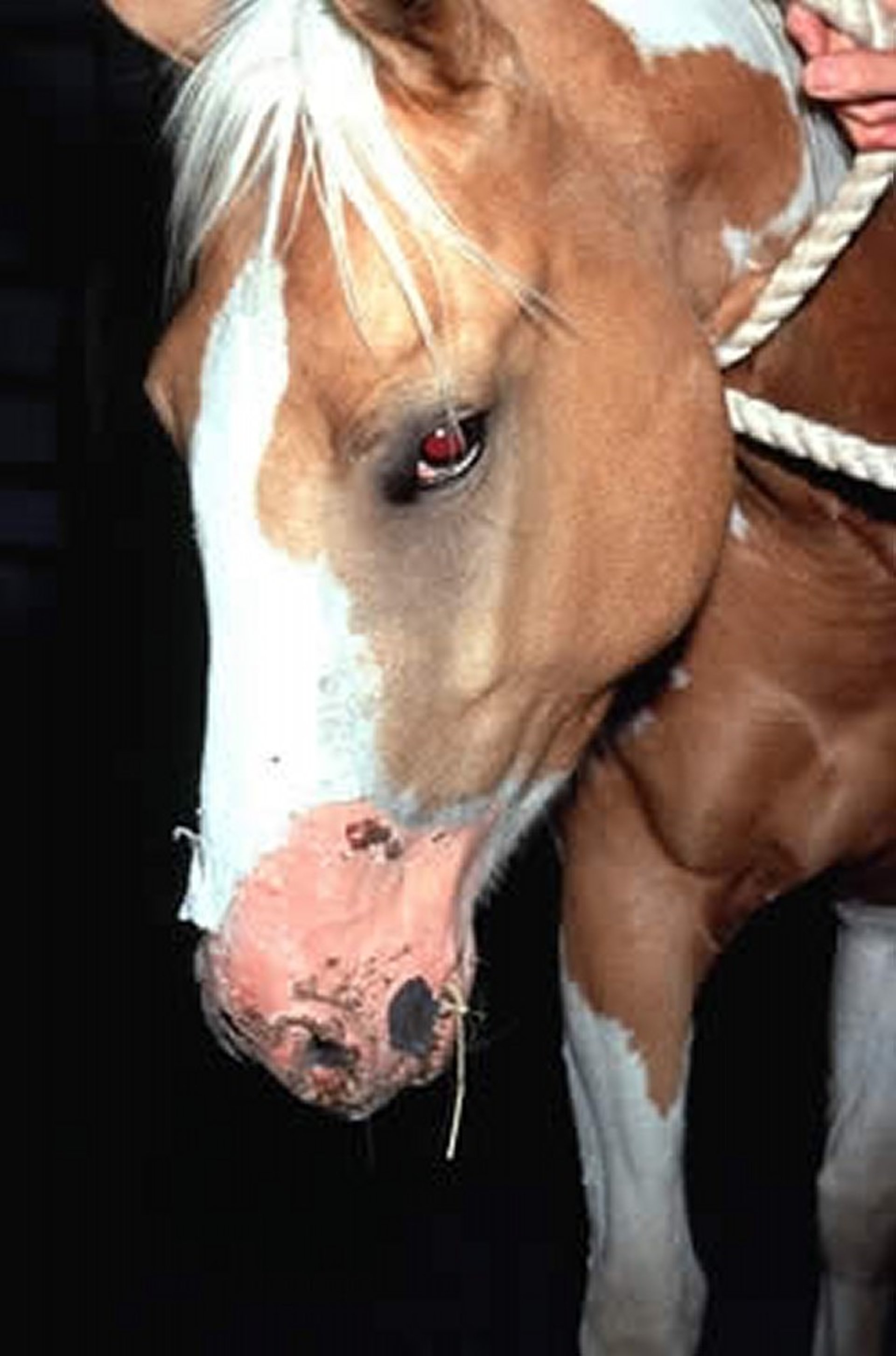
The cracking and peeling of the skin on this adult horse are the result of photosensitization.
Courtesy of Dr. Stephen White.
The time interval between exposure to a photodynamic agent and the onset of clinical signs of photosensitization depends on the type of agent, its dose, and the length of time the animal is exposed to sunlight.
Photosensitization is typically classified into four categories defined by the source of the photodynamic agent:
type I: primary photosensitization
type II: aberrant pigment metabolism photosensitization
type III: secondary (hepatogenous) photosensitization
type IV: idiopathic photosensitization
A wide range of chemicals, including some that are fungal and bacterial in origin, may act as photosensitizing agents. However, most compounds that are implicated in photosensitization in animals are plant derived.
Photosensitization occurs worldwide and can affect any species, but it is most commonly observed in cattle, sheep, goats, and horses.
Primary Photosensitization (Type I)
Primary photosensitization occurs when a photodynamic agent is ingested, injected, or absorbed through the skin. The agent enters the systemic circulation in its native form, where it results in skin cell membrane damage after the animal is exposed to UV light. Examples of primary photosensitizing agents include hypericin (from Hypericum perforatum[St. John’s wort]) and fagopyrin (from Fagopyrum esculentum [buckwheat]).
Plants in the families Apiaceae (also known as Umbelliferae) and Rutaceae contain photoactive furocoumarins (psoralens), which lead to photosensitization in production animals and poultry. For example, Ammi majus (bishop's-weed) and Cymopterus watsonii (spring parsley) can produce photosensitization in cattle and sheep, respectively. Ingestion of A majus and Ammi visnaga seeds can produce severe photosensitization in poultry.
Plant species in the genera Trifolium, Medicago (clovers and alfalfa), Erodium, Polygonum, and Brassica (mustards) have been incriminated in primary photosensitization. Many other plants (eg, Cynodon dactylon [Bermuda grass]) have been suspected; however, the toxins responsible have not been identified.
In addition, exposure to coal tar derivatives, such as polycyclic aromatic hydrocarbons, tetracyclines, and some sulfonamides, can lead to primary photosensitization. Phenothiazine anthelmintics have been reported to lead to primary photosensitization in cattle, sheep, goats, and swine.
Aberrant Pigment Metabolism Photosensitization (Type II)
Type II photosensitization, due to aberrant pigment metabolism, occurs in both cattle and cats. In this syndrome, the photosensitizing porphyrin agents are endogenous pigments that arise from inherited or acquired defective functions of enzymes involved in heme synthesis. Bovine congenital erythropoietic porphyria and bovine erythropoietic protoporphyria (see Cutaneous Manifestations of Multisystemic and Metabolic Defects in Animals) are the most commonly reported diseases in this category.
Secondary (Hepatogenous) Photosensitization (Type III)
Secondary, or type III, photosensitization—also known as hepatogenous photosensitization—is by far the most common type of photosensitization observed in production animals. It results from impaired biliary excretion of phylloerythrin (a porphyrin), which is a normal by-product of chlorophyll metabolism.
Secondary photosensitization may be a sequela of any hepatocellular dysfunction or cholestasis; it is not related to phototoxin ingestion.
Phylloerythrin, but not chlorophyll, is normally absorbed into the circulation and is effectively excreted by the liver into the bile, following the same general pathway as bilirubin metabolism. Failure to excrete phylloerythrin increases its circulating concentrations. When phylloerythrin reaches the skin, it can absorb and release light energy, initiating a phototoxic reaction.
Phylloerythrin has been incriminated as the phototoxic agent in the following conditions: common bile duct occlusion; facial eczema; lupinosis; congenital photosensitization of Southdown and Corriedale sheep; and poisoning by numerous plants, including the following:
Tribulus terrestris (puncture vine)
Lippia rehmannii
Lantana camara
several Panicum spp (kleingrass, broomcorn millet, witchgrass)
Cynodon dactylon
Myoporum laetum (ngaio)
Narthecium ossifragum (bog asphodel)
Photosensitization associated with phylloerythrin accumulation can occur in animals that have liver damage associated with various toxic substances (notably, these are hepatobiliary toxins, not direct phototoxins):
pyrrolizidine alkaloids (eg, from Senecio spp, Cynoglossum spp, Heliotropium spp, and Echium spp)
cyanobacteria (Microcystis spp, Oscillatoria spp)
Nolina spp
Agave lechuguilla (lechuguilla)
Holocalyx glaziovii
Kochia scoparia (Mexican fireweed)
Tetradymia spp (horsebrush or rabbitbrush)
Brachiaria brizantha
Brassica napus
Trifolium pratense (red clover) and Trifolium hybridum (alsike clover)
Medicago sativa
Ranunculus spp
phosphorus
carbon tetrachloride
Photosensitization was been reported in a cow with cholestasis and Anaplasma centrale infection. It was hypothesized that intrahepatic cholestasis had occurred secondary to overwhelming RBC breakdown and bilirubin metabolism within the liver (1).
Idiopathic Photosensitization (Type IV)
Photosensitization in which the pathogenesis is unknown or the photodynamic agent is not identified is classified as idiopathic, or type IV.
Outbreaks of photosensitization have been reported in cattle exposed to water-damaged alfalfa hay, moldy straw, and orchard grass hay containing foxtails. These cases were suspected to be hepatogenous in origin. Ranunculus bulbosus (bulbous buttercup) has also been presumed to result in hepatogenous photosensitization.
Other plants associated with photosensitization include winter wheat (in cattle), Medicago spp), Brassicaspp, and Kochia scoparia. Many of these plants are believed to be agents of type I photosensitization. Forages such as oats, wheat, and red clover have been suspected in cases of photosensitization and may be associated with specific environmental conditions such as heavy rainfall.
Clinical Findings and Lesions of Photosensitization in Animals
Dermatological signs associated with photosensitization are similar regardless of the type of photosensitization.
Photosensitive animals are photophobic immediately when exposed to sunlight and appear agitated and uncomfortable. They may scratch or rub lightly pigmented, exposed areas of skin (eg, ears, eyelids, muzzle).
Furocoumarin toxicity has been associated with uveitis, as well as with skin lesions in affected animals.
Lesions caused by photosensitization initially appear in white-haired, nonpigmented, or hairless areas such as the nose and udder. However, high concentrations of plant-derived photoactive toxins or phylloerythrin coupled with very bright sunlight can induce lesions on pigmented skin.
Erythema associated with photosensitization develops rapidly and is soon followed by edema. If exposure to light stops at this stage, the lesions soon resolve. When exposure is prolonged, lesions may progress to include vesicle and bulla formation, serum exudation, ulceration, scab formation, and skin necrosis.
In the final stage of photosensitization, skin is sloughed. In cattle, and especially in deer, exposure of the tongue while licking may result in glossitis, characterized by ulceration and deep necrosis. Regardless of coat color, cattle may develop epiphora, corneal edema, and blindness.
Depending on what initially caused the photosensitizing agent to accumulate, other clinical signs of photosensitization may be present. For example, if the photosensitization is hepatogenous, icterus may be present.
In cases of bovine congenital erythropoietic porphyria, discoloration of dentin, bone (and other tissues), and urine often accompanies the skin lesions. In bovine erythropoietic protoporphyria, photodermatitis is the sole clinical sign observed (see Cutaneous Manifestations of Multisystemic and Metabolic Defects in Animals).
Diagnosis of Photosensitization in Animals
Presumptive diagnosis: signalment and clinical signs
Definitive diagnosis: measurement of porphyrin in blood, urine, and feces
Diagnosis of photosensitization is based on clinical signs, evidence or history of exposure to photosensitizing agents or hepatotoxins, and characteristic lesions. Photophobia in combination with erythema and edema of hairless, nonpigmented areas of skin strongly suggests the disease. The length of time between exposure to photodynamic or hepatotoxic agents and the onset of clinical signs can vary from several hours to 10 days.
Clinical signs, alterations in serum biochemical measurements (including increased activities of sorbitol dehydrogenase, gamma-glutamyl transferase, and alkaline phosphatase, as well as increased direct bilirubin concentration), and gross or histological signs of liver disease all help support a diagnosis of hepatogenous photosensitization.
A presumptive diagnosis of porphyria is based on signalment (sex, breed, age) combined with clinical signs. A definitive diagnosis can be made by measuring porphyrin concentrations in blood, feces, and urine. Porphyrin concentrations are elevated only in cases of abnormal phylloerythrin excretion.
Treatment of Photosensitization in Animals
Palliative and supportive care
The prognosis is poor for patients with hepatogenous photosensitization and porphyria; however, it is generally good for patients with primary photosensitization. The severe stress of photosensitization and extensive skin necrosis can be highly debilitating and cause death.
Photosensitization is treated primarily with palliative measures. While the condition persists, patients should be shaded fully or, preferably, housed, and they should be allowed to graze only during darkness.
Corticosteroids, administered parenterally in the early stages of photosensitization, may be helpful. Secondary skin infections and suppurations should be treated with basic wound management techniques, and fly strike should be prevented.
The skin lesions resulting from photosensitization heal remarkably well, even after extensive necrosis.
Key Points
Photosensitization is due to the production or ingestion of photodynamic agents, which lead to increased susceptibility to skin damage after exposure to sunlight.
Secondary (type III) photosensitization, due to abnormal hepatic metabolism of phylloerythrin, is the most common type observed in production animals.
For More Information
Collet MG. Photosensitisation diseases of animals: classification and a weight of evidence approach to primary causes. Toxicon X. 2019;3:100012. doi:10.1016/j.toxcx.2019.100012
Also see pet health content regarding photosensitization in dogs and in cats.
References
Anton A, Solcan G. A case study of photosensitivity associated with Anaplasma spp. infection in cattle. Animals (Basel). 2022;12(24):3568. doi:10.3390/ani12243568


