


Fatigue during high intensity exercise is determined by the limitations of anaerobic metabolism that fuels these activities. The heat produced by muscles at full exertion, as well as the ambient temperature, can result in a syndrome of exertional heat illness. Clinical signs of this type of exercise-induced heat stress include endotoxemia and cerebral edema secondary to alterations in systemic circulation. The diagnosis of exertional heat illness is based on clinical signs of heat stress and changes in mentation. Treatment is centered on external cooling, anti-inflammatory medications, and supportive care.
Energetics of Exercise and Fatigue in Animals
The contribution of aerobic or anaerobic energy pathways during exercise depends on the duration and energy demands of the event. In short, during intense exercise lasting 20–30 seconds (eg, Quarter horse races [400 m], some Greyhound races), 60% of energy demand is supplied by anaerobic sources. For intense exercise at maximal speeds for a longer duration (eg, Standardbred or Thoroughbred races [1,600–2,100 m] lasting 1–3 minutes), the energy supply is estimated to be 20%–30% anaerobic. In contrast, events lasting many hours (eg, endurance races for horses, camels, and dogs) have >90% of energy demands met by aerobic sources.
During brief, high-intensity exercise, fatigue is secondary to engagement of muscle fibers that heavily rely on anaerobic metabolism. The higher the intensity, the greater the anaerobic demand. Fatigue is the result of an increase in hydrogen ions, lactate, inorganic phosphate, ammonia, and ADP, and a decrease in ATP, phosphocreatine, and pH in active muscle cells. Clinically, fatigue is initially identified by a decrease in exercise intensity or a drop in the maximal speed.
As anaerobic metabolism increases, lactate production is directly correlated to the percentage of type IIB muscle fibers present and corresponds to the accumulation of protons in the muscle tissue. Intracellular acidosis caused by lactate accumulation has a negative feedback effect on the glycolytic enzymes required for energy production and mitochondrial respiration, resulting in a decline in ATP concentration. Lack of ATP prevents calcium recycling through the sarcoplasmic reticulum, resulting in accumulation of calcium in the sarcoplasm and slowing of the relaxation phase of muscle contraction. Acidosis also interferes with excitation-contraction coupling by interfering with calcium binding to troponin C, reducing the ability of the muscles to contract. Unfortunately, there is no correlation between muscle lactate concentration and placement in a race, or plasma lactate concentration and performance indexes.
A decrease in muscle ATP after maximal exercise has been noted in conjunction with high muscle lactate concentration. For high-intensity exercise (eg, a Thoroughbred race lasting 2 minutes), intramuscular stores of ATP can decrease 14%–50%. Depletion of ATP varies by muscle fiber type. In type I fibers, depletion is negligible, whereas in type IIB fibers, ATP loss is significant. Low levels of ATP impair optimal functioning for muscle contraction, reuptake of calcium by the sarcoplasmic reticulum, and sodium-potassium exchange. Fatigue is associated with depletion of phosphocreatine stores and accumulation of ADP and inorganic phosphate. A correlation between stride length and muscle ADP accumulation has been seen at the time of fatigue.
Muscular Fatigue in Animals
Increased ADP concentration results in accumulation of adenosine monophosphate, inosine monophosphate, allantoin, ammonia, and uric acid in horses. In treadmill studies, the decrease in muscle ATP during intense exercise is correlated with an increase in plasma uric acid concentration 30 minutes after exercise. Running time during treadmill tests is correlated with uric acid concentration after exercise. Significant but low correlations have also been found between racing performance of Standardbred pacers and uric acid concentration after a race.
Ammonia accumulation in plasma is correlated to decreased ATP levels and increased muscle lactate levels. It has been postulated that ammonia accumulation in the plasma may contribute to fatigue. However, infusion with ammonium acetate during treadmill exercise until fatigue did not significantly affect time to fatigue, suggesting plasma ammonia levels do not have a role in fatigue during intense exercise.
Similar to ATP depletion, muscle glycogen concentration decreases up to 30% after a single exercise bout and by as much as 50% with repeated bouts of intense exercise. Again, depletion varies between muscle fiber types, with greater depletion seen in type IIB muscle fibers. Glycogen depletion may play a role in fatigue, in that horses that perform repeated bouts of exercise before an anaerobic exercise session may be at increased risk of fatigue because of the slow rate of glycogen repletion in this species.
During high-intensity exercise, the normal equilibrium between release of potassium from recruited muscle and uptake by inactive muscle fibers is lost, resulting in a continual increase of extracellular potassium until the onset of fatigue. Changes in the ratio of intracellular to extracellular potassium across the sarcolemma alter the resting membrane potential and decrease sarcolemma excitability and the ability to generate an action potential. Reduced excitability contributes to reduced calcium release by the sarcoplasmic reticulum (a process that requires ATP) and a consequent reduction in the force of muscle contraction. This loss of force may relate to the idea of an inherent safety mechanism, in that plasma potassium level rapidly declines after cessation of exercise by reuptake into the now inactive muscle.
Intracellular acidosis as a result of lactate accumulation has been blamed for the decrease in efficiency or force of muscle contractions. However, in vitro research has demonstrated a protective effect of lactic acidosis or hydrogen ions in maintaining sarcolemma function and muscle force production in the face of potassium shifts within the cell and plasma associated with intense exercise.
Thermoregulation and Fatigue in Animals
Fatigue during high-intensity exercise is also influenced by environmental conditions. Intense exercise in hot conditions is associated with earlier onset of fatigue, due to increased blood flow to the skin for thermoregulation at the expense of cardiac output and oxygen delivery to the exercising muscle. Attenuation of normal increases in muscle blood flow during exercise in hot environments has been suggested as a contributor to the onset of fatigue. There is also a central effect of high temperatures, resulting from increased blood temperature at the level of the hypothalamus. Early onset of fatigue in hot conditions is thought to be a protective response to avoid heat stroke.
Exertional Heat Illness in Animals
In racing Thoroughbreds performing at maximal intensity, a form of exercise-induced heat illness has shown parallels with human syndromes of heat stress.
Etiology
The key clinical feature of exertional heat illness is the inability to effectively dissipate heat that is produced rapidly due to an abnormally high rate of endogenous heat production caused during a race. Risk factors for horses include:
environmental temperature (summer months, track location, time of day for the race)
sand track surfaces (which radiate heat)
increasing race distance
Anhydrosis should also be considered as a risk factor but has not been directly linked to exertional heat illness in clinical cases.
Pathophysiology
Clinical signs of exertional heat illness are secondary to the inability to dissipate heat produced at a high rate, resulting in a progressive rise in core body temperature. Initially as the thermoregulatory mechanisms are engaged, blood flow is shunted from the gastrointestinal system to the skin in an effort to increase heat dissipation. If core temperatures continue to rise unabated, intestinal ischemia will occur, resulting in the release of endotoxin into the circulatory system. Activation of the systemic inflammatory response, as well as the coagulation cascade, will occur and may progress to multi-organ failure or death.
Neuronal injury can also result from exertional heat illness. A reduction in blood flow to the brain occurs directly from hyperthermia, as well as secondary to hypovolemia. Hypoperfusion may lead to cerebral ischemia, edema, and alterations in the blood-brain barrier. Clinical progression of neurologic signs in horses is similar to that noted in humans with exertional heat illness. These include, progressing from mild to severe, the loss of normal reflexes, altered mental status, combativeness, collapse, seizures and coma.
Clinical Signs

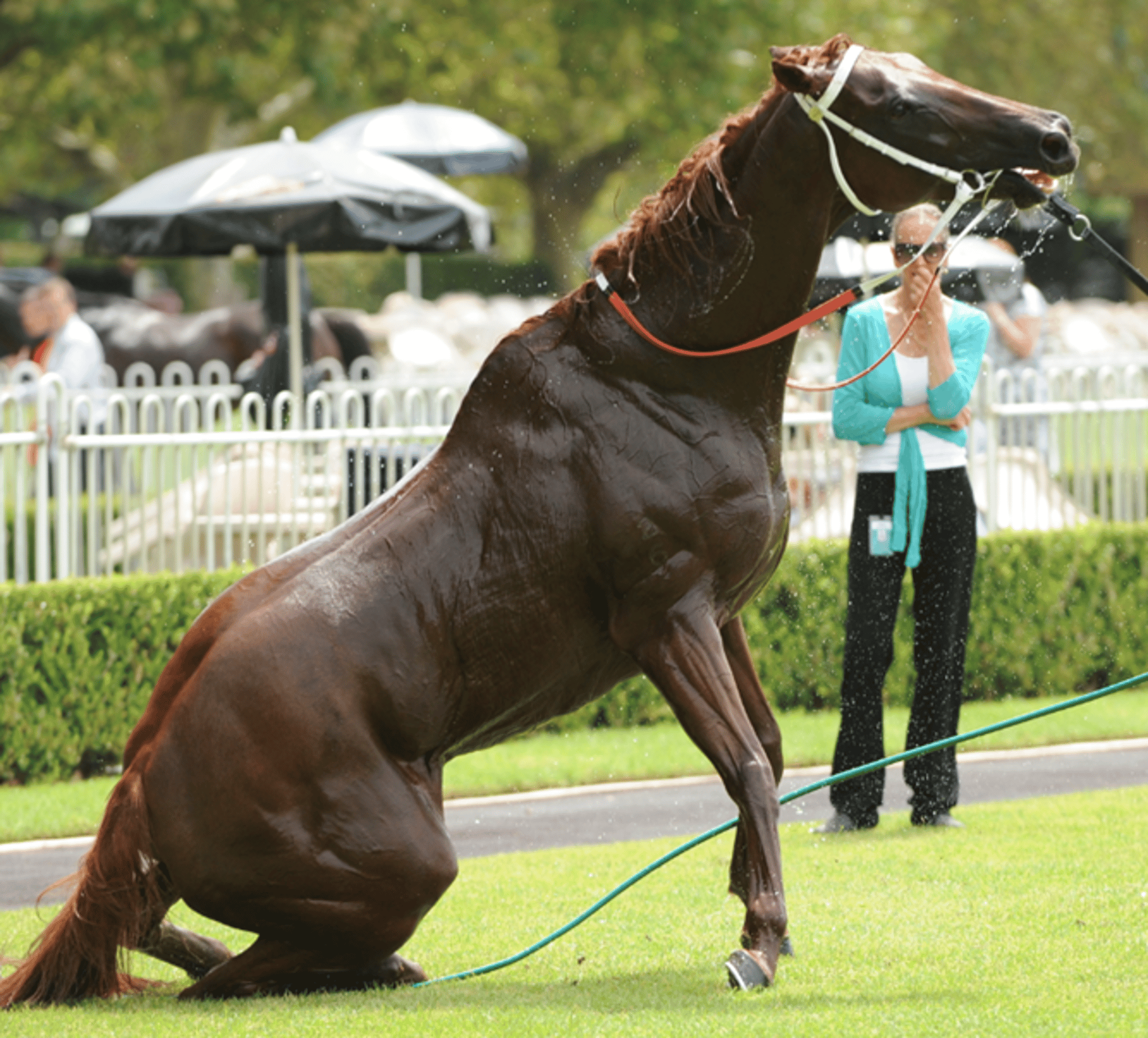
Courtesy of Dr. Meg Brownlow.


Courtesy of Dr. Meg Brownlow.
The clinical features of exertional heat illness in racehorses have been defined with four levels, progressing in increasing severity:*
Level 1: Normal mentation; showing signs of distress. Skin is hot and profusely sweating. Recovery after a race is slow, with persistent tachypnea >60-100 breaths/min and tachycardia >150 beats/min. Dilated nostrils, with exaggerated respiratory effort.
Level 2: Normal mentation; irritable and uncooperative (head shaking, kicking out). Subtle gait abnormalities.
Level 3: Altered mentation; signs of depression/disorientation. Unpredictable, and dangerous to handle. Ataxic or reluctant to move.
Level 4: Severe mentation changes; obtunded with signs of central blindness, progressing to stupor, coma, convulsions, and death. May fall, rear, or crash into objects. Clinical signs of systemic endotoxemia (hyperemic mucous membranes with a toxic line and tachycardia).
Diagnosis
Clinical signs are more important in diagnosis of exertional heat illness than determination of core temperature. Measurement with a rectal thermometer is often hampered in these horses, because fatigue may result in a lack of muscle tone in the rectum, causing a falsely low reading. In addition, heat may be stored in muscle tissues during high intensity exercise due to ineffective heat dissipation, causing rectal temperatures to lag behind the core body temperature. Finally, the horse’s behavior may preclude safely obtaining an accurate measurement.
Treatment
External and internal cooling therapies
Sedation
Restoration of the circulating fluid volume
Rapid cooling
Treatment of exertional heat illness is centered on rapid and aggressive external cooling to maximize heat transfer and reduce the core body temperature. Shade should be provided, as well as cool water to drink. Milder cases may respond to a misting fan to increase evaporative and convective cooling, but an enclosed room would be ideal to provide air conditioning to allow further control and lowering of the ambient temperature. For horses with signs of moderate to severe exertional heat illness, the skin should be hosed with cool water, which must be removed intermittently with a sweat scraper to prevent trapping of heat at the level of the skin. Internal cooling methods, such as gastric and rectal lavage, may also be instituted if it is safe to intubate. Treatment should continue until the rectal temperature is < 38.5°C (101.3°F). After this, the horse should be closely monitored for rebound hyperthermia because the circulation removes additional heat from muscle stores.
Sedation
Sedation may be needed to allow for treatment of exertional heat illness, and α2-agonists are indicated for the safety of both patient and handlers. Side effects of these medications include bradycardia, reduced cardiac output, and alterations in blood pressure, so doses should be carefully titrated and a reversal agent kept on hand.
Fluid therapy
The hydration status of the horse should be identified, because it is often variable in acute heat illness. Fluids should be titrated to the horse’s needs, including electrolyte supplementation where indicated. If cerebral edema is suspected based on changes in mentation, treatment with hypertonic saline or mannitol may be justified, with adjustments in fluid therapy to maintain normovolemia.
Emerging concepts in treatment of exertional heat illness involve attempts to modulate the exaggerated acute phase response. Although heat-induced cellular necrosis and direct endothelial damage cannot be prevented, there are possibilities for modulation of the consequences of heat injury. Mucosal injury due to gastrointestinal ischemia and reperfusion may indicate treatment with polymixin B or hyperimmune plasma. Although endogenous levels of corticosteroids are increased due to adrenal activation by cytokines, adrenal exhaustion may occur. Dexamethasone has been shown to have a positive effect on cerebral ischemia, vascular tone, and inflammation caused by heat stroke. Coagulopathies are often difficult to identify with current coagulation testing, but if noted clinically or suspected, treatment with low-molecular-weight heparin and plasma may reduce complications. Protective heat shock protein production can be stimulated by nonsteroidal medications and aspirin, which may also treat systemic inflammation and the secondary effects of endotoxemia.
Key Points
The inherent limitations of anaerobic metabolism mean that an animal’s highest attainable speed cannot be maintained for >30–40 seconds before fatigue occurs.
A clinical correlation between markers of fatigue (eg, lactate) and performance outcomes cannot be made.
The syndrome of exertional heat illness, characterized by tachypnea, tachycardia, and changes in mentation, has been observed in racing Thoroughbreds that race in hot conditions.
Treatment of exertional heat illness should focus on rapid external cooling with cool water hosing and sweat scraping, as core body temperature may continue to rise after the race.
Additional therapies may include sedation, intravenous fluids, NSAIDs, and glucocorticoids.
For More Information
*Adapted from Aust Vet J. 2016 Jul;94(7):240-7. doi: 10.1111/avj.12454. Exertional heat illness: a review of the syndrome affecting racing Thoroughbreds in hot and humid climates. Brownlow MA1, Dart AJ2, Jeffcott LB3


