


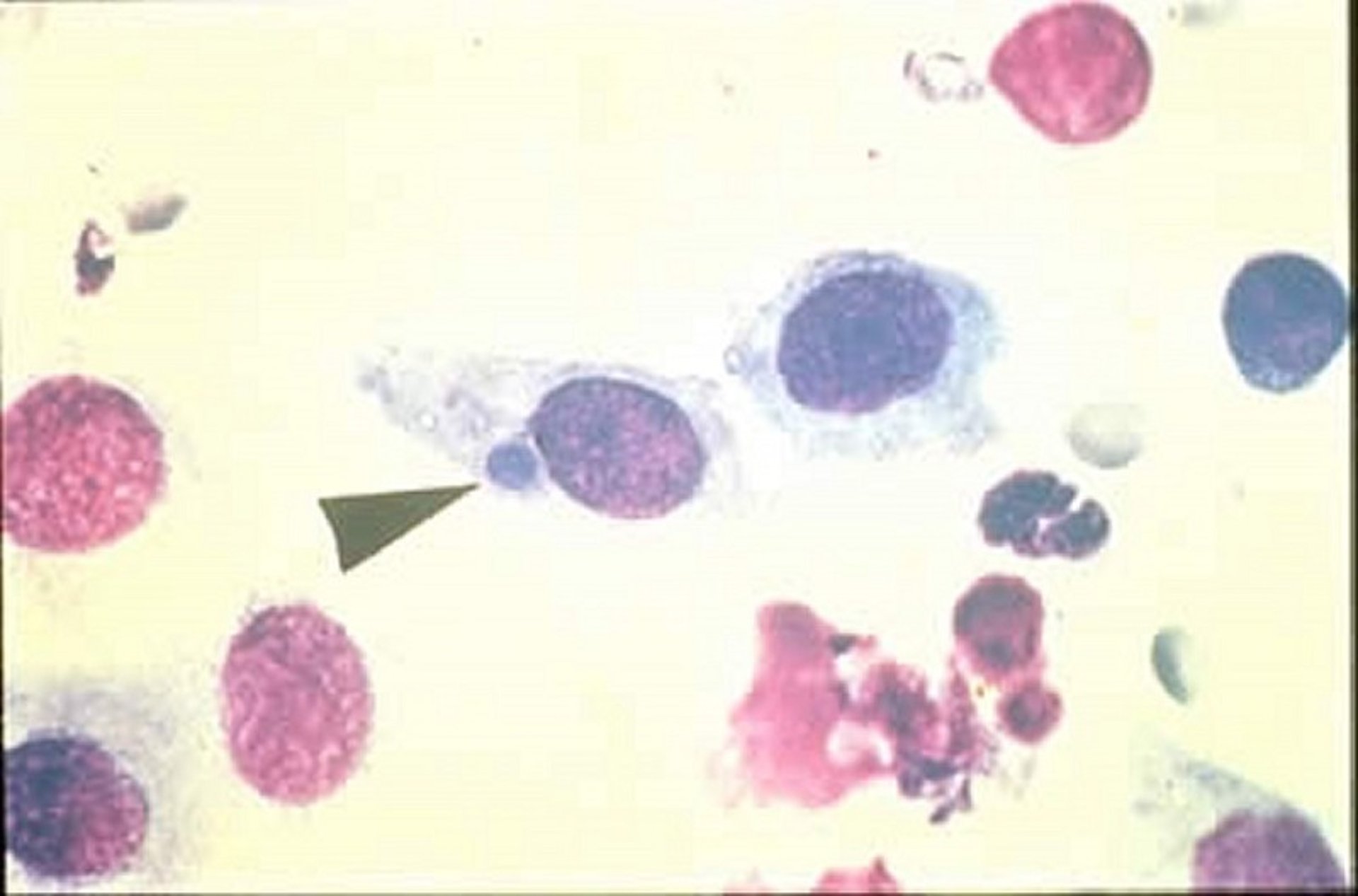
Chlamydial keratoconjunctivitis, cytological image, cat
Photomicrograph of a conjunctival cytological sample from a cat, showing inclusion bodies from chlamydial keratoconjunctivitis. The arrowhead points to a stained intracellular inclusion (adjacent to the cell's nucleus). Chlamydial intracellular inclusions typically range in size from 15 to 30 mcm.
Courtesy of Dr. Glenn A. Severin.
In these topics