


Inguinal hernia, ram, ultrasonograph
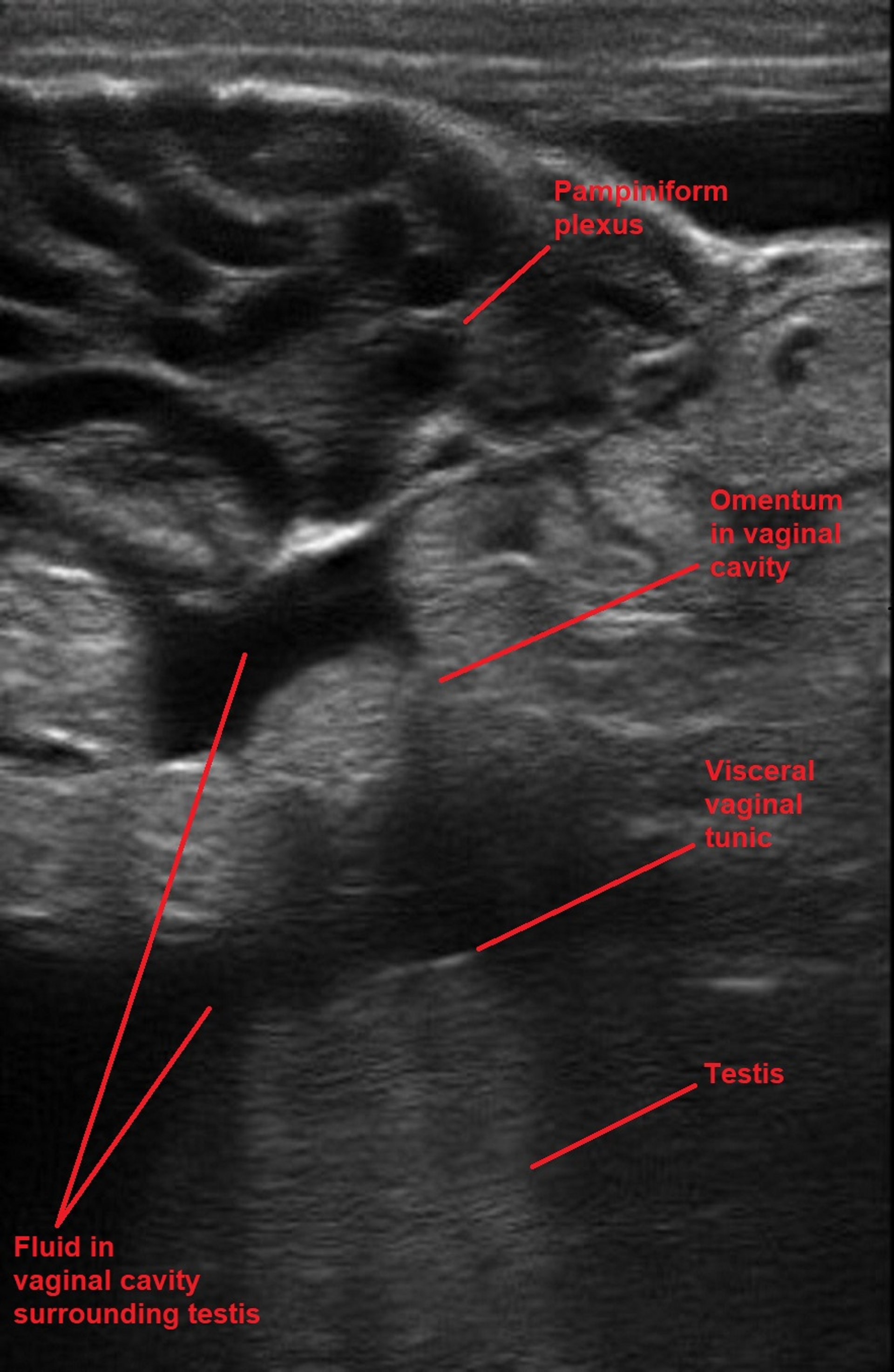
Ultrasonographic image of the pampiniform plexus and dorsal scrotum of a ram with an inguinal hernia. The vessels of the pampiniform plexus are dilated, and fluid and omentum are present within the vaginal cavity surrounding the testis. The top of the image is dorsal and the bottom is ventral. The probe is oriented longitudinally.
Courtesy of Dr. Jennifer Roberts.
In these topics